In Situ Lipid Interactions of an Anticancer Metal Complex
Edward C. Lant, Archana C. Jadhav, Annabel Sumeray, Gustavo F. Trindade, Luca Craciunescu, Andrew W. Prentice, Juliusz A. Wolny, Jaspreet S. Grewal, Robert Dallmann, Guy J. Clarkson, Ann M. Dixon, Volker Schünemann, Ian S. Gilmore, Martin J. Paterson, Maria Harkiolaki

TL;DR
A new anticancer metal complex interacts with cell lipids, showing strong effects on membranes and lipid structures in cancer cells.
Contribution
The discovery of unexpected lipid targeting and supramolecular interactions of a Rh(III) anticancer complex.
Findings
The Rh(III) complex accumulates in plasma membranes and shows intense luminescence in cancer cells.
The complex induces lipid droplet architecture remodeling and interacts strongly with glycerophosphorylcholine lipids.
Multimodal imaging and DFT modeling reveal deep penetration into lipid-rich tissues.
Abstract
An integrated multimodal imaging workflow of cryogenic super-resolution fluorescence microscopy and soft X-ray tomography, Orbitrap secondary ion mass spectrometry, and inductively coupled plasma-mass spectrometry has revealed the unexpected targeting of a half-sandwich cyclopentadienyl Rh(III) phenylazopyridine anticancer complex to cellular lipid membranes and lipid droplets. The complex accumulates in plasma membranes with a surprisingly intense switch-on luminescence in living cancer cells, drives remodeling of lipid droplet architecture, and penetrates deeply into lipid-rich tissue environments. DFT modeling shows strong supramolecular interactions between the complex and glycerophosphorylcholine lipids.
Genes, proteins, chemicals, diseases, species, mutations and cell lines named across the full text — each resolved to its canonical identifier and authoritative record.
Click any figure to enlarge with its caption.
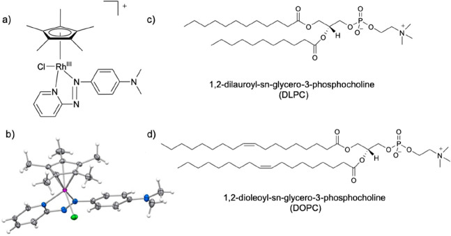 Figure 1
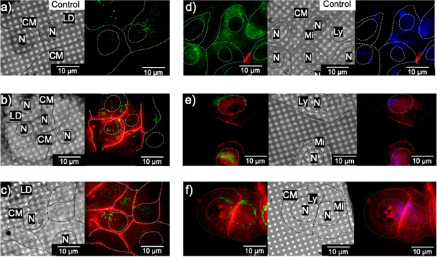 Figure 2
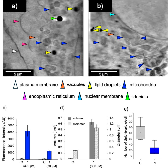 Figure 3
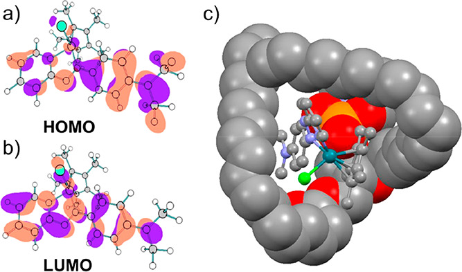 Figure 4
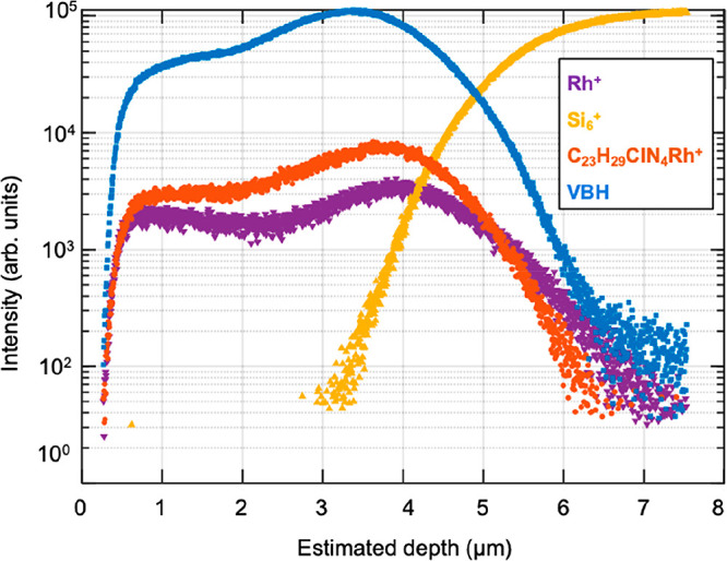 Figure 5
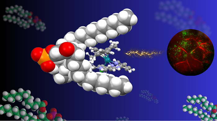 Figure 6
- —Bruker BioSpin10.13039/100009018
- —Engineering and Physical Sciences Research Council10.13039/501100000266
- —Engineering and Physical Sciences Research Council10.13039/501100000266
- —Engineering and Physical Sciences Research Council10.13039/501100000266
- —Engineering and Physical Sciences Research Council10.13039/501100000266
- —Leverhulme Trust10.13039/501100000275
- —Bundesministerium f?r Bildung und Forschung10.13039/501100002347
- —Anglo American Platinum LimitedNA
- —Future Innovation in Targeted TherapiesNA
- —UK National Measurement SystemNA
Peer Reviews
No public reviews on file for this paper yet. If you reviewed it on a platform where reviews are public (OpenReview, ICLR, NeurIPS, ICML), you can paste yours below so the community can read it here.
Videos
No videos yet. Explain this paper in a talk, walkthrough, or lecture? Add one.
Taxonomy
TopicsMetal complexes synthesis and properties · Lipid Membrane Structure and Behavior · Cancer Research and Treatment
Metal-based therapeutic agents, in particular organometallic complexes, offer promising opportunities for the discovery of novel drugs. ?,? Progress is hampered by the need to identify their molecular targets and modes of action. ?−? ? Many metallodrugs are pro-drugs which undergo ligand exchange and redox reactions in complicated biological media.? Hence mapping the chemical species which are their active pharmacophores is important. Recent advances in multimodal and integrative analytical approaches provide the means to interrogate these species and their interactions with unprecedented spatial, chemical, and mechanistic resolution. ?,? These include high resolution luminescence imaging, soft- and hard-X-ray imaging,? secondary ion mass spectrometry, ?,? and inductively coupled plasma-mass spectrometry.? Important is the emergence of methods to study intact cryo-fixed cells since chemical fixatives and other treatments can affect metal speciation.? The combination of advances in metal speciation together with genomics and proteomics (“metallomics”) offers major advances in systems pharmacology for metallodrugs. ?−? ?
Here we study a low-spin pseudo-octahedral 4d^6^ Rh(III) complex coordinated to a π-bonded η^5^-cyclopentadienyl, a N,N-chelated phenylazopyridine and monodentate chlorido ligands, [(η^5^-Cp*)Rh(Me_2_ N-phenylazopyridine)Cl]^+^, 1 (Figurea). The complex exhibits micromolar cytotoxic potency across multiple cancer cell lines, with low toxicity toward normal cells, and no cross-resistance with cisplatin.? The azo-bond is locked into the trans-E configuration (Figureb), unlike the free ligand. The parent phenylazopyridine ligand is a nonemissive azo dye and a safe anti-infective drug (Phenazopyridine).?
To elucidate the intracellular chemistry of 1 and mode of action, we implemented a multimodal correlative imaging workflow combining super-resolution cryogenic structured illumination microscopy (cryoSIM), soft X-ray tomography (SXT) on vitrified cancer cells, inductively coupled plasma-mass spectrometry (ICPMS), and Orbitrap secondary ion mass spectrometry (OrbiSIMS) for depth-resolved molecular profiling in tissue,? complemented by DFT modeling to characterize supramolecular lipid interactions.
cryoSIM enables visualization of emissive organic dyes and metal complexes in vitrified cells, while SXT allows high-resolution, label-free assessment of organelle morphology. Applying both modalities to the same biological system can afford complementary insights into the localization of drugs and structural outcomes. Human A549 lung adenocarcinoma cells were treated with complex 1, plunge-frozen, and imaged under cryogenic conditions at beamline B24 (Diamond Light Source) using cryoSIM for correlative analysis. In selected experiments, organelle-specific dyes provided additional subcellular context (Table S1). Surprisingly, complex 1 exhibited intense membrane-associated luminescence (Figurec), absent in untreated controls (Figured). Figuref shows the dose-dependent intensity of the red fluorescence from the complex localized in the cell membranes.
Strikingly, even at a high concentration of 300 μM (10 × IC_50_, 1 h treatment), 1 did not induce detectable structural damage, and treated cells retained morphology comparable to that of untreated controls (X-ray mosaics in Figure S1; cryoSXT in Movies S1 and S2; compare cryoSXT Movie S1 for control cells, with Movie S2 for treated cells). Consistent behavior was observed across independent experiments at IC_50_ concentration (30 μM), confirming a reproducible and selective interaction of 1 with lipid-rich membrane domains. While no major ultrastructural alterations were detectable during the first hour of exposure, the emergence of punctate red luminescence indicated localized accumulation of the complex, suggesting the initiation of early molecular events that may precede downstream cytotoxic responses occurring on longer time scales.
The downstream metabolic consequences were investigated by quantifying lipid droplet (LD) morphology. LDs play crucial roles in cell proliferation.? They contain mostly triacyl glycerols, and also cholesteryl esters with embedded surface proteins including lipid droplet metabolism enzymes. Designs for luminescent metal complexes as stains for LDs have been reported, e.g., in refs ? and ?.
Lipid droplet-rich fractions were isolated from A549 cells treated with complex 1, and Rh content was determined by ICPMS (details in Section S2.15, Figure S2). Accumulation of Rh was ca. 3-fold higher at 5 × IC_50_ compared to 1 × IC_50_ (Figure S2). LipidSpot staining, widefield fluorescence imaging and FIJI analysis revealed significant enlargement of LDs in Rh(III)-treated cells relative to controls, with increases in LD diameter of ca. 3.7-fold and volume of ca. 48-fold, but a ca. 4-fold decrease in LD number for cells treated with 1 (Figured,e, SI Section S2.15). LDs can grow by acquiring lipids from the ER, or by fusing with other lipid droplets dependent on cell death-inducing DNA fragmentation factor-like effector (CIDE) proteins.? The marked increase in droplet diameters and volumes induced by complex 1 are consistent with fusion or coalescence of smaller droplets into fewer, enlarged lipid stores. Notably the Rh species in the LDs did not give rise to detectable luminescence.
Remarkably, for a cytotoxic anticancer compound, cryogenic soft X-ray studies on vitrified A549 cells treated with complex 1 (10 × IC_50_, 300 μM) revealed that their ultrastructure is almost indistinguishable from controls. Tomogram reconstructions clearly resolved the nucleus, mitochondria, lipid droplets, endosomes, and endoplasmic reticulum, with little evidence of rupture or organelle-loss. Across multiple preparations, 1-treated cells consistently preserved intact architecture suitable for semiquantitative analysis. The luminescence observed for complex 1 in cells was unexpected. Luminescent half-sandwich Rh(III) complexes are rare.? Typically they lack emission due to thermally accessible metal-centered (MC) states.? TD-DFT calculations on 1 (gas phase) showed that the S_1_ state is primarily π → π* on the Me_2_N-azpy unit with ≤30% MLCT character (Figurea,b). Simulated emission at ∼650 nm matches the experimental maximum, supporting azopyridine as the emissive center (Figures S3–S6). While parent phenylazopyridine is nonfluorescent,? substituents such as NMe_2_ in 1 can influence electron donation to the NN bond. The stereochemistry of the NN bond can have a major effect on its chemical and optical properties.? For the free ligands, photoisomerization from the ground-state trans to excited state cis NN configuration plays a role in the emission. The CNNC dihedral angle modulates the S_0_–S_1_ gap, impacting emission. Free ligands undergo trans–cis photoisomerization, but in complex 1, the azo bond is locked in the trans (E) configuration, which stabilizes the emission behavior.
Interactions of 1 with the model membrane phospholipids dioleoylphosphatidylcholine (DOPC, Figuresc, and S7; solvent growth: Movies S3 and S4) and dilauroylphosphatidylcholine (DLPC, Figure S8) were explored by DFT modeling For DLPC, in a hydrophilic model (Figure S8a), the lipid quaternary ammonium substituent interacts with the chlorido ligand of 1 (N–Cl = 4.96 Å), and phosphate oxygens contact C3/C4 pyridine protons (2.17 and 2.53 Å). Together with lipid chain interactions, this results in a formation energy of 50 kJ/mol. In contrast, in the hydrophobic model (Figure S8b), the lipid chains wrap around Cp* and azopyridine rings, giving a more favorable formation energy of 70 kJ/mol. The DOPC supramolecular complex was even more stable (119 kJ/mol), with lipid chains nearly in contact around the Rh complex (C15–C17 = 4.3 Å) and a phosphate group near the Me_2_ N-phenyl unit (C4–PO = 3.8 Å; PO–N = 3.73 Å, Figure S7).
We investigated the penetration of complex 1 into veal brain homogenate (VBH) as a lipid-rich surrogate that mirrors the structural complexity and fluidity of native tissue, using OrbiSIMS. This combines the depth spatial precision of SIMS (<10 nm) with the ultrahigh mass resolution and accuracy of Orbitrap detection (R = 240,000), an even more powerful combination than time-of-flight (ToF)-SIMS.? This can provide unambiguous identification of intact metal complexes, their fragments, and endogenous biomolecules.?
A solution of 1 deposited onto veal brain homogenate on a silicon wafer was dried, and analyzed using a 20 keV Ar^+^ cluster ion beam optimized to minimize surface damage while preserving molecular ion signal.? Depth-profiling revealed persistent and colocalized signals for both [Rh]^+^ and intact 1 across the full tissue homogenate thickness, sectioned with 10 μm thickness (Figures, S9, and S10, Table S2). Additional signals for choline-containing lipids (VBH, [C_5_H_14_NO]^+^, m/z 104.1070) (Table S3) and the underlying silicon substrate ([Si_6_]^+^) served as depth benchmarks. Sputter-yield volumes were calibrated using an organic standard (Irganox).?
Ion dose–response analysis showed that the intensity of these ions changes as the sample is increasingly sputtered. Following an initial transient region attributed to stabilization of the surface charge, fragment Rh^+^ and 1 ions were detected at a high-intensity throughout the homogenate identified by the choline headgroup ions. At the interface with the silicon wafer their intensities fall sharply with a concomitant rise in Si_6_ ^+^. The similar ion intensity depth profiles for 1 and the VBH marker ions indicate that 1 is uniformly distributed throughout the depth of the homogenate tissue. Importantly, intact 1 was consistently detected across the homogenate depth, indicating that the complex remains chemically stable and structurally intact under these biologically relevant, lipid-rich conditions.
This work has revealed the multitargeting of a chelated half-sandwich Rh(III) complex to lipid membranes and lipid droplets in human lung cancer cells, and provided insights into its strong molecular lipid interactions. Lipids represent underexplored targets for metallo-anticancer drugs. We envisage that machine learning incorporating the type of data reported here will eventually pave the way for the application of artificial intelligence to aid metallodrug design as it has done for targeted organic drugs.? However, the complexity of both the thermodynamics and kinetics of metallodrug activation and speciation are formidable concepts for current AI technology.
Supplementary Material
The reference list from the paper itself. Each links out to its DOI / PubMed record.
- 1Mjos K. D.Orvig C.Metallodrugs in Medicinal Inorganic Chemistry Chem. Rev.201411484540456310.1021/cr 400460 s 24456146 · doi ↗ · pubmed ↗
- 2Gasser G.Ott I.Metzler-Nolte N.Organometallic Anticancer Compounds J. Med. Chem.201154132510.1021/jm 100020 w 21077686 PMC 3018145 · doi ↗ · pubmed ↗
- 3Casini A.Pöthig A.Metals in Cancer Research: Beyond Platinum Metallodrugs ACS Cent. Sci.202410224225010.1021/acscentsci.3c 0134038435529 PMC 10906246 · doi ↗ · pubmed ↗
- 4Xiong X.Liu L.-Y.Mao Z.-W.Zou T.Approaches towards Understanding the Mechanism-of-Action of Metallodrugs Coord. Chem. Rev.202245321431110.1016/j.ccr.2021.214311 · doi ↗
- 5Anthony E. J.Bolitho E. M.Bridgewater H. E.Carter O. W. L.Donnelly J. M.Imberti C.Lant E. C.Lermyte F.Needham R. J.Palau M.Sadler P. J.Shi H.Wang F.–X.Zhang W.–Y.Zhang Z.Metallodrugs Are Unique: Opportunities and Challenges of Discovery and Development Chem. Sci.20201148128881291710.1039/D 0SC 04082 G 34123239 PMC 8163330 · doi ↗ · pubmed ↗
- 6Scalese G.Kostenkova K.Crans D. C.Gambino D.Metallomics and Other Omics Approaches in Antiparasitic Metal-Based Drug Research Curr. Opin. Chem. Biol.20226710212710.1016/j.cbpa.2022.10212735248865 · doi ↗ · pubmed ↗
- 7C Marchi R.Harkiolaki M.Sadler P. J.A Correlative X-ray Bioimaging Triad for Metals in Biomedical Research Chem. Biomed. Imaging 202510.1021/cbmi.5c 00093 PMC 1301432941889468 · doi ↗ · pubmed ↗
- 8Jia F.Zhao X.Zhao Y.Advancements in To F-SIMS Imaging for Life Sciences Front. Chem.202311123740810.3389/fchem.2023.123740837693171 PMC 10483116 · doi ↗ · pubmed ↗