Extract of Polygala tenuifolia, Angelica tenuissima, and Dimocarpus longan improve skin wound healing in streptozotocin- induced diabetic mouse
Hayan Jeong, Hyo-Jin Chong, Yejin Jo, Jungtae Na, Yeon Jae Jang, Su Young Moon, Jangho So, In Ho Jung, Ok Nam Park, Bong-Gun Ju

TL;DR
A cream containing extracts from three plants improves wound healing in diabetic mice by reducing inflammation and promoting tissue repair.
Contribution
WIN-1001X cream promotes wound healing in diabetic mice by suppressing inflammation and enhancing tissue regeneration.
Findings
WIN-1001X cream reduced neutrophil and monocyte infiltration and pro-inflammatory cytokine gene activation in diabetic mouse skin.
The cream promoted M1 to M2 macrophage polarization and increased anti-microbial peptide gene expression.
WIN-1001X cream enhanced cell proliferation, granulation tissue formation, myofibroblast transition, and keratinocyte differentiation.
Abstract
Chronic skin wounds caused by diabetes, peripheral artery disease, pressure ulcers, and venous insufficiency do not fully recover anatomically and functionally. We previously found that topical application of 3% WIN-1001X cream reduces skin inflammation. In this study, we investigated whether 3% WIN-1001X cream alleviates chronic skin wounds exhibiting prolonged and extensive inflammation using streptozotocin-induced diabetic mouse. WIN-1001X contained 20% ethanol extracts of three botanical drugs: Polygala tenuifolia Willd., Angelica tenuissima Nakai, and Dimocarpus longan Lour. Streptozotocin-induced diabetic mice were used as a chronic wound model. After making full-thickness excision wounds were made on shaved dorsal skin, 3% WIN-1001X cream was topically applied daily for 12 days. The wound area was measured, and histology was performed to detect granulation tissue and collagen…
Genes, proteins, chemicals, diseases, species, mutations and cell lines named across the full text — each resolved to its canonical identifier and authoritative record.
Click any figure to enlarge with its caption.
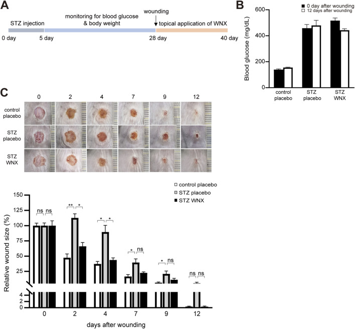 FIGURE 1
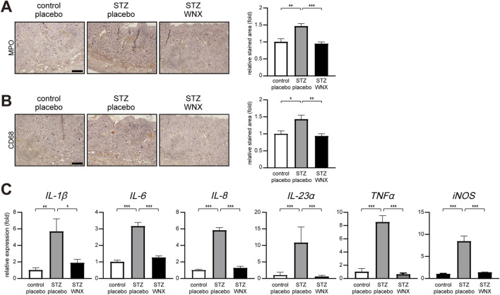 FIGURE 2
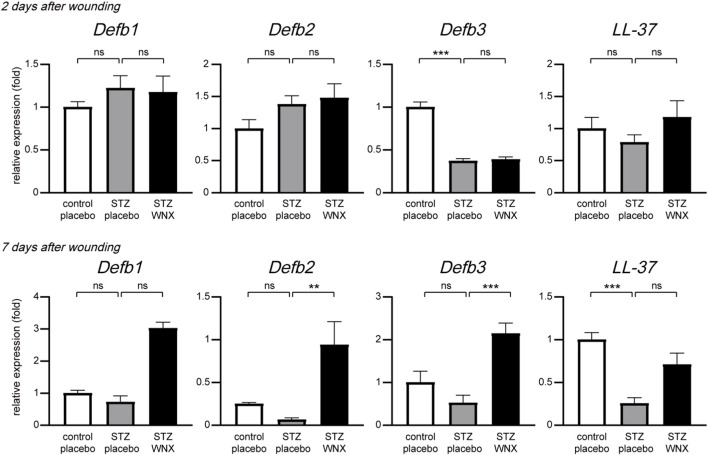 FIGURE 3
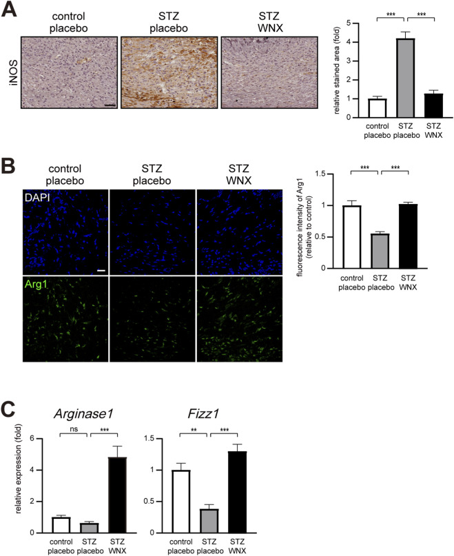 FIGURE 4
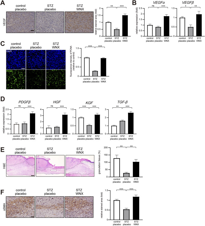 FIGURE 5
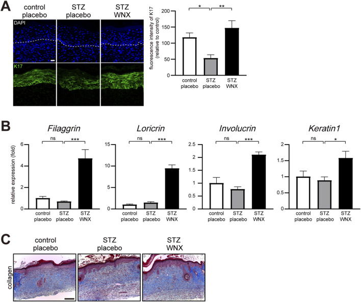 FIGURE 6
Peer Reviews
No public reviews on file for this paper yet. If you reviewed it on a platform where reviews are public (OpenReview, ICLR, NeurIPS, ICML), you can paste yours below so the community can read it here.
Videos
No videos yet. Explain this paper in a talk, walkthrough, or lecture? Add one.
Taxonomy
TopicsWound Healing and Treatments · Natural Compound Pharmacology Studies · Pressure Ulcer Prevention and Management
Introduction
1
Skin wounds heal naturally through four stages: hemostasis, inflammation, proliferation, and dermal remodeling (Pastar et al., 2014; Rodrigues et al., 2019; Wilkinson and Hardman, 2020). In hemostasis, platelets bind to the extracellular matrix (ECM) in the vessel wall and form blood clots to prevent hemorrhage. They also secrete various chemokines and growth factors, which are important for the recruitment of immune cells to the wound site and the stimulation of resident cells such as fibroblasts and keratinocytes. Then, signals from damaged tissues and necrotic cells, as well as bacterial components, evoke immune responses by activating immune cells including mast cells, neutrophils, macrophages, and T cells. These cells play critical role in phagocytosis and the production of cytokines and growth factors for cell proliferation, migration, and angiogenesis. In the proliferation phase, proliferated fibroblasts synthesize disorganized collagen, keratinocytes migrate from the wound edges to form epithelial layer, and endothelial cells also migrate from existing blood vessels into the wound site, contributing to granulation tissue formation, re-epithelialization, and neovascularization. In the final remodeling phase, granulation tissues matures into permanent scar. Additionally, type III reticular collagen is replaced with type I fibrillar collagen.
Chronic wounds refer to wounds that do not fully recover anatomically and functionally within 3 months during the wound healing process (Han and Ceilley, 2017; Berthiaume and Hsia, 2022). Chronic wounds are induced by conditions such as diabetes, peripheral artery disease, pressure ulcers, and venous insufficiency. In particular, diabetic wounds exhibit nerve damage, reduced blood flow, and impaired immune responses (Patel et al., 2019; Burgess et al., 2021). Consequently, diabetic skin wounds impede the normal phases of wound healing, including hemostasis, inflammation, proliferation, and tissue remodeling. In the inflammation phase, the M1 macrophage phenotype predominates and plays a key role in the chronic nature of diabetic wounds (Aitcheson et al., 2021; Burgess et al., 2021; Sim et al., 2022). Impairment of the transition process from M1 to M2 macrophages reduces the rate of wound healing, leading to impaired wound closure, impaired angiogenesis, and reduced collagen deposition (Aitcheson et al., 2021; Sim et al., 2022). Although various drugs for chronic skin wounds, including growth factors, have been developed, addressing these wounds remains challenging due to their complicated features of wound healing (Han and Ceilley, 2017; Spampinato et al., 2020).
WIN-1001X is a 20% ethanol extract of Polygala tenuifolia, Angelica tenuissima, and Dimocarpus longan combined in a 1:1:1 ratio (Li et al., 2021). It is also a modified Korean traditional botanical drug formula ‘Chungsimyeolda-tang’ which has been well described in the historic text ‘Dongui Sasang Shinpyun’ (Shim et al., 2008). In this study, we investigated whether WIN-1001X alleviates chronic skin wounds induced in streptozotocin (STZ)-induced diabetic.
Materials and methods
2
Preparation of WIN-1001X
2.1
WIN-1001X is composed of 20% ethanol extracts of three botanical drugs; Polygala tenuifolia Wildenow [Polygalaceae; Polygalae radix], Angelica tenuissima Nakai [Apiaceae; Angelicae tenuissimae radix], and Dimocarpus longan Loureiro [Sapindaceae; Longan arillus] which were purchased from Booyoung Pharmacy (Seoul, Korea). These three medicinal botanical drugs were mixed in a weight ratio of 1:1:1. The identification and authentication of these botanical drugs were verified by KGC Yebon Co., Ltd. (Cheongju-si, Korea), and specimens were stored in the Medihelpline Research Center plant specimen room (Seoul, Korea). The mixture was reflux-extracted for 3 h with 6 times the weight of the raw material in 20% ethanol (v/v) and then filtered. The residue was subsequently reflux-extracted for another 3 h with 4 times the weight of the raw material in 20% ethanol (v/v) and filtered. The combined filtrates were concentrated under reduced pressure and lyophilized to obtain the final dry extract. The drug-to-extract ratio (DER) is 3:1, meaning 1 g of the final extract (WIN-1001X) corresponds to 3 g of the initial raw botanical drug mixture. The preparation of WIN-1001X was conducted at KGC Yebon Co., Ltd. The analytical results for WIN-1001X are provided in Supplementary Figures 2–6.
UPLC-MS analysis
2.2
To confirm chemical compositions and consistent preparation of WIN-1001X from batch to batch, the UPLC-MS analysis was performed using a single quadrupole mass spectrometer (MSD, Agilent 6120, Santa Clara, CA, USA) coupled with Waters ACQUITY ultra performance liquid chromatography (UPLC) system (Waters, Milford, USA). An ACQUITY BEH C18 column (3.0 × 150 mm, 1.7 µm) was used and the mobile phase was composed of solvent A (0.2% acetic acid in H2O) and solvent B (0.2% acetic acid in acetonitrile). The elution gradient proceeded as follows: 0–3 min, 5% B; 3–53 min, 28% B; 53–55 min, 34% B; 55–75 min, 38% B; 75–76 min, 70% B; 76–80 min, 70% B. The flow rate is maintained at 0.5 mL/min and injection volume is 10 µL. The column oven is maintained at a temperature of 30 °C. The mass spectrometry parameters were as follows: positive ion mode; gas temperature, 350 °C; drying gas, 12 L/min; capillary voltage, +4,000 V; m/z range, 200–1,500. The quantitation of each major metabolite was made using a Photodiode Array (PDA) detector set at 320 nm.
Preparation of WIN-1001X cream
2.3
The WIN-1001X cream contained 3% (w/w) of WIN-1001X, respectively. Polyethylene glycol 400 was added as a base, and WIN-1001X was dissolved at 72∼78 °C for 20 min. Then, with the addition of other base ingredients and preservatives, the manufacturing process followed oil phase preparation, cooling, filling and packaging processes in accordance with the manufacturing method of ointment in the General Regulations of the Korean Pharmacopoeia. The 3% WIN-1001X cream was formulated with the following composition (w/w): WIN-1001X (3.0%), propylene glycol (8.0%), heavy liquid paraffin (5.0%), cetanol (2.5%), stearyl alcohol (5.0%), stearic acid (5.0%), isopropyl myristate (4.0%), sorbitan monostearate (3.0%), polysorbate 60 (3.0%), methyl parahydroxybenzoate (0.1%), and propyl parahydroxybenzoate (0.05%). The mixture was homogenized to ensure uniform dispersion. The preparation of WIN-1001X cream was conducted by Cires Pharmaceutical Inc. (Hwaseong-si, Korea).
Animal model of chronic skin wound
2.4
Adult male C57BL/6J mice (7 weeks old, DBL, Korea) were maintained in a temperature-controlled room (23 °C) at 55% humidity, with a 12-h light-dark cycle. The Committee for Experimental Animal Research at Sogang University approved the animal experiments (IACUCSGU2021_11). Diabetes was induced by intraperitoneal injection of streptozotocin (ALX-380-010-G001, ENZO) for 5 days. Streptozotocin was dissolved in 0.5 M sodium citrate buffer (pH 4.5). Whole blood from the mouse tail vein was measured using a blood glucose monitoring system (Accu-CheK Rerforma, Roche). On day 25, mice with a blood glucose value ≥300 mg/dL, were defined as streptozotocin-induced diabetic mice. On day 28, the mouse was anesthetized with 2,2,2-tribromoethanol and a full-thickness excision wound was made on the shaved dorsal skin using a 6 mm biopsy punch (Kai Industries) (n = 6 mice). WIN-1001X cream was topically applied daily for 12 days. All animal experiments were indeed performed three times independently, with n = 6 mice per group in each iteration. Data analysis was performed in a blinded manner. Samples were coded, and investigators were unaware of the treatment groups during measurement and histological evaluation.
Quantitative PCR
2.5
Total RNA was extracted from mouse skin wounds using Tri-RNA Reagent (TR118, Favorgen). First-strand cDNA synthesis was performed with PrimeScript RT master mix (RR036A, Takara). The resulting cDNAs were subjected to real-time PCR using qPCR 2x Premix SYBR (RT500M, Enzynomics) with a QuantStudio 1 Real-Time PCR System (Applied Biosystems). The PCR conditions were 10 min at 95 °C, followed by 40 cycles of 95 °C for 15 s and 64 °C for 40 s. Expression data were calculated from the cycle threshold (Ct) value using the ΔCt method of quantification. 18s rRNA was used for normalization. Oligonucleotides are listed in Supplementary Table 1.
Histology
2.6
Mouse dorsal skin samples were immediately fixed with 10% neutral buffered formalin (0144, Medilab, Korea) and left overnight at 4 °C. The samples were dehydrated, embedded in paraffin, and sectioned at 5 μm. The tissue sections were deparaffinized with xylene twice for 10 min each. Rehydration of sections was serially performed with 100%, 95%, 70%, 50% ethanol, followed by tap water. Tissue sections were stained with hematoxylin and eosin. Mast cells and collagen were stained with toluidine blue and Masson’s trichrome stain, respectively.
Immunohistochemistry
2.7
Rehydrated tissue sections were autoclaved in sodium citrate buffer (pH 6.0) for 10 min. After cooling, sections were washed in PBST (0.1% Triton X-100 in PBS) for 5 min and blocked with 5% BSA in 0.1% PBST for 1 h. For immunofluorescence, tissue sections were incubated with anti-arginase1 (66129-1-Ig, Proteintech), anti-PCNA (ab15497, Abcam), and anti-cytokeratin 17 (sc-393002, Santa Cruz Biotechnology) antibodies overnight at 4 . A secondary antibody conjugated to Cy5 (ab6563, ab6564, Abcam) was used. For DAB staining immunohistochemistry, tissue sections were incubated with anti-MPO (PA5-16672, Invitrogen), anti-iNOS (BD 610329, BD Bioscience), anti-VEGF (sc-152, Santa Cruz Biotechnology), and anti-αSMA (sc-32251, Santa Cruz Biotechnology) antibodies. HRP/DAB (ABC) detection IHC kit (ab64264, Abcam) was used. For DAB immunostaining, tissue sections were incubated with anti-MPO (PA5-16672, Invitrogen) and anti-iNOS (BD 610329, BD bioscience), anti-VEGF (sc-152, Santa Cruz Biotechnology), anti-αSMA (sc-32251, Santa Cruz Biotechnology) antibodies overnight at 4 . Then, the HRP/DAB (ABC) detection IHC kit (ab64264, Abcam) was used according to the manufacturer’s instructions. Expression was quantified using ImageJ software (NIH, Bethesda, MD, USA).
Statistical analysis
2.8
All animal experiments were indeed performed independently three times, with n = 6 mice per group in each experiment. All quantitative data were presented as the mean ± standard error of the mean (S.E.M.) from three independent experiments. Statistical differences among multiple groups were analyzed by one-way or two-way analysis of variance (ANOVA) followed by Dunnett’s post hoc test. Statistical significance was defined as *P ≤ 0.05, **P ≤ 0.01, and ***P ≤ 0.005. All p-values indicated in the figures are listed in Supplementary Table 2.
Results
3
3% WIN-1001X cream accelerates chronic skin wound healing
3.1
We previously found that topical application of WIN-1001X reduces skin inflammation (manuscript in preparation). Thus, we further tested whether WIN-1001X cream alleviates chronic skin wounds that exhibit prolonged and extensive inflammation (Rosique et al., 2015; Zhao et al., 2016; Burgess et al., 2021). We used streptozotocin (STZ)-induced diabetic mice with fasting blood glucose levels exceeding 300 mg/dL as a chronic skin wound model (Figures 1A,B). For topical application, different percentages of WIN-1001X cream (WNX) were prepared and tested the efficacy of WIN-1001X cream in chronic skin wound healing (Supplementary Figure 1). We finally selected 3% WIN-1001X cream for further study. Although diabetic mouse treated with placebo cream (STZ placebo) showed delayed skin wound healing compared with normal skin wounds treated with placebo cream (control placebo), topical application of 3% WIN-1001X cream (STZ WNX) accelerated skin wound healing in diabetic mice (Figure 1C).
*3% WIN-1001X cream accelerates chronic skin wound healing. (A) Experimental scheme. After diabetes was induced by intraperitoneal injection of streptozotocin (STZ), full-thickness excisional wounds were created in the shaved dorsal skin. Three percent of WIN-1001X cream (WNX) was topically applied every day for 12 days after wounding (see Materials and Methods). (B) Fasting blood glucose levels was measured at 0 and 12 days after wounding. Mice with fasting blood glucose levels exceeding 300 mg/dL were included for this study. (C) Topical application of 3% WIN-1001X cream accelerates chronic skin wound healing in diabetic mice (n = 6/per group). Normal wounds topically applied with placebo cream were used as controls. The wound was photographed and representative images are shown. The wound area was quantified using the ImageJ software. All data represent mean ± S.E.M. Statistical significance was indicated as *P ≤ 0.05, **P ≤ 0.01, and **P ≤ 0.005.
3% WIN-1001X cream suppresses infiltration of immune cells in chronic skin wounds
3.2
Given the extensive and prologned inflammatory phase in diabetic skin wounds (Rosique et al., 2015; Zhao et al., 2016; Burgess et al., 2021), we next examined the infiltration of immune cells into the skin wound. The infiltration of MPO (myeloperoxidase)-positive neutrophils was increased in diabetic skin wounds compared with normal skin wounds at 2 days after wounding. However, topical application of 3% WIN-1001X cream reduced the infiltration of neutrophils in diabetic skin wounds (Figure 2A). The infiltration of CD68-positive monocytes and macrophages in diabetic skin wounds treated with 3% WIN-1001X cream was also reduced compared with diabetic skin wounds treated with placebo cream at 2 days after wounding (Figure 2B). Consistently, the topical application of 3% WIN-1001X cream suppressed the upregulation of the gene expression of pro-inflammatory cytokines, including IL-1β, IL-6, IL-8, IL-23α, and TNFα as well as iNOS in diabetic skin wounds at 2 days after wounding (Figure 2C).
*3% WIN-1001X cream suppresses skin inflammation in chronic skin wounds. (A,B) Topical application of 3% WIN-1001X cream (WNX) suppresses infiltration of neutrophils and macrophages in chronic skin wounds (n = 6/group). At 2 days after topical application of 3% WIN-1001X cream, skin tissues were harvested. Skin sections were immunostained with anti-MPO (myeloperoxidase) and anti-CD68 antibodies for the detection of neutrophils and macrophages, respectively. Normal wounds topically applied with placebo cream were used as controls. Expression was quantified using the ImageJ software. Representative images are shown. Scale bar, 50 μm. (C) Topical application of 3% WIN-1001X cream suppresses activation of proinflammatory cytokines and iNOS genes in chronic skin wounds (n = 6/per group). At 2 days after topical application of 3% WIN-1001X cream, skin tissues were harvested. Transcripts of IL-1β, IL-6, IL-8, IL-23α, TNFα, iNOS, and 18S rRNA were quantified using real-time RT-PCR (n = 6). All data represent mean ± S.E.M. Statistical significance was indicated as *P ≤ 0.05, **P ≤ 0.01, and **P ≤ 0.005.
3% WIN-1001X cream upregulates gene expression of anti-microbial peptides in chronic skin wounds
3.3
Microbial infection delays wound healing and often results in serious outcomes such as surgical debridement (Frykberg and Banks, 2015; Negut et al., 2018). WIN-1001X has no direct antimicrobial activity against bacteria including Staphylococcus aureus and Pseudomonas aeruginosa (data not shown). The skin has antimicrobial activity as part of the early stage of the immune defense system through the expression of antimicrobial peptides (Yamasaki and Gallo, 2008; Rademacher et al., 2021). We thus tested whether 3% WIN-1001X cream regulates the gene expression of antimicrobial peptides in diabetic skin. At 2 days after wounding, the topical application of 3% WIN-1001X cream had no effect on the expression of Defb1, Defb2, Defb3, and LL-37 genes in diabetic skin wounds. However, upregulation of Defb2, Defb3, and LL-37 gene expression was observed in 3% WIN-1001X cream-treated diabetic skin wounds at 7 days after wounding (Figure 3).
*3% WIN-1001X cream upregulates gene expression of antimicrobial peptides in chronic skin wounds. Topical application of 3% WIN-1001X cream upregulates the gene expression of antimicrobial peptides in chronic skin wounds (n = 6/group). At 2 and 7 days after topical application of 3% WIN-1001X cream, skin tissues were harvested. Transcripts of Defb1, Defb2, Defb3, LL-37, and 18S rRNA were quantified using real-time RT-PCR (n = 6). All data represent mean ± S.E.M. Statistical significance was indicated as *P ≤ 0.05, **P ≤ 0.01, and **P ≤ 0.005.
3% WIN-1001X cream promotes M1 to M2 macrophage polarization in chronic skin wounds
3.4
Classically activated M1 macrophages, which promote inflammation, could be polarized into alternatively activated M2 macrophages for successful wound repair (Ferrante and Leibovich, 2012; Louiselle et al., 2021). The prolonged existence of M1 macrophages is one of the characteristics of chronic wound healing. Thus, we examined the existence of M1 and M2 macrophages at 7 days after wounding by immunostaining with anti-iNOS (inducible NOS) and anti-Arg1 (Arginase 1) antibodies, respectively. The increased level of iNOS-positive M1 macrophages was maintained in diabetic skin wounds compared with normal skin wounds at 7 days after wounding (Figure 4A). However, 3% WIN-1001X cream treatment restored the normal level of M1 macrophages in diabetic skin wounds (Figure 4A). Additionally, 3% WIN-1001X cream maintained the normal level of Arg1-positive M2 macrophages in diabetic skin wounds at 7 days after wounding (Figure 4B). Consistent with immunohistochemical results, 3% WIN-1001X cream induces gene expression of markers of M2 macrophages such as Arg1 and Fizzl (Figure 4C).
*3% WIN-1001X cream promotes M1 to M2 macrophage polarization in chronic skin wounds. (A) Topical application of 3% WIN-1001X cream suppresses excessive infiltration of M1 macrophage in chronic skin wounds (n = 6/per group). At 2 days after topical application of 3% WIN-1001X cream, skin tissues were harvested. Skin sections were immunostained with anti-iNOS antibody for the detection of M1 macrophages. Normal wounds applied with placebo cream were used as controls. Expression was quantified using the ImageJ software. Representative images are shown. Scale bar, 50 μm. (B,C) Topical application of 3% WIN-1001X cream promotes infiltration of M2 macrophages in chronic skin wounds (n = 6/group). At 7 days after topical application of 3% WIN-1001X cream, skin tissues were harvested. Skin sections were immunostained with anti-Arg1 (Arginase 1) antibody for the detection of M2 macrophages. Normal wounds applied with placebo cream were used as controls. Expression was quantified using the ImageJ software. Representative images are shown. Scale bar, 50 μm. Transcripts of Arg1, Fizz1, and 18S rRNA were quantified using real-time RT-PCR (n = 6). All data represent mean ± S.E.M. Significance values were *P ≤ 0.05, **P ≤ 0.01, and **P ≤ 0.005.
3% WIN-1001X cream promotes cell growth in chronic skin wounds
3.5
VEGF (vascular endothelial growth factor) plays a role in angiogenesis in wound healing through endothelial cell proliferation, migration, differentiation, and survival (Bao et al., 2009; Johnson and Wilgus, 2014). We found that topical application of 3% WIN-1001X upregulated VEGF expression in diabetic skin wounds compared with the placebo control at 7 days after wounding (Figures 5A,B). Additionally, the transition from inflammation to proliferation is also a critical process for successful wound healing (Landén et al., 2016). We examined the cell proliferation of keratinocytes and fibroblasts by immunostaining using anti-PCNA (proliferating cell nuclear antigen) antibody. Topical application of 3% WIN-1001X promoted keratinocytes and fibroblasts proliferation in diabetic skin wounds compared with placebo-treated diabetic skin wounds at 7 days after wounding (Figure 5C). Moreover, 3% WIN-1001X cream activated the gene expression of growth factors including PDGFβ, HGF, KGF and TGFβ, which are responsible for cell proliferation in diabetic skin wounds compared with diabetic skin wounds treated with placebo cream (Figure 5D). An increase in granulation tissue formation was observed in 3% WIN-1001X cream-treated diabetic skin wounds compared with placebo cream-treated diabetic skin wounds at 7 days after wounding (Figure 5E). Additionally, we observed that 3% WIN-1001X cream promoted myofibroblast formation in granulation tissue (Figure 5F).
*3% WIN-1001X cream promotes cell proliferation in chronic skin wounds. (A,B) Topical application of 3% WIN-1001X cream upregulates VEGF expression in chronic skin wounds (n = 6/per group). At 7 days after topical application of 3% WIN-1001X cream, skin tissues were harvested. Skin tissue sections were immunostained with anti-VEGF antibody. Expression was quantified using the ImageJ software. Representative images are shown. Transcripts of VEGFα, VEGFβ, and 18S rRNA were quantified using real-time RT-PCR (n = 6). (C) Topical application of 3% WIN-1001X cream increases cell proliferation in chronic skin wounds (n = 6/per group). At 7 days after topical application of 3% WIN-1001X cream, skin tissues were harvested. Skin sections were immunostained with anti-PCNA antibody. Normal wounds applied with placebo cream were used as controls. Expression was quantified using the ImageJ software. Representative images are shown. Scale bar, 50 μm. (D) Topical application of 3% WIN-1001X cream upregulates the gene expression of growth factors in chronic skin wounds (n = 6/per group). At 7 days after topical application of 3% WIN-1001X cream, skin tissues were harvested. Transcripts of PDGFβ, HGF, KGF, TGFβ, and 18S rRNA were quantified using real-time RT-PCR (n = 6). (E) Topical application of 3% WIN-1001X cream promotes the formation of granulation tissues in chronic skin wounds (n = 6/per group). At 7 days after topical application of 3% WIN-1001X cream, skin tissues were harvested. Skin sections were stained with hematoxylin and eosin. Normal wounds applied with placebo cream were used as controls. The area of granulation tissues was quantified using the ImageJ software. Representative images are shown. Scale bar, 100 μm. (F) Topical application of 3% WIN-1001X cream promotes myofibroblast differentiation in chronic skin wounds (n = 6/per group). At 7 days after topical application of 3% WIN-1001X cream, skin tissues were harvested. Skin sections were immunostained with anti-αSMA (smooth muscle actin α) antibody. Expression was quantified using the ImageJ software. Representative images are shown. Scale bar, 50 μm. All data represent mean ± S.E.M. Statistical significance was indicated as *P ≤ 0.05, **P ≤ 0.01, and **P ≤ 0.005.
3% WIN-1001X cream promotes re-epithelialization and collagen deposition in chronic skin wounds
3.6
Immunostaining with an anti-K17 (keratin 17) antibody showed that topical application of 3% WIN-1001X cream promoted re-epithelialization as a result of epithelial keratinocyte migration over the wound bed in chronic skin wounds at 7 days after wounding (Figure 6A). We further examined the expression of genes related to keratinocyte differentiation. Topical application of 3% WIN-1001X cream upregulated Filaggrin, Loricrin, Involucrin, and Keratin 1 gene expression in chronic skin wounds at 7 days after wounding (Figure 6B). We also found that topical application of 3% WIN-1001X cream promoted collagen deposition, as observed in tissue sections stained with Masson’s trichrome (Figure 6C).
*3% WIN-1001X cream promotes reepithelialization and tissue remodeling in chronic skin wounds. (A) Topical application of 3% WIN-1001X cream promotes reepithelialization in chronic skin wounds (n = 6/per group). At 7 days after topical application of 3% WIN-1001X cream, skin tissues were harvested. Skin sections were immunostained with anti-K17 antibody. Expression was quantified using the ImageJ software. Representative images are shown. Scale bar, 50 μm. (B) Topical application of 3% WIN-1001X cream promotes keratinocyte differentiation in chronic skin wounds (n = 6/per group). At 7 days after topical application of 3% WIN-1001X cream, skin tissues were harvested. Transcripts of Filaggrin, Loricrin, Involucrin, Keratin 1, and 18S rRNA were quantified using real-time RT-PCR (n = 6). (C) Topical application of 3% WIN-1001X cream promotes collagen synthesis in chronic skin wounds (n = 6/per group). At 12 days after topical application of 3% WIN-1001X cream, skin tissues were harvested. Skin sections were stained with Masson’s trichrome. Expression was quantified using the ImageJ software. Representative images are shown. Scale bar, 100 μm. All data represent mean ± S.E.M. Statistical significance was indicated as *P ≤ 0.05, **P ≤ 0.01, and **P ≤ 0.005.
Discussion
4
WIN-1001X is a 20% ethanol extract of Polygala tenuifolia, Angelica tenuissima, and Dimocarpus longan combined in a 1:1:1 ratio (Li et al., 2021). We recently found that topical application of WIN-1001X reduces skin inflammation (manuscript in preparation). In this study, we further investigated whether WIN-1001X alleviates chronic skin wounds that exhibit prolonged and extensive inflammation as well as other abnormal healing processes (Rosique et al., 2015; Zhao et al., 2016; Burgess et al., 2021). In fact, a persistent inflammation state disrupts normal skin wound healing progress and increases scar formation (Eming et al., 2007; Wilgus, 2020). Thus, the transition from the inflammation stage to the proliferation stage may be a key therapeutic intervention for chronic skin wounds.
Our results demonstrated that 3% WIN-1001X cream suppressed extensive inflammation by suppressing the of infiltration of neutrophils and monocytes and cytokine gene expression such as including IL-1β, IL-6, IL-8, IL-23α, and TNFα compared with placebo- treated chronic wounds. In addition, we found that 3% WIN-1001X cream promoted pro-inflammatory M1 to anti-inflammatory M2 macrophage polarization, which is required for successful wound repair (Ferrante and Leibovich, 2012; Louiselle et al., 2021). Additionally, oral and topical toxicity tests were conducted for WIN-1001X, and the data confirmed its safety (Supplementary Table 3). Indeed, extracts of each plant have been reported to have anti-inflammatory activity. The root extract of Polygala tenuifolia suppressed lipopolysaccharide (LPS)-stimulated upregulation of iNOS, COX-2, TNFα, and IL-1β in BV2 microglial cells via inhibition of IκB-α degradation (Cheong et al., 2011). The extract of Angelica tenuissima exhibited anti-inflammatory effects by suppressing of calcium release, as well as p38MAPK and STAT3 phosphorylation in LPS-treated RAW264.7 macrophages (Kim et al., 2022). In addition, the extracts of Dimocarpus longan inhibited LPS-induced degradation of IκBα and the activation of NF-κB, AP-1, Akt, and MAP kinases in RAW264.7 macrophages (Kunworarath et al., 2016).
We also have identified active metabolites of WIN-1001X using UPLC-MS analysis (Supplementary Figure 7). Interestingly, previous have demonstrated that some of the active metabolites of Polygala tenuifolia, Angelica tenuissima, and Dimocarpus longan have anti-inflammatory effects. For instance, tenuigenin, tenuifoliside A, tenuifolin, and senegenin isolated from Polygala tenuifolia suppressed inflammation by various mechanisms such as the inhibition of NF-κB and NLRP3 inflammasome or by activation of NRF2-HO1 signaling (Yuan et al., 2012; Kim et al., 2013; Lv et al., 2016; Wang et al., 2016; Lu et al., 2017; Wang et al., 2017; Li et al., 2017; Chen and Jia, 2020). In addition, the 3-O-(3,4,5-trimethoxy-cinnamoyl), 6′-O-(p-methoxybenzoyl) sucrose ester from Polygala tenuifolia inhibited expression of the iNOS, COX-2, TNF-α, IL-1β, and IL-6 (Son et al., 2022). Onjisaponin B of WIN-1001X also reduces the level of TNF-α, IL-1β, and IL-6 in LPS-treated PC12 cells (Li et al., 2018; Li et al., 2021). Decursin and (Z)-ligustilide from Angelica tenuissima extract also have anti-inflammatory effects in skin wound healing. Decursin decreased LPS-induced oxidative stress and inflammation through suppression of activation of the NF-κB pathway (Zhu and Dong, 2023). (Z)-ligustilide also suppressed activation of the NF-κB pathway via inhibition of gene expression and signaling (Wang et al., 2010; Chung et al., 2012; Choi et al., 2018).
Topical application of 3% WIN-1001X cream further promoted cell proliferation, granulation tissue formation, myofibroblast formation, and collagen deposition in skin wound healing. Although the exact active metabolite of WIN-1001X responsible for these wound healing process is not currently known, polygalaxanthone III, a xanthone glycoside of Polygala tenuifolia has been reported to promote skin wound healing induced by yeast infection through the STAT3 pathway (Tsujimoto et al., 2019; Yang et al., 2022). Additionally, decursin improves keratinocyte wound healing by upregulating the expression of genes encoding extracellular matrix remodeling proteins and growth factors (Han et al., 2018).
In conclusion, we studied the efficacy of 3% WIN-1001X cream in chronic skin wounds using streptozotocin-induced diabetic mice. Specifically, 3% WIN-1001X cream suppressed skin inflammation by decreasing cytokine gene expression and immune cell infiltration, and by increasing macrophage polarization. It also promoted cell proliferation, granulation tissue formation, and myofibroblast transition. Furthermore, 3% WIN-1001X cream promoted keratinocyte re-epithelialization and differentiation as well as increased collagen deposition in chronic skin wounds. Thus, our results suggest that 3% WIN-1001X cream may help alleviate chronic skin wounds, for which currently no effective therapeutics exist.
Conclusion
5
3% WIN-1001X cream suppressed skin inflammation by decreasing cytokine gene expression and immune cell infiltration, and by increasing macrophage polarization. It also promoted cell proliferation, granulation tissue formation, and myofibroblast transition. Furthermore, 3% WIN-1001X cream promoted keratinocyte re-epithelialization and differentiation as well as increasing collagen deposition in chronic skin wounds. Thus, our results suggest that 3% WIN-1001X cream may help alleviate chronic skin wounds.
The reference list from the paper itself. Each links out to its DOI / PubMed record.
- 1Aitcheson S. M. Frentiu F. D. Hurn S. E. Edwards K. Murray R. Z. (2021). Skin wound healing: normal macrophage function and macrophage dysfunction in diabetic wounds. Molecules 26 (16), 4917. 10.3390/molecules 26164917 34443506 PMC 8398285 · doi ↗ · pubmed ↗
- 2Bao P. Kodra A. Tomic-Canic M. Golinko M. S. Ehrlich H. P. Brem H. (2009). The role of vascular endothelial growth factor in wound healing. J. Surg. Res. 153 (2), 347–358. 10.1016/j.jss.2008.04.023 19027922 PMC 2728016 · doi ↗ · pubmed ↗
- 3Berthiaume F. Hsia H. C. (2022). Regenerative approaches for chronic wounds. Annu. Rev. Biomed. Eng. 24, 61–83. 10.1146/annurev-bioeng-010220-113008 35226819 · doi ↗ · pubmed ↗
- 4Burgess J. L. Wyant W. A. Abdo Abujamra B. Kirsner R. S. Jozic I. (2021). Diabetic wound-healing science. Med. Kaunas. 57 (10), 1072. 10.3390/medicina 57101072 34684109 PMC 8539411 · doi ↗ · pubmed ↗
- 5Chen S. Jia J. (2020). Tenuifolin attenuates Amyloid-β42-Induced neuroinflammation in microglia through the NF-κB signaling pathway. J. Alzheimers Dis. 76 (1), 195–205. 10.3233/JAD-200077 32444542 · doi ↗ · pubmed ↗
- 6Cheong M. Lee S. Yoo H. Jeong J. Kim G. Kim W. (2011). Anti-inflammatory effects of Polygala tenuifolia root through inhibition of NF-κB activation in lipopolysaccharide-induced BV 2 microglial cells. J. Ethnopharmacol. 137 (3), 1402–1408. 10.1016/j.jep.2011.08.008 21856398 · doi ↗ · pubmed ↗
- 7Choi E. S. Yoon J. J. Han B. H. Jeong D. H. Lee Y. J. Kang D. G. (2018). Ligustilide attenuates vascular inflammation and activates Nrf 2/HO-1 induction and, NO synthesis in HUVE Cs. Phytomedicine 38, 12–23. 10.1016/j.phymed.2017.09.022 29425644 · doi ↗ · pubmed ↗
- 8Chung J. W. Choi R. J. Seo E. K. Nam J. W. Dong M. S. Shin E. M. (2012). Anti-inflammatory effects of (Z)-ligustilide through suppression of mitogen-activated protein kinases and nuclear factor-κB activation pathways. Arch. Pharm. Res. 35 (4), 723–732. 10.1007/s 12272-012-0417-z 22553066 · doi ↗ · pubmed ↗