Skull base reconstruction with nasoseptal flap in oncologic cases: can narrow-band imaging help? How we do it
M Stavrakas, Y Kanaan, R Al-Ashqar, H Khalil, S Muquit

Abstract
Genes, proteins, chemicals, diseases, species, mutations and cell lines named across the full text — each resolved to its canonical identifier and authoritative record.
Click any figure to enlarge with its caption.
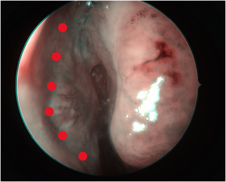 Figure 1
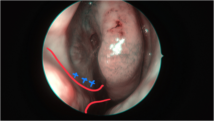 Figure 2
Peer Reviews
No public reviews on file for this paper yet. If you reviewed it on a platform where reviews are public (OpenReview, ICLR, NeurIPS, ICML), you can paste yours below so the community can read it here.
Videos
No videos yet. Explain this paper in a talk, walkthrough, or lecture? Add one.
Taxonomy
TopicsHead and Neck Surgical Oncology · Cleft Lip and Palate Research · Meningioma and schwannoma management
In cases in which the tumour involves the superior septum, careful planning of the flap and clear edges are of paramount importance (Figure 1). Our standard practice is to obtain tissue samples from macroscopically clear areas for frozen sections. This is an essential step to ensure avoidance of tumour seeding at the area of reconstruction. Following narrow-band imaging (NBI) mapping of the septum and nasal cavity, we take a biopsy from the superior edge of the proposed flap (Figure 2) for frozen section. Dual confirmation of the tumour-free edge helps us to proceed with safe reconstruction. The flap is raised following the standard technique, extending to the nasal floor up to the inferior meatus, ensuring Hasner's valve remained intact. On follow-up scans of our cases, there was no evidence of any residual disease related to the flap.
The principle behind NBI lies in the use of specific narrow-bandwidth filters that illuminate tissue with light at wavelengths corresponding to the peak absorption of haemoglobin.^1^ More specifically, NBI utilises narrow-band spectrum optical filters to emit light at two specific wavelengths, 415nm (blue) and 540nm (green).^2^ By highlighting vascular structures and mucosal patterns, NBI facilitates the identification of lesions, inflammatory changes and neoplastic growths with remarkable precision. One of the notable advantages of NBI is its ability to differentiate between benign and malignant lesions.^3^ NBI technology can represent a useful adjunct in the planning of anterior skull base reconstruction, assisting towards optimal flap viability and tumour-free edges. Further studies and larger study samples are required to draw safe conclusions about this technique.
The reference list from the paper itself. Each links out to its DOI / PubMed record.
- 1Dalgorf DM, Harvey RJ. Navigated endoscopic surgery for sinonasal tumors: a paradigm shift in surgical technique. Otolaryngol Clin North Am 2017; 50: 593–609.
- 2Olympus. Narrow band imaging: Transforming the world of endoscopy. https://www.olympus.co.uk/medical/en/stories-detail/2022-04-11/Narrow-Band-Imaging-Transforming-the-World-of-Endoscopy.html (cited April 2025).
- 3Kanaan Y, Khalil H, Geronatsios K, Stavrakas M. The added value of narrow band imaging in Sinonasal tumour resection and surveillance: Our experience. Am J Otolaryngol 2025; 46: 104534.39653616 10.1016/j.amjoto.2024.104534 · doi ↗ · pubmed ↗