Comparative histopathology of climbing perch, Anabas testudineus challenged with Aeromonas veronii via IM and IP routes
Md. Ali Reza Faruk, Fatema Jahan, Maliha Farzana, K. M. Abdul Halim, Tamanna Tabassum, Tanvir Rahman, Salman Shahriar Nibir, Ishrat Zahan Anka

TL;DR
This study compares the effects of different bacterial loads on the health of climbing perch infected with Aeromonas veronii via two injection routes.
Contribution
The study provides new insights into the histopathological effects of varying Aeromonas veronii concentrations in fish.
Findings
Higher bacterial loads caused more severe clinical signs and mortality in fish.
Histopathological changes like vacuolation and necrosis were more pronounced at higher bacterial concentrations.
Intraperitoneal injection resulted in higher mortality than intramuscular injection.
Abstract
The focal purpose of this investigation was to contrast the pathological changes from different histological observations of Anabas testudineus artificially infected with Aeromonas veronii. Intramuscular (IM) and intraperitoneal (IP) injection routes were used to challenge fish with three different bacterial concentrations, including 2.1 × 10⁵, 2.1 × 10⁵, and 2.1 × 10⁴cfu ml−1 of bacteria to investigate the effects of various loads of A. veronii on the histopathological alterations in the skin-muscle, liver, and intestine of A. testudineus during 13 days of post challenge test. Two replicates (n = 10) were used for each of the IM and IP groups, corresponding to the three aforementioned bacterial loads, in the challenge test for this study. The highest bacterial challenge (2.1× 10⁴ cfu ml−1) groups from both IP and IM produced some prominent clinical signs, e.g., red spots, ulcers, and…
Genes, proteins, chemicals, diseases, species, mutations and cell lines named across the full text — each resolved to its canonical identifier and authoritative record.
Click any figure to enlarge with its caption.
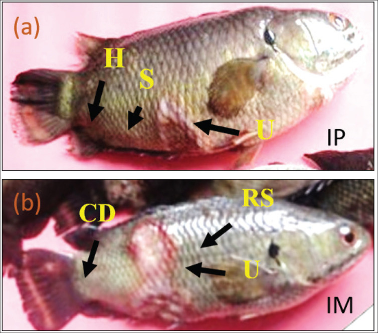 Figure 1
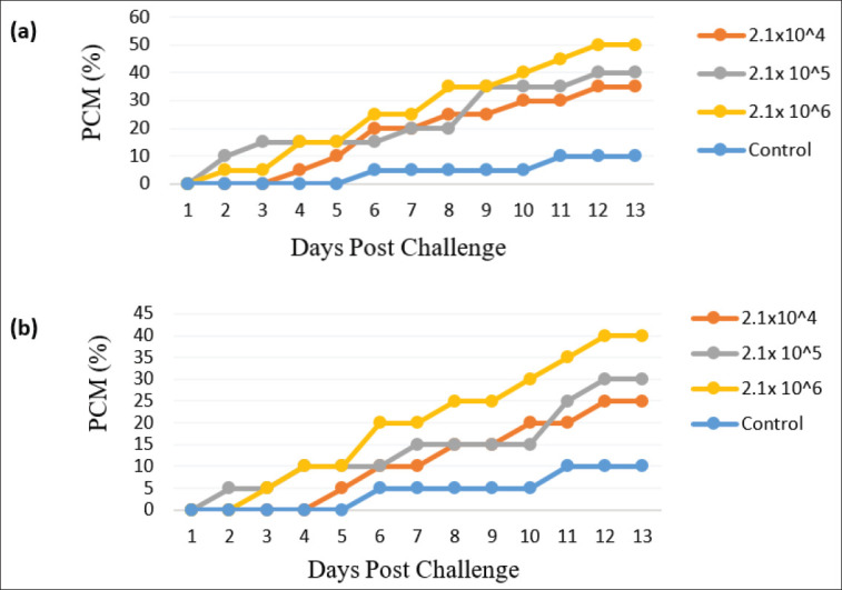 Figure 2
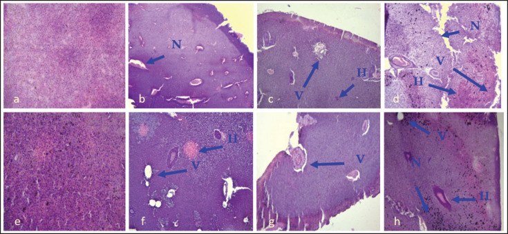 Figure 3
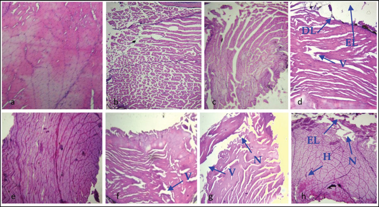 Figure 4
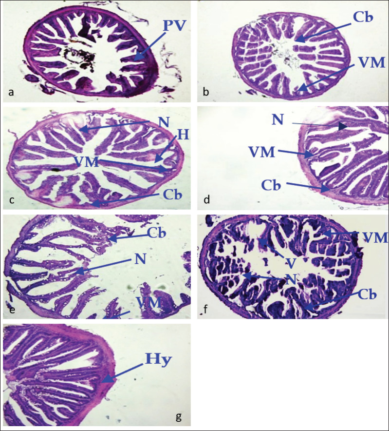 Figure 5
| Fish groups (Code) | Replicate | Challenged fish (number) | Challenge dose (Cfu ml − 1)/ PS |
|---|---|---|---|
| IM | 1 | 10 | 2.1 × 10⁴ |
| 2 | 10 | 2.1 × 10⁴ | |
| IM | 1 | 10 | 2.1 × 10⁵ |
| 2 | 10 | 2.1 × 10⁵ | |
| IM | 1 | 10 | 2.1 × 10⁶ |
| 2 | 10 | 2.1 × 10⁴ | |
| IP | 1 | 10 | 2.1 × 10⁴ |
| 2 | 10 | 2.1 × 10⁴ | |
| IP | 1 | 10 | 2.1 × 10⁵ |
| 2 | 10 | 2.1 × 10⁵ | |
| IP | 1 | 10 | 2.1 × 10⁶ |
| 2 | 10 | 2.1 × 10⁶ | |
| CF | 1 | 10 | PS |
| 2 | 10 | PS |
Peer Reviews
No public reviews on file for this paper yet. If you reviewed it on a platform where reviews are public (OpenReview, ICLR, NeurIPS, ICML), you can paste yours below so the community can read it here.
Videos
No videos yet. Explain this paper in a talk, walkthrough, or lecture? Add one.
Taxonomy
TopicsAquaculture disease management and microbiota · Myxozoan Parasites in Aquatic Species · Invertebrate Immune Response Mechanisms
Introduction
The total production of the world’s fisheries and aquaculture was recorded at 223.2 million tons in 2022, according to a recent report by [1]. There, 84% of the total inland production was obtained from aquaculture, with the majority (70%) contributed by the production of Asian countries. Among the major aquaculture-producing countries in Asia, Bangladesh has secured 2nd place for the world’s freshwater fish production (13.22 lakh tons, comprising 11.7% of the global total production). Bangladesh has also held the 5th position in a row for five times, although it has dropped (in Asia) from 3^rd^ to 5^th^ regarding the world’s total farmed aquatic animals’ harvesting [1,2]. Freshwater fish farming is a very common practice in Bangladesh. Among the many freshwater aquaculture finfish species in Bangladesh, the climbing perch (Anabas testudineus), also known as koi, has become the preferred and most profitable species for aquaculture farmers. It is due to this fish’s higher growth [3], comparatively short culture duration, high survival with its unique respiratory support [4], high market demand, and better nutritional value compared to native species [5]. Thus, this species has been contributing to the country’s overall increasing aquaculture demand.
A new variety of A. testudineus, commonly known as Thai koi, was introduced in Bangladesh from Thailand in 2002 and has opened a new scope for fish farmers, which was first reported in Mymensingh district, mostly through pond culture, and the production of this species was also found as profitable among farmers [6,7]. The production of A. testudineus was recorded at 46,457 Metric tons (MTs) in 2018 and 57,244 MT in 2022 [8,9], which clearly demonstrates the increasing farming trend of this species in pond aquaculture in Bangladesh. As a consequence of the rapid intensification of fish farming, subsequent risk factors have been faced by aqua farmers throughout the production process. Throughout the culture, A. testudineus can be subjected to various unfavorable challenges, which can lead to stress and disease outbreaks. Major disease incidence occurs in aquaculture due to the intensification and extension of farming practices [10], and as a result, disease can hinder the economic gain of many aquatic species at various scales [11]. In farmed koi in Bangladesh, diseases were reported for example, the disease called epizootic ulcerative syndrome (EUS), followed by some cases of tail and fin rot, and also some symptoms related to gill damage, skin erosion [12], and also the presence of some pathogenic bacteria [13] has been reported from both the indigenous and exotic A. testudineus.
Among different bacterial pathogens, the genus Aeromonas, from the Aeromonadaceae family, has garnered significant attention worldwide over the past few years due to its association with a high mortality rate in infected fish species [14]. Diverse Aeromonas spp. have been recognized for causing infection and diseases in fish [15] as a primary pathogen in different farmed fishes [16], and among many Aeromonads, Aeromonas veronii has been reported as a most virulent pathogen with a high percentage of mortality in fish (e.g., observed in Nile Tilapia) [17]. Aeromonas veronii was also detected as a significant pathogen from the aquaculture fish species (i.e., European seabass) [18] and for different freshwater ornamental fish species [19]. More recently, A. veronii infection in fish has been found to have an increased rate of infection, as well as very similar symptoms and histological observations, similar to those typically observed in A. hydrophila infections [20]. Several strains of A. veronii have been increasingly isolated from diseased fish (e.g., striped catfish [21], Largemouth bass [22]), and a range of diverse clinical signs and symptoms have been detected in infected fish. The common clinical signs for A. veronii infection have been detected as a form of ulcer, exophthalmia and hemorrhage, abdominal distention, and fin rot or tail rot in fish, whereas the symptoms are not always evenly shown with potential pathogenicity in different fish infected with diverse strains or isolates of A. veronii [20]. So, there is still scope for research considering the diversity of clinical signs and histological pathology in farmed fish infected with A. veronii. This type of information can be useful for better understanding the virulence of this pathogen and for comparing the state of histological lesions across infected fish. However, the information has not been explored adequately in aquaculture fish species in Bangladesh.
Recently, A. veronii has been isolated from diseased climbing perch* A*. testudineus in Bangladesh [23]. However, comparative histopathological studies using different challenge routes in A.* testudineus* from Bangladesh are scarce, and this type of comparative infection study in aquaculture species has yet to be explored in this context. This sector requires greater attention, not only for the health management aspects of this fish to prevent production losses due to bacterial infection, but also to ensure the welfare of the farmed fish species. Experimental infection can shed light on the mechanisms involved in the virulence and pathology of A. veronii in Thai koi, a susceptible host species. Considering this, the present study was set to compare visible pathological variations in different organs of Thai koi, A. testudineus, artificially infected with A. veronii via two different challenge routes.
Materials and Methods
Ethical approval
This study on climbing perch (A. testudineus) challenged with A. veronii was approved by the Ethics and Safety Review Committee (ESRC) of BAURES (Approval No. ESRC/57/FISH/2025).
Bacterial preparation
Laboratory stock of A. veronii bacteria, previously isolated from diseased Thai koi have been tested in this study. The bacteria culture was done in Tryptic Soy Agar (TSA) agar media for 24 h at 25^°^^C^. Bacterial calculation was determined by following the serial dilution method and termed as colony forming unit (cfu ml^−1^). This cfu ml^−1^ unit was calculated from previously cultured bacterial suspension following the drop count method (Supplementary Information S1).
Experimental infection
For this study, A. testudineus (average size of 85 ± 1.72 gm) were obtained from the healthy fish stocks from a commercial hatchery and farm named Sarnatala Hatchery located at Mymensingh. Duplicate groups of fish were deposited in 30 L tanks. Each tank was aerated, and fish were acclimatized for 4 days prior to being released into the tank. Throughout the experimental period, a commercially pelleted diet was provided to the fish at a rate of 2–3 times daily. To maintain good water quality, approximately one-third of the tank water was replaced daily. Dead fish were removed if observed, and siphoning was performed as needed to remove debris from the bottom of the tank.
Two routes (intramuscular, IM, and intraperitoneal, IP injections) were used in this experiment to perform the challenge test. The experiment was conducted following the ethical approval given by BAURES (ESRC/57/FISH/2025). The challenge was done by injecting the fish with three different concentrations of A. veronii, including 2.1 × 10⁴, 2.1 × 10⁵, and 2.1 × 10⁴cfu ml^−1^. Two replicate fish groups (n = 10 each) were subjected to a challenge test via IM and IP routes by injecting the three bacterial loads mentioned above in this study (Table 1). A randomized distribution was followed to assign the fish individuals to their respective experimental study groups. Details of the experimental set-up for each challenge test group are mentioned in Table 1. However, no statistical test was performed in this study, given the comparatively small sample size (n = 10), which resulted in limited statistical power, and the nature of the data analysis.
The injection site for fish was selected as below the left dorsal fin, and injection was done, taking 0.1 ml of bacterial suspension (from low to high dose with a view to comparing the gradients of pathological changes in fish), which has been isolated and stocked previously in the laboratory. Alongside, two corresponding groups of fish as a control group (Table 1) were also included in this challenge test, and these naive fish were injected with 0.1 ml of sterile physiological saline (PS) (0.85% NaCl) following the same protocol that was used to inject the bacterial treatment groups. After that, fish were monitored for 13 consecutive days after the challenge test. Mortality and morbidity were recorded daily. Alongside, any unusual behavior and clinical signs or symptoms of the fish have been monitored daily. Water temperature and pH were also recorded and monitored during the post-challenge observation.
Sampling
After 13 days of the post-challenge period, fish from all experimental groups were euthanized using a standardized laboratory protocol to facilitate sampling. Necroscopy involved both the gross external and internal examinations prior to taking the samples of the target organs. The collected sample consisted of skin, muscle, liver, and intestine, and was preserved in 10% buffered formalin until it was processed for further histopathological examination.
Histological observation
The samples fixed with 10% buffered formalin were first prepared for dehydration, then cleared, and subsequently infiltrated in an Automatic Tissue Processor (Shandon, Citadel 1000). After that, the samples were embedded with melted wax and sectioned using a microtome (Leica Jung Rm 2035) at a thickness of 5 µm. The sections were then placed on glass slides and set on a hot plate to dry at 37°C for 1 min. Then, staining was performed with hematoxylin and eosin, and the sections were mounted with Canada balsam as follows [24]. Finally, the photographs of the histological slides were taken using Carl Zeiss Microscopy, GmbH, Axiocam ERc 5s. The histological photographs were then carefully assessed following a blinding process to avoid bias effects in the observation. All the data and images were then processed for further comparative analysis.
Results
Throughout the study period, the water temperature ranged from 27°C to 31°C, and the water pH ranged from 7.5 to 8.4 (Supplementary Tables S2, S3). After injection, each group of IM and IP-injected fish showed irregular movement and loss of balance. The posterior end of the body had a lesion that extended up to the caudal fin region. Anal region and the fin bases were found red. Ulcers and radish spores like clinical signs were also observed in group having IP injection challenge route at a dose of 2.1 × 10⁴ cfu ml^−1^. In the IM fish group injected with 2.1 × 10^6^ CFU/ ml, the fish showed some identical clinical signs, including rough skin with reduced mucus, and the caudal region of a few fish was found deformed (Fig. 1a, b). In the next step, mortality was recorded, and histological observations were performed on some selected organs (i.e., liver, skin-muscle, and intestine); however, the study lacks molecular confirmation of infection following the challenge test.
Clinical pathology observed in A. testudineus at a challenge dose of 2.1 × 10⁶ cfu ml−1 from a) IP-injected and b)IM-injected fish, where H = Hemorrhage, S = Spores, U = Ulcer, CD = Caudal deformity, and RS = Rough Skin in the experimental koi fish.
Mortality
A total of 25 fish died from the IP challenge group, and a total of 19 fish died from the IM-challenged fish during the experimental period. In control, just 2 fish died (Supplementary Tables S4, S5). Cumulative mortality (CM) at bacterial concentration of 2.1 × 10⁴, 2.1 × 10⁶ and 2.1 × 10⁶cfu ml^−1^ were then calculated respectively to find out the highest percent cumulative mortality (PCM %), and it was recorded in 13^th^ days of post challenge at 2.1 × 10^6^ cfu ml^−1^dose and lowest was in the control group (Fig. 2).
Percent cumulative mortality (PCM) of A. testudineus after 13 days post challenge with different concentrations of A. veronii, at a dose of 2.1 × 10⁶, 2.1 × 10⁶ and 2.1 × 10⁶ cfu ml−1 fish following IP (a) and IM (b) injection method.
Pathological changes in the liver by IP and IM methods
In the case of IP injection, the section of the liver was almost normal in the control group (Fig. 3a, e). In both IP and IM injected group, necrosis (N), vacuums (V), and hemorrhage (H) were found in 2.1 × 10⁴and 2.1 × 10⁵cfu ml^−1^ injected fish (Fig. 3b, c, f, g). In case of 2.1 × 10⁶cfu ml^−1^ injected group (both IP and IM), more pathological changes were observed (Fig. 3d, h).
Section of liver of A. testudineus control group (H & E 120x magnification). Here, (a) and (e) are the sections of normal liver in control group, Figure (b) and (f) are demonstrating the challenge dose of 2.1 × 10⁵ cfu ml−1 from IP and IM group respectively, the histopathology observations include vacuums (V), necrosis (N) and hemorrhage (H) indicated with arrows. Section c and g) represent the dose of 2.1 × 10⁵ cfu ml-1 injected group having V, N and H, and section (d) and (h) are showing the dose of 2.1 × 10⁶ cfu ml−1 having V, N and H in IP and IM injected fish, respectively.
Pathological changes in skin-muscle from IP and IM challenge groups
The skin-muscle interface had a normal structure in the control group (Fig. 4a, e). In 2.1 × 10⁵cfu ml^−1^ IP and IM groups, fish skin-muscle had loss of epidermis and dermis (EL, DL) and necrosis (N) (Fig. 4b, f). In 2.1 × 10⁵ cfu ml^−1^ injection groups (both IP and IM), vacuums (V), and necrosis (N) were present (Fig. 4c, g). In 2.1 × 10⁶ cfu ml^−1^ challenge group, both IP and IM injected fish showed the loss of epidermis and dermis, necrosis (N), and vacums (V) in the observed histological slides (Fig. 4d, h).
Sections of skin-muscle of A. testudineus (H & E 120x magnification). Section (a) and (e) show the skin-muscle of fish from control group having normal structure; Sections (b) to (d) belongs to IP-challenged fish and (f–h) belongs to IM-challenged fish. Here, respective arrows indicate loss of epidermis and dermis (EL, DL) and necrosis (N), vacuums (V), and hemorrhage (H) in skin-muscle
Pathological changes in the intestine by the IP and IM methods
In the control group, histological sections of the intestine were almost normal (Fig. 5g) with minor hypertrophy (Hy). In case of 2.1 × 10⁴ cfu ml^−1^ challenge dose, IP injection caused the partial loss of villi (PV) (Fig. 5a) and IM injected group was revealed with a partial missing of villi (VM), clubbed (Cb) villi, and necrosis (N) (Fig. 5d). On the other hand, in case of 2.1 × 10⁵ cfu ml^−1^ injection group, partial loss of villi and clubbing were recorded in IP injected fish (Fig. 5b), and the partial loss of villi, necrosis, and clubbing were recorded in IM injected fish (Fig. 5e). In 2.1 × 10^−6^ cfu ml^−1^injection group, VM, clubbed (Cb) villi, hemorrhage (H) and necrosis (N) were documented from both IP (Fig. 5c) and IM (Fig. 5f) challenged fish in this study.
Sections of intestine of A. testudineus (H & E 120x magnification). Section (g) was from the control group, and sections (a), (b), and (c) were from IP-injected fish and sections (d), (e), and (f) show the IM-injected fish groups’ intestine. Here, the arrows are directed to pathological signs, minor hypertrophy (Hy), partial loss of villi (PV) partial missing of villi (VM), clubbed (Cb) villi and necrosis (N), and hemorrhage (H).
Discussion
This experimental study was set to explore the pathogenic influences of A. veronii in climbing perch, A. testudineus through the comparison of histological observations in some specific organs such as skin-muscle, liver, and intestine under different routes of infection, consisting IP and IM with different doses- 2.1 × 10⁶, 2.1 × 10⁶,and 2.1 × 10⁶ cfu ml^−1^ using experimental infection method.
Before experimental infection, the fish used in these experiments appeared to be bright and healthy. After injection*,* the challenged fish groups exhibited abnormal appearances and behavior. Clinically, external signs in fish can be evident as restlessness and erratic movement, which may be caused by stress [25]. In this study, the clinical signs observed in the infected fish included red coloration in the anal region and fin bases, reduced skin mucus, and skin lesions in the high-dose (2 × 10⁶ cfu ml^−1^) bacterial challenge groups. Infected fish with pale body coloration and fin loss were recorded in another aquaculture species, stinging catfish Heteropneustes fossilis, in a study by [26]; however, due to EUS infection.
In an experimental pathogenesis test of A. hydrophila in shing, as the major clinical sign, hemorrhagic lesions were observed at the injection site, a hyperemic anal region, and hyperemic fin bases. Grayish-white lesions were detected on the caudal area of the experimental fish [27]. In another study, hemorrhagic lesions and reddish spots were observed in naturally infected exotic carp, Barbodes gonionotus [28]. This type of comparative histological study, related to experimental infection, can be used to inform further observations by providing insights into the pattern of disease signs and transmission routes in aquaculture candidates.
In this study, the highest number of dead fishes was recorded in IP-injected fish from 2.1 × 10⁶ challenge group, and the highest percent cumulative mortality (PCM%) was recorded from 2.1 × 10⁶cfu ml^−1^ dose in the IP-injected fish group, and the lowest was in the control group. The highest mortality in the IP-injected fish could be a result of the pathogenic effect of A. veronii on fish. However, with the increase in bacterial concentrations, mortality can also be increased, which might be the case in the present study.
The present investigation was done to examine the effect of various dilutions of A.* veronii* on the histopathological alterations in the liver, skin-muscle, and intestine of A. testudineus. At the end of the certain post challenge period (13 days), pathological signs were observed in the fish organs from 2.1 × 10^−^5cfu ml^−1^ injection groups (IP and IM) compared to the other bacterial suspension (2.1 × 10^−^5 cfu ml^−1^ and 2.1 × 10^−^5cfu ml^−1^). The control group had almost the normal structure in all the organs. For histological observation of skin-muscle, 2.1 × 10^−^5cfu ml^−1^ injection showed vacuum in IM group, and 2.1 × 10^−^5 cfu ml^−1^ group were observed with vacuum and necrosis in IM injected fish as features of histopathology. In skin-muscle, IP and IM injected group had partial loss of epidermis and dermis lost, necrosis, and vacuums in 2.1 × 10⁶ cfu ml^−1^ bacterial suspension. Skin hemorrhages and necrosis were also observed in Cyprinus carpio after A. hydrophila infection [29]. The possible explanation could be that Aeromonas sp. produces toxins and extracellular products (e.g., hemolysin, protease, and elastase), which can cause severe necrosis and vacuolation sometimes in the liver and skin-muscle [30]. Necrotic lesions of the skin and muscle, along with inflammation and hemorrhage in walking catfish infected with A. hydrophila through the IM challenge test, have also been observed by [31]. The findings from the present study will have direct or practical implications regarding understanding the pathogenic nature of A. veronii. Thus, it can also be useful to identify critical control points for disease incidence and overall aquaculture health management, and most importantly, for disease prevention.
The liver of the control fish was almost normal, and no pathological changes were observed. But with the increased dilutions of A. veronii, pathologies in organs were increased, like hemorrhage, necrosis, and vacums. The 2.1 × 10⁶ cfu ml^−1^ injected group revealed more prominent pathological changes (hemorrhage, necrosis and vacuums) than the 2.1 × 10⁶ and 2.1 × 10⁶ injected group. Liver of A. testudineus at 2.1 × 10⁶ cfu ml^−1^ injection groups had some cells with vacuums and severe necrosis.
At the cellular level, another observation related to chronic infections caused by A. hydrophila has also been reported by [32], including dermal histological changes such as the presence of inflammatory cells, tissue necrosis, and cell degeneration in Nile tilapia, Oreochromis niloticus. In addition, authors [30] found various hemorrhages in the interstitial tissues of visceral organs and some skin hemorrhages on the ventral surface of the body and anal regions.
Mildly affected liver with hemorrhages, necrosis, and vacuoles of A. testudineus have also been observed by [31]. The most severe histopathological damage caused by Aeromonas sp. was also observed in the functional epithelium of the liver and kidneys, followed by the intestines [29]. In addition, they observed various hemorrhages, partial loss of villi, and necrosis, as we recorded in the interstitial tissues in the present study. The results revealed that Aeromonas sp.may have negative effects on the intestinal tissues of fish, which can further cause a reduction in nutrient absorption and can ultimately retards the growth and normal physiology of fish [24]. However, the present study had some limitations, including a small sample size, a lack of molecular confirmation for virulence gene detection, and the absence of quantitative scoring for the observed lesions.
Conclusion
Aeromonas veronii is increasingly gaining importance as a serious emerging pathogen for the aquaculture and ornamental fish industries. This research was conducted to observe the effects of A. veronii on clinical and histological changes of the organs of A.* testudineus*. The main findings of this study revealed that freshwater fish are the most susceptible to the pathogenic effects of Aeromonas, as several Aeromonas species are commonly found in diverse freshwater environments. Our results clearly demonstrate that characteristic histopathological variations in the liver, muscle, and intestine can serve as a reliable biomarker for evaluating Aeromonas bacterial disease in farmed A.* testudineus*. We recommend routine histopathological screening in cultured A. testudineus populations for early detection of Aeromonas infections and further research into mitigation strategies towards comprehensive aquaculture health management and timely policy interventions.
The reference list from the paper itself. Each links out to its DOI / PubMed record.
- 1FAO The State of World Fisheries and Aquaculture 2024: blue transformation in action FAOFAO Rome, Italy 2024 https://doi.org/10.4060/cd 0683 en
- 2FAO Fish Stat: global production by production source 1950–2022 FAO, Rome, Italy 2024 https://www.fao.org/fishery/en/statistics/software/fishstatj
- 3Mumu UH Hossain MN Sustainable semi-intensive aquaculture of climbing perch (Anabas testudineus) in Mymensingh region, Bangladesh: a comparative study Asian J Fish Aquat Res 2021111822 https://doi.org/10.9734/ajfar/2021/v 11i 130193
- 4Vikas PA Subramannian S Evaluation of Anabas testudineus farming in natural ponds and impact on subsequent crop Indian J Ecol 202249621602170
- 5Mondal S Hasan R Nur As-ad U Comparative study on the nutritional composition of native koi and cultured Thai koi (Anabas testudineus) in Bangladesh Int J Fish Aquat Stud 2019722514 https://www.fisheriesjournal.com
- 6Mohona R Nishat N Hossain M An economic study on Vietnam koi farming in some selected areas of Muktagacha Upazila in Mymensingh district J Agric Food Environ 2022322933 https://doi.org/10.47440/JAFE.2022.3206
- 7Begum S Sarker MA Mukta MZN Adoption of Thai koi by the fish farmers in a selected area of Mymensingh district Bangladesh J Ext Educ 20203217386
- 8Do F.Yearbook of Fisheries Statistics of Bangladesh 2021–22Fisheries Resources Survey System (FRSS), Department of Fisheries, Ministry of Fisheries and Livestock, Dhaka, Bangladesh 202239139 https://fisheries.portal.gov.bd/site/download/42836060-aa 5e-491d-8309-cf 750886813 b