Texture Analysis of Kidney MRI Using Diffusion-Weighted Imaging and Intravoxel Incoherent Motion for Classifying the Severity of Chronic Kidney Diseases
Hirokazu Shimizu, Keita Nagawa, Yuki Hara, Yuya Yamamoto, Tsutomu Inoue, Hirokazu Okada, Kaiji Inoue, Eito Kozawa

TL;DR
This study explores using MRI texture analysis with diffusion-weighted imaging to classify the severity of chronic kidney disease, finding that ADC-based models perform best.
Contribution
The study demonstrates that texture analysis of ADC maps outperforms IVIM-derived parameters in classifying CKD severity.
Findings
ADC map-based QDA models achieved the highest AUC of 0.851 for CKD severity classification.
Texture features from the left kidney showed slightly better results than those from the right kidney.
IVIM-derived parameters did not outperform ADC-based models in classifying renal dysfunction.
Abstract
Introduction Chronic kidney disease (CKD) is a significant global health concern, and noninvasive imaging biomarkers for chronic kidney disease have been investigated using magnetic resonance imaging (MRI), including diffusion‑weighted imaging (DWI), blood oxygenation level-dependent (BOLD) imaging, arterial spin labeling (ASL), and T1 mapping; however, there are currently no widely accepted imaging biomarkers for the non-invasive assessment of renal dysfunction severity. DWI and intravoxel incoherent motion (IVIM), when combined with texture analysis (TA), provide a promising approach for the non-invasive assessment of renal microstructure. This study investigated the utility of TA applied to DWI/IVIM-derived maps for assessing the severity of renal dysfunction. Materials and methods We retrospectively analyzed kidney MRI data from 68 patients with CKD who underwent DWI-IVIM. Data…
Genes, proteins, chemicals, diseases, species, mutations and cell lines named across the full text — each resolved to its canonical identifier and authoritative record.
Click any figure to enlarge with its caption.
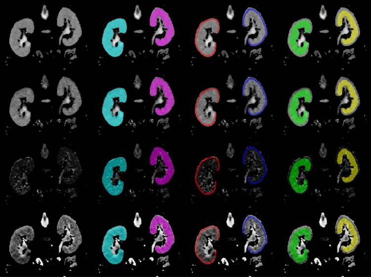 Figure 1
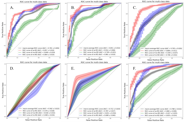 Figure 2
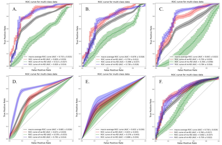 Figure 3
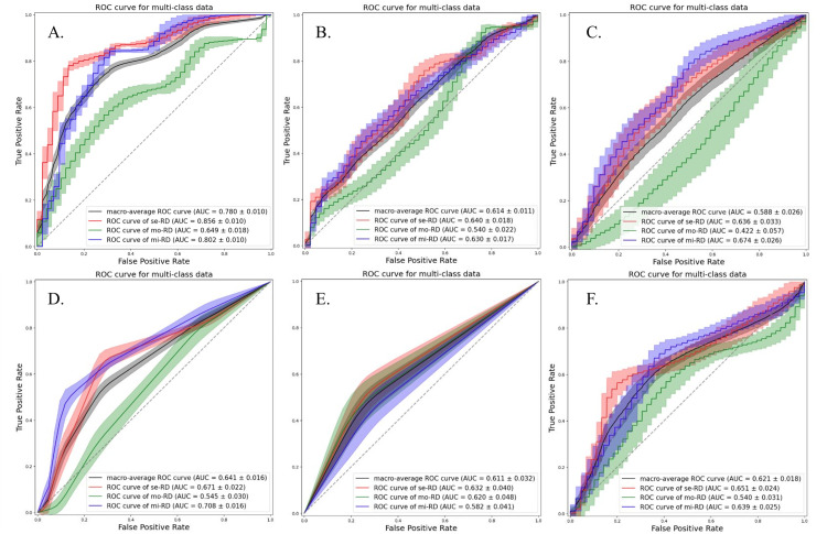 Figure 4
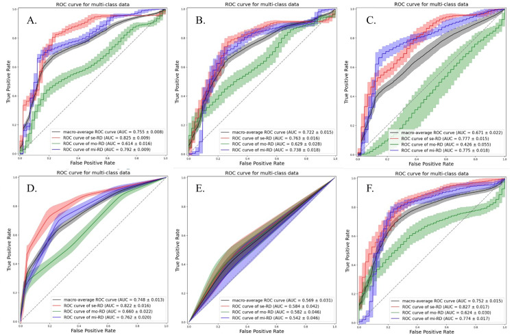 Figure 5
 Figure 6
Peer Reviews
No public reviews on file for this paper yet. If you reviewed it on a platform where reviews are public (OpenReview, ICLR, NeurIPS, ICML), you can paste yours below so the community can read it here.
Videos
No videos yet. Explain this paper in a talk, walkthrough, or lecture? Add one.
Taxonomy
TopicsMRI in cancer diagnosis · Radiomics and Machine Learning in Medical Imaging · Renal and Vascular Pathologies