Spinal cord versus brain imaging biomarkers of multiple sclerosis trajectory combining 7T and 3T MRI
Alessandro Miscioscia, Constantina A Treaba, Elena Barbuti, Valeria T Barletta, Jacob A Sloane, Eric C Klawiter, Julien Cohen-Adad, Paolo Gallo, Patrizia Pantano, Caterina Mainero

TL;DR
This study compares brain and spinal cord MRI biomarkers in multiple sclerosis to determine which best predict disability and disease progression.
Contribution
The study identifies cortical lesions as the strongest predictor of disease progression independent of relapse activity in multiple sclerosis.
Findings
Cortical lesion volume was the strongest independent predictor of progression independent of relapse activity.
Spinal cord atrophy primarily explains current disability, while brain white matter atrophy and paramagnetic rim lesions predict future disability trajectory.
Non-rim white matter lesions and cervical spinal cord atrophy mainly reflect current disability levels.
Abstract
In multiple sclerosis, different types of lesions and their localization can have varying effects on clinical disability and disease progression. Ultra-high field 7-Tesla MRI improves the visualization of cortical, especially subpial, lesions and of white matter lesions with a paramagnetic rim that are associated with smoldering inflammation. Spinal cord atrophy is also a critical determinant of clinical disability in multiple sclerosis, but its importance relative to paramagnetic rim and cortical lesions in predicting neurological disability and its progression remains unclear. In this longitudinal study, we aimed to identify the most relevant predictors of both the baseline Expanded Disability Status Scale status and 4-year progression independent of relapse activity in a heterogeneous multiple sclerosis cohort. One-hundred-twelve patients (83 relapsing-remitting and 29 secondary…
Genes, proteins, chemicals, diseases, species, mutations and cell lines named across the full text — each resolved to its canonical identifier and authoritative record.
Click any figure to enlarge with its caption.
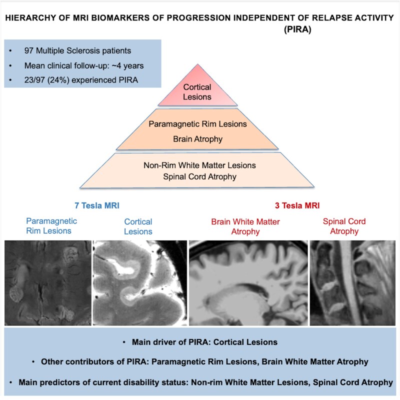 Figure 1
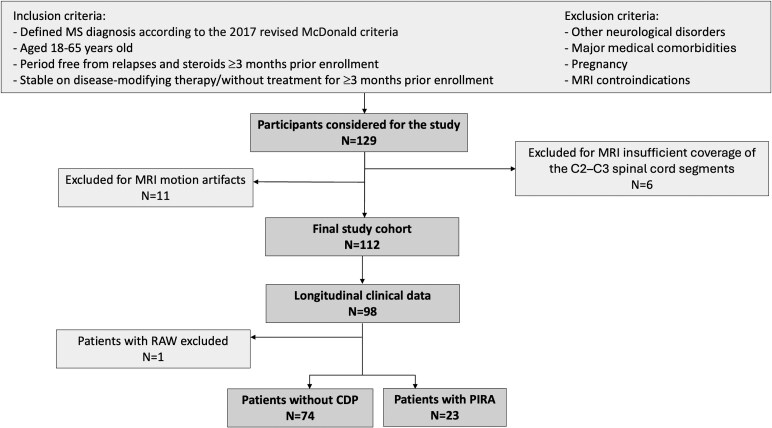 Figure 2
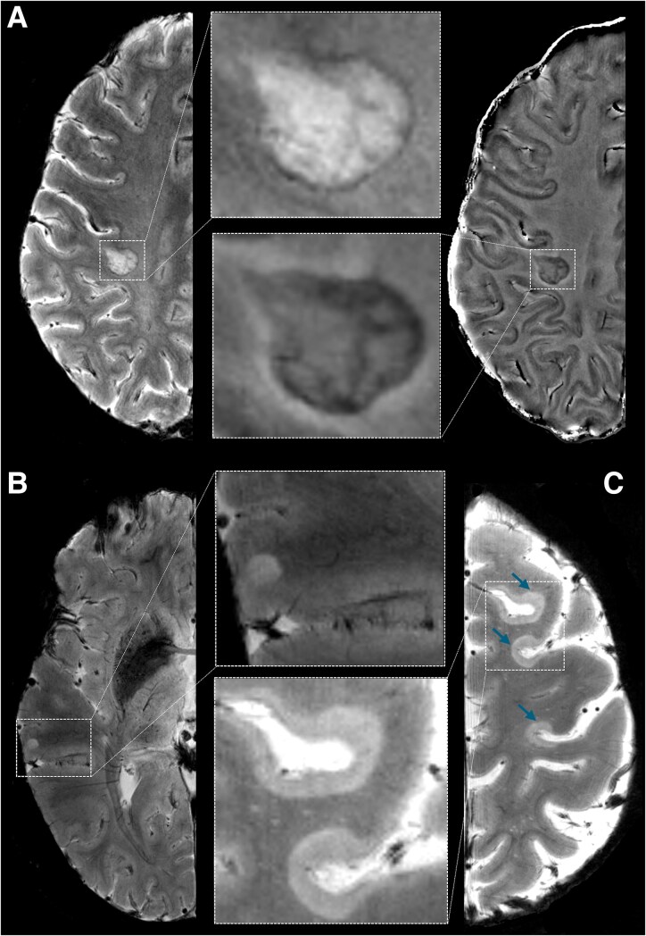 Figure 3
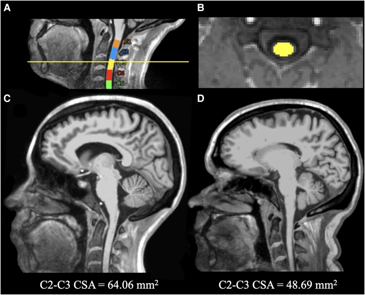 Figure 4
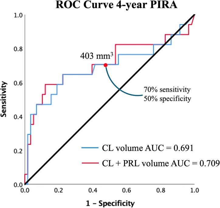 Figure 5
Peer Reviews
No public reviews on file for this paper yet. If you reviewed it on a platform where reviews are public (OpenReview, ICLR, NeurIPS, ICML), you can paste yours below so the community can read it here.
Videos
No videos yet. Explain this paper in a talk, walkthrough, or lecture? Add one.
Taxonomy
TopicsMultiple Sclerosis Research Studies · Amyotrophic Lateral Sclerosis Research · Peripheral Neuropathies and Disorders