Clinical and histopathological characterization of metastatic lobular breast cancer: lessons learned from post-mortem tissue donation programs
Gitte Zels, Karen Van Baelen, Alexander CC Chang, Anirudh Pabba, Maxim De Schepper, Marion Maetens, François Richard, Josephine Van Cauwenberge, Tatjana Geukens, Kristien Borremans, Amena Mahdami, Ha Linh Nguyen, Sophia Leduc, Patrick Neven, Hans Wildiers, Vincent Vandecaveye

TL;DR
This study examines metastatic lobular breast cancer using post-mortem tissue donations to better understand tumor characteristics and improve detection methods.
Contribution
The study provides new insights into intra-patient heterogeneity and detection accuracy in metastatic lobular breast cancer using post-mortem data.
Findings
Metastases showed lower ER and PR expression and higher KI67 compared to primary tumors.
HER2-low metastases were common across patients but varied in proportion.
Imaging and pathology had moderate agreement in detecting metastases, highlighting limitations in current methods.
Abstract
While primary invasive lobular carcinoma (ILC) is well characterized, metastatic ILC remains understudied. Within the post-mortem tissue donation programs, UPTIDER (Belgium) and Hope for Others (USA), we first aimed to explore intra-patient heterogeneity of key prognostic and predictive markers (stromal tumor-infiltrating lymphocytes (sTIL), estrogen receptor (ER), progesterone receptor (PR), human epidermal growth factor receptor 2 (HER2) and KI67). Secondly, we compared detection of the metastases by pathology on autopsy samples versus pre-mortem imaging. In total, 306 metastases from 12 patients were collected at autopsy (median: 27 per patient). Both primary tumors (n = 15) and metastases (n = 232) had low sTIL levels, with a median of 2% (range: 0.67–6.67%) and 0.67% (range: 0–13.33%), respectively. Regression models showed lower ER- and PR-expression in metastases (respectively, n…
Genes, proteins, chemicals, diseases, species, mutations and cell lines named across the full text — each resolved to its canonical identifier and authoritative record.
Click any figure to enlarge with its caption.
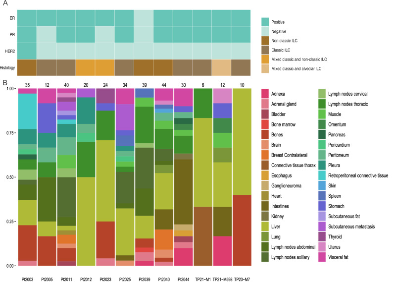 Figure 1
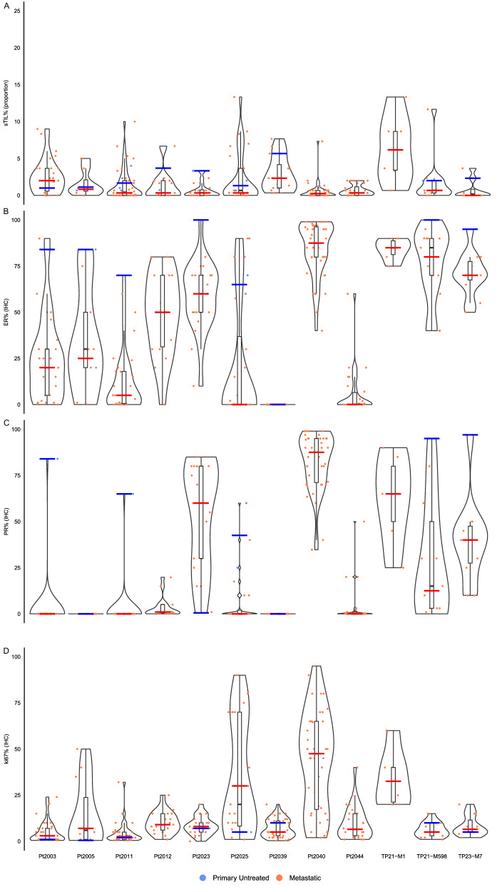 Figure 2
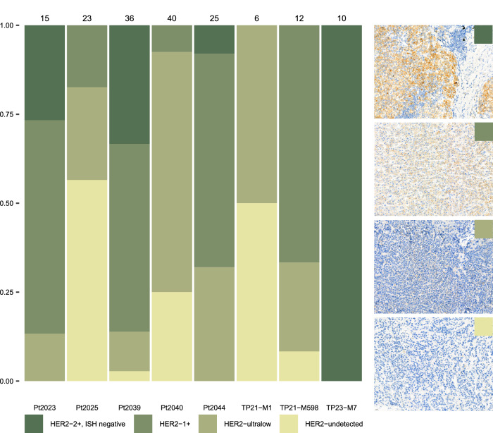 Figure 3
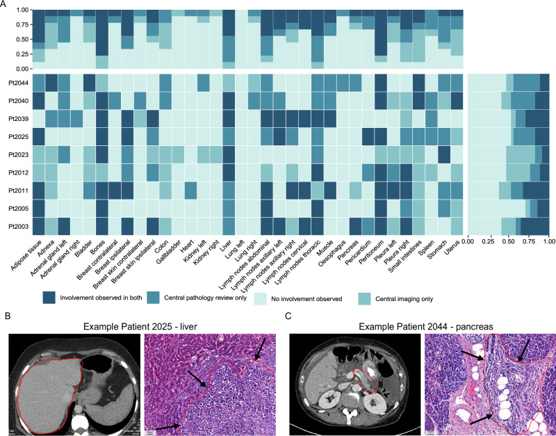 Figure 4
Peer Reviews
No public reviews on file for this paper yet. If you reviewed it on a platform where reviews are public (OpenReview, ICLR, NeurIPS, ICML), you can paste yours below so the community can read it here.
Videos
No videos yet. Explain this paper in a talk, walkthrough, or lecture? Add one.
Taxonomy
TopicsBreast Cancer Treatment Studies · Breast Lesions and Carcinomas · Metastasis and carcinoma case studies