Contextual dimensions of pediatric tuberculosis imaging: radiation exposure, access, and system capacity in high- and low-resource settings
Isabelle Munyangaju, Andreas Jahnen, Ridwaan Esmail, Benedita José, Jacinta Adrigwe, Criménia Mutemba, Patricia Pérez, José Miguel Escudero Fernández, Antoni Soriano-Arandes, Maria Espiau, Begoña Santiago Garcia, Alicia Hernanz-Lobo, Ángel Lancharro-Zapata, Aleix Soler-Garcia

TL;DR
This study compares pediatric tuberculosis imaging practices and radiation exposure in high-resource Spain and low-resource Mozambique to improve safe and equitable diagnosis.
Contribution
The study provides empirical evidence on imaging practices and radiation doses in pediatric tuberculosis across different resource settings.
Findings
Spanish children received multiple chest X-rays and CT scans, with lung doses below diagnostic reference levels.
Mozambican children had fewer X-rays and much lower radiation doses due to limited equipment and protocols.
Spain showed structured dose optimization, while Mozambique faced challenges with equipment and non-physician interpretation.
Abstract
Pediatric tuberculosis diagnosis relies heavily on imaging, yet access, equipment standards, and dose monitoring differ widely across health systems. Evidence describing how these contextual factors influence imaging use and radiation exposure in children remains scarce. To describe pediatric tuberculosis imaging practices and estimated radiation doses across two distinct resource settings, Spain (hospital-based, high-resource) and Mozambique (primary care-based, low-resource), to inform strategies for safe, equitable, and context-appropriate imaging. A descriptive mixed-methods study combined retrospective data of children (<16 years) diagnosed with tuberculosis (Spain 2015–2021; Mozambique 2018–2021) with complementary surveys of imaging providers. In Spain, chest X-ray and computed tomography parameters were extracted from digital imaging and communications in medicine files to…
Genes, proteins, chemicals, diseases, species, mutations and cell lines named across the full text — each resolved to its canonical identifier and authoritative record.
Click any figure to enlarge with its caption.
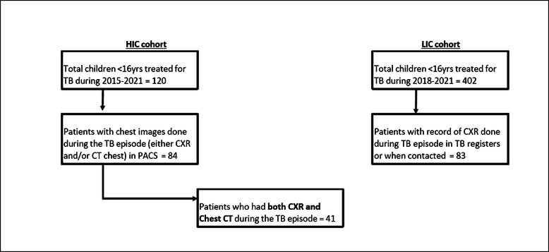 Figure 1
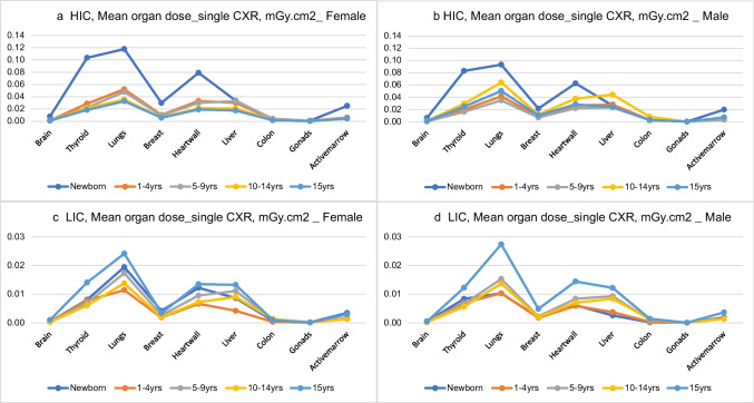 Figure 2
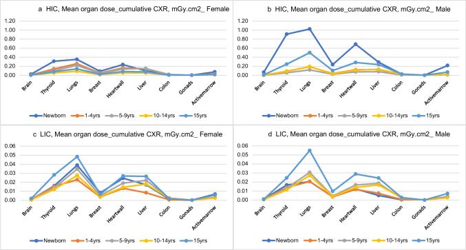 Figure 3
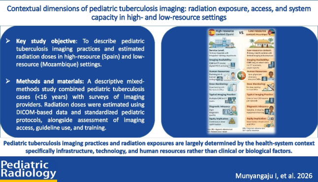 Figure 4
Peer Reviews
No public reviews on file for this paper yet. If you reviewed it on a platform where reviews are public (OpenReview, ICLR, NeurIPS, ICML), you can paste yours below so the community can read it here.
Videos
No videos yet. Explain this paper in a talk, walkthrough, or lecture? Add one.
Taxonomy
TopicsUltrasound in Clinical Applications · Radiation Dose and Imaging · Radiation Detection and Scintillator Technologies