Comprehensive deep learning-assisted multi-condition analysis of knee MRI studies improves resident radiologist performance
Roman Vuskov, Alexander Hermans, Martin Pixberg, Jonas Müller-Hübenthal, Andreas Brauksiepe, Eric Corban, Malin Cubukcu, Julia Nowak, Aleksandar Kargaliev, Marc von der Stück, Robert Siepmann, Christiane Kuhl, Daniel Truhn, Sven Nebelung

TL;DR
A deep-learning model for knee MRI analysis improves accuracy and efficiency of resident radiologists in detecting various knee conditions.
Contribution
A 3D slice transformer network for multi-tissue, multi-condition knee MRI analysis that enhances resident radiologists' diagnostic performance.
Findings
The model achieved an AUC of at least 0.85 for 8 conditions and 0.75 for 18 conditions.
Model assistance improved accuracy and sensitivity for inexperienced residents and increased inter-reader agreement.
Reading times for experienced residents were reduced by 10% with model assistance.
Abstract
Developing a deep-learning model for automated multi-tissue, multi-condition knee MRI analysis and assessing its clinical potential. This retrospective dual-center study included 3121 MRI studies from 3018 adults, who underwent routine knee MRI examinations at a radiologic practice (2012–2019). Twenty-three conditions across cartilage, menisci, bone marrow, ligaments, and other soft tissues were manually labeled. A 3D slice transformer network was trained for binary classification and evaluated in terms of the area under the receiver operating characteristic curve (AUC), sensitivity, and specificity using a five-fold cross-validation and an external test set of 448 MRI studies (429 adults) from a university hospital (2022–2023). To assess differences in diagnostic performance, two inexperienced and two experienced radiology residents read 50 external test studies with and without model…
Genes, proteins, chemicals, diseases, species, mutations and cell lines named across the full text — each resolved to its canonical identifier and authoritative record.
Click any figure to enlarge with its caption.
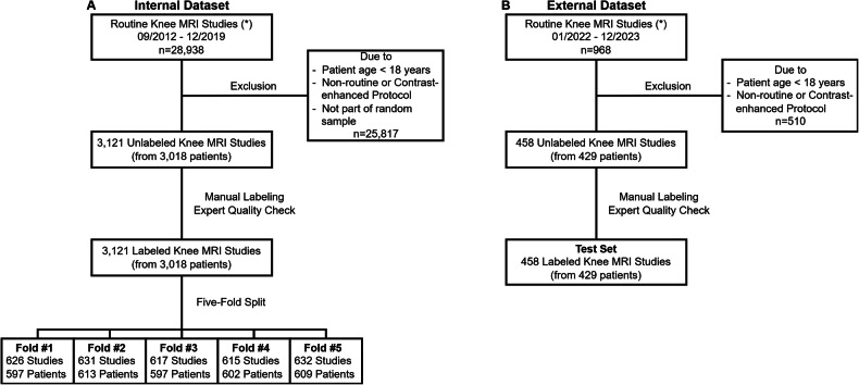 Figure 1
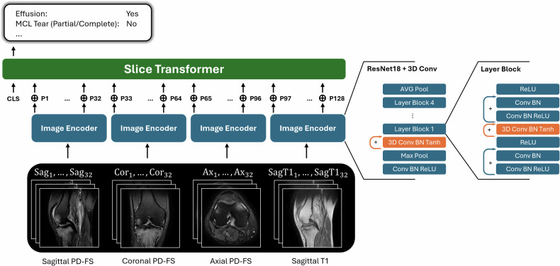 Figure 2
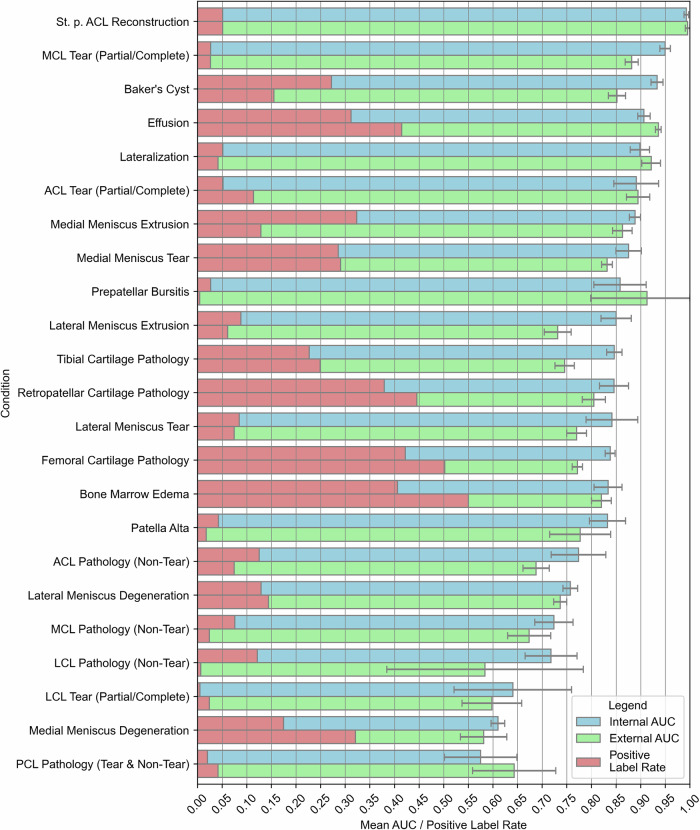 Figure 3
 Figure 4
 Figure 5
 Figure 6
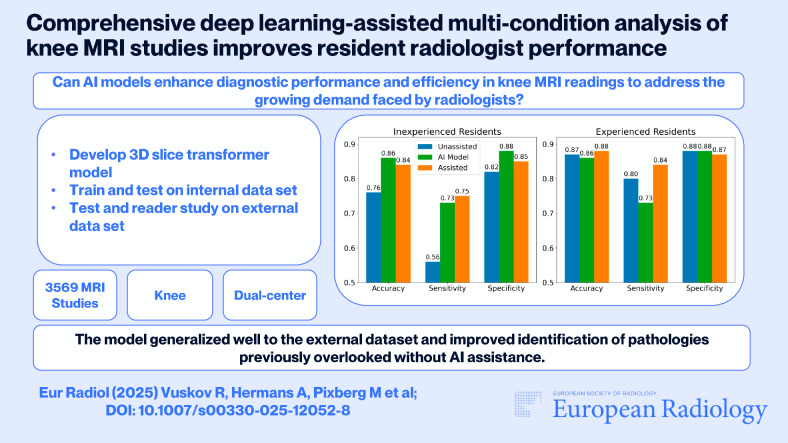 Figure 7
Peer Reviews
No public reviews on file for this paper yet. If you reviewed it on a platform where reviews are public (OpenReview, ICLR, NeurIPS, ICML), you can paste yours below so the community can read it here.
Videos
No videos yet. Explain this paper in a talk, walkthrough, or lecture? Add one.
Taxonomy
TopicsRadiomics and Machine Learning in Medical Imaging · Artificial Intelligence in Healthcare and Education · Advanced X-ray and CT Imaging