Loss of AMPK potentiates inflammation by activating the infammasome in a preclinical mouse model of TBI
Mohammad Ejaz Ahmed, Hamid Suhail, Mohammad Nematullah, Benoit Viollet, Shailendra Giri, Abdullah Shafique Ahmad

TL;DR
This study shows that loss of AMPK after traumatic brain injury worsens inflammation and brain damage in mice, suggesting that restoring AMPK activity could be a new treatment strategy.
Contribution
The study identifies AMPKα1 as a key regulator of neuroinflammation and tissue damage after TBI, offering a novel therapeutic target.
Findings
AMPK loss after TBI correlates with worsened behavioral deficits and increased NLRP3 inflammasome activation.
AMPKα1-KO glial cells show heightened inflammatory responses compared to wild-type cells.
Restoring AMPKα1 activity may help reduce neuroinflammation and brain injury damage.
Abstract
Traumatic brain injury (TBI) is a major cause of mortality and long-term neurological disabilities. Adenosine monophosphate-activated protein kinase (AMPK), a key cellular energy sensor, plays a critical role in maintaining energy homeostasis. Loss of AMPK phosphorylation following TBI impairs the restoration of cellular energy homeostasis and promotes inflammation. In this study, we investigated whether post-TBI loss of AMPK worsens functional impairments, amplifies inflammation, and exacerbates tissue damage in a mouse model of TBI. Adult male C57BL/6 wild-type (WT) and (AMPKα1-KO) mice were subjected to TBI or sham surgery. Behavioral assessments were performed at 24 h post-TBI, followed by mice were anesthetized, and their brains were rapidly collected for histological and biochemical analyses. To further support our findings, mixed glial cells isolated from WT and AMPKα1-KO pups…
Genes, proteins, chemicals, diseases, species, mutations and cell lines named across the full text — each resolved to its canonical identifier and authoritative record.
Click any figure to enlarge with its caption.
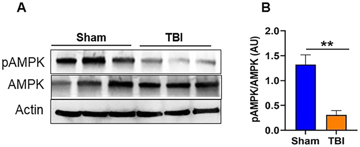 Figure 1
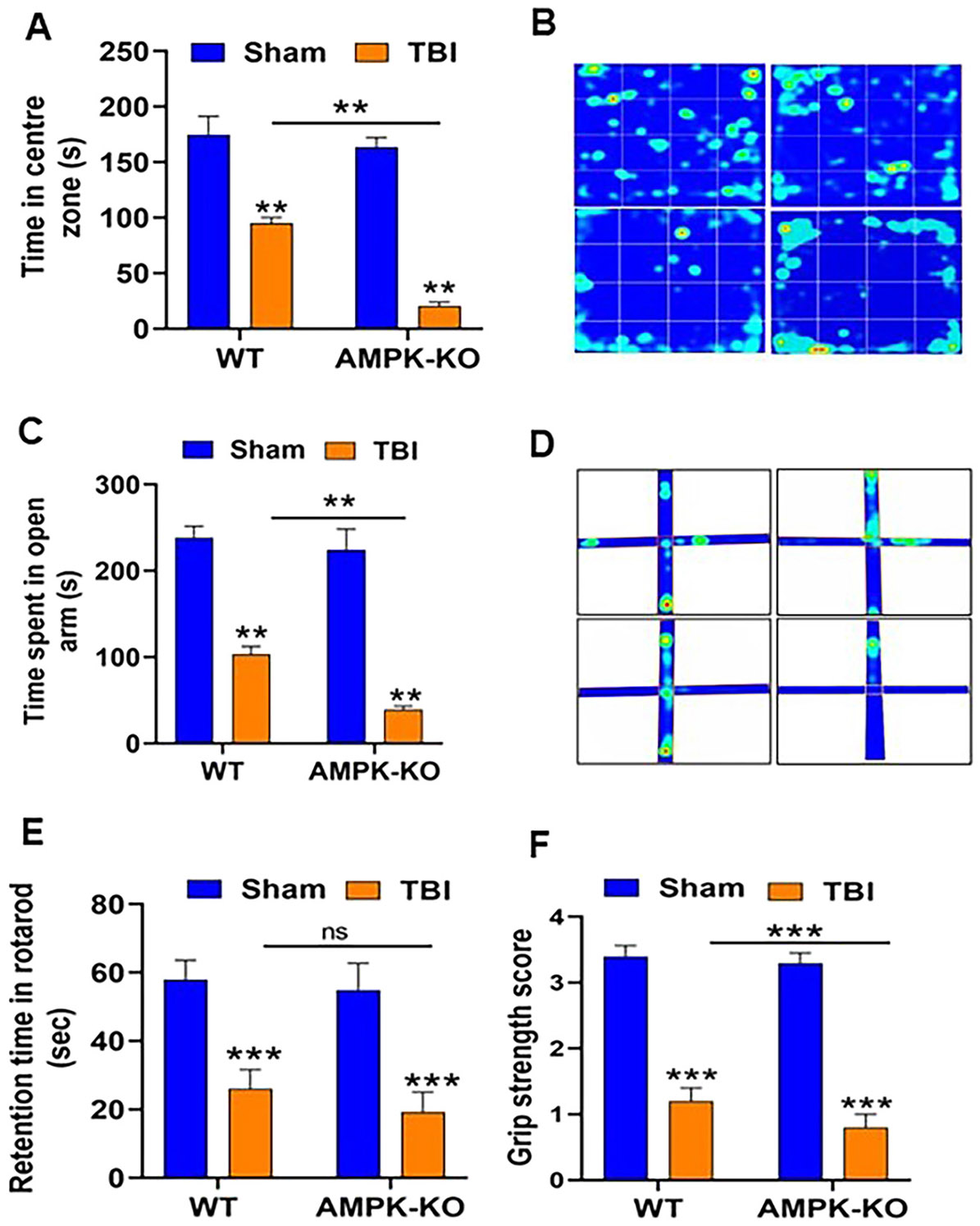 Figure 2
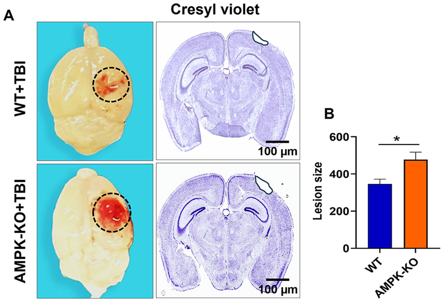 Figure 3
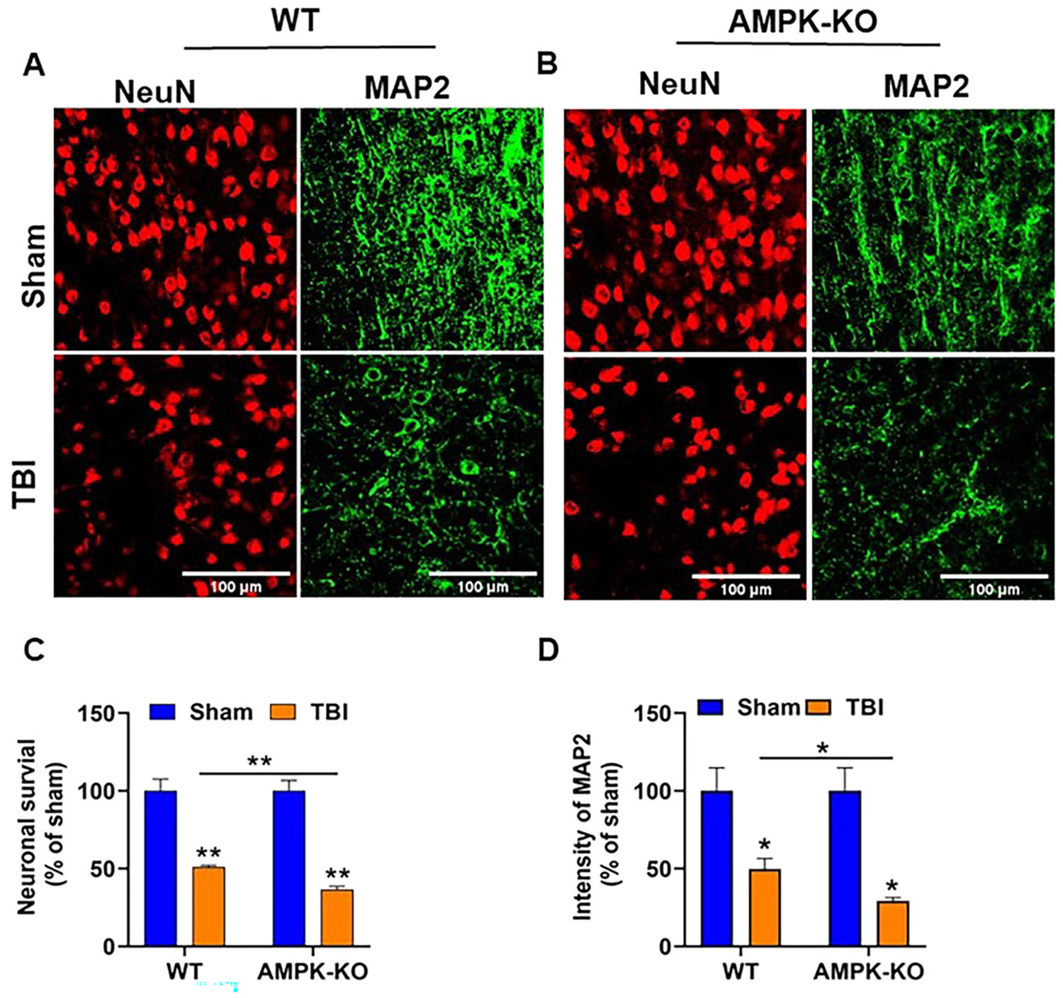 Figure 4
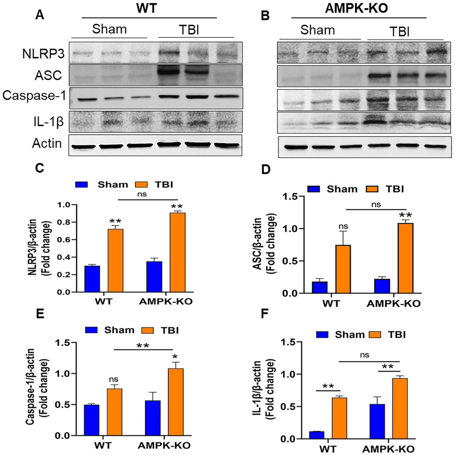 Figure 5
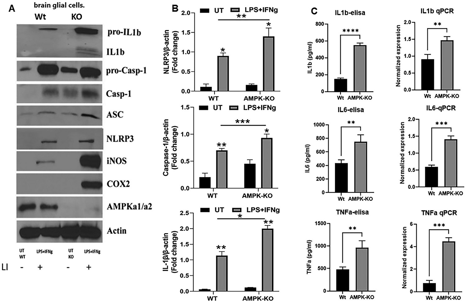 Figure 6
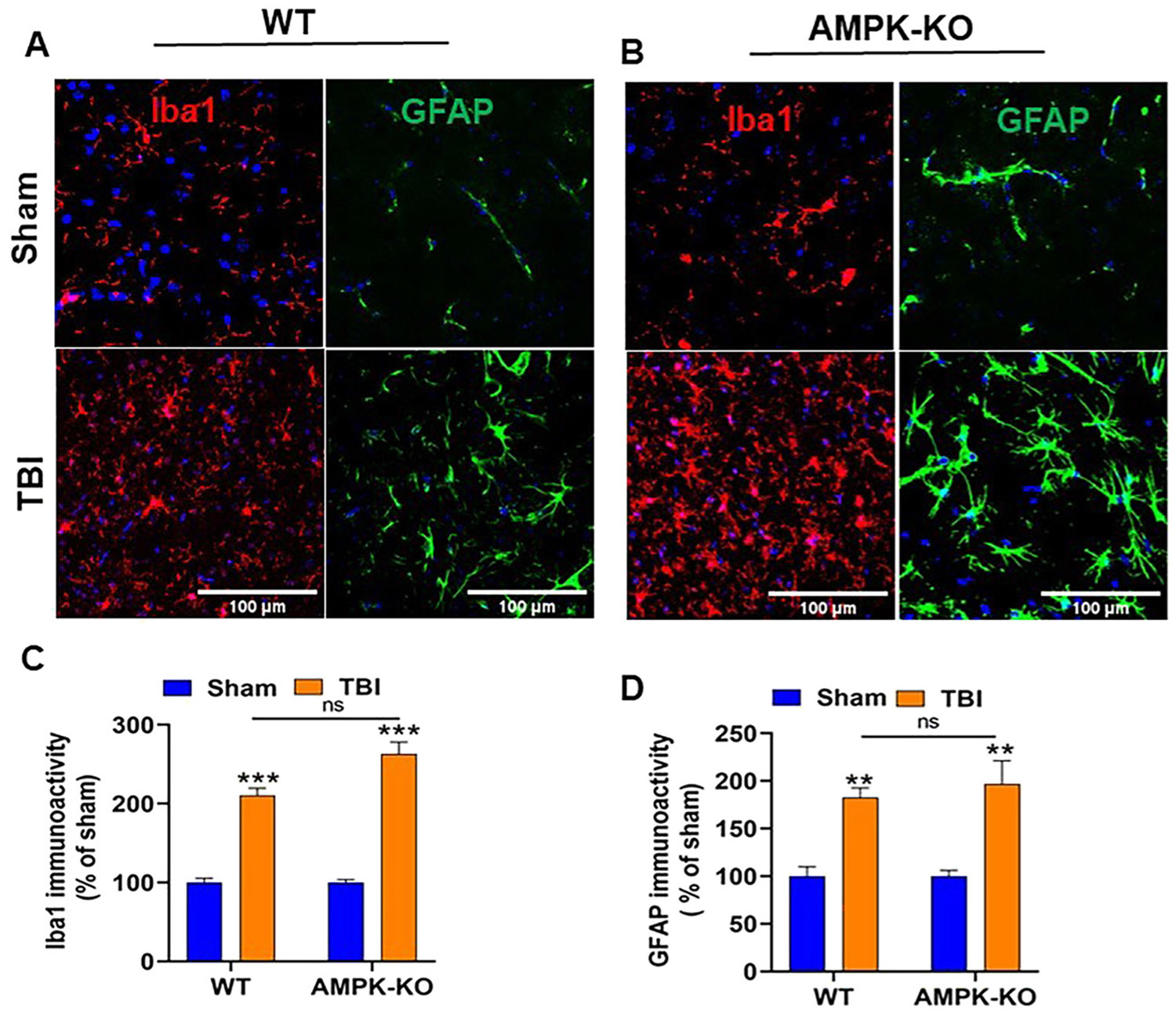 Figure 7
Peer Reviews
No public reviews on file for this paper yet. If you reviewed it on a platform where reviews are public (OpenReview, ICLR, NeurIPS, ICML), you can paste yours below so the community can read it here.
Videos
No videos yet. Explain this paper in a talk, walkthrough, or lecture? Add one.
Taxonomy
TopicsMetabolism, Diabetes, and Cancer · Cancer, Hypoxia, and Metabolism · Traumatic Brain Injury and Neurovascular Disturbances
Introduction
Traumatic brain injury (TBI) remains a major contributor to mortality and long-term disability in the United States and worldwide. It results from external mechanical forces that damage brain tissue, triggering a cascade of secondary events, including inflammation and neuronal death. TBI imposes significant emotional and financial burdens on patient families, often requiring long-term care [1, 2]. Despite advances in clinical care, accurately diagnosing TBI severity and implementing effective treatments remain challenging due to the diverse nature of the injury and the complexity of the secondary process. These challenges underscore the urgent need for improved diagnostic tools and therapeutic strategies to enhance outcomes for TBI patients.
Both preclinical and clinical studies have demonstrated that TBI leads to a long-lasting reduction in glucose metabolism, which adversely affects brain function [3-5]. Recent advances in metabolic analysis indicate that pathways regulating energy metabolism play a critical role in controlling neuroinflammation after TBI. AMPK is a serine/threonine kinase that acts as a central cellular energy sensor, and is rapidly activated in response to metabolic stress, hypoxia, excitotoxicity, and altered AMP/ATP or ADP/ATP ratios [6-8] and protect cells against energy depletion by inhibiting anabolic pathways and promoting ATP-generation and increase AMP/ATP ratio [9, 10]. Following TBI, loss of AMPKα1 activity contributes to impaired bioenergetic homeostasis, which in turn amplifies inflammatory response and neuronal vulnerability [11]. Thus, AMPKα1 represents a key molecular link between metabolic dysfunction and neuroinflammation. Following TBI, AMPKα1 activation typically acts to restore cellular energy balance by promoting ATP-generating pathways and regulating energy consumption [12-14].
Recently, AMPK and the NLRP3 inflammasome have attracted significant attention from both preclinical and clinical researchers. The involvement of AMPK in the progression and pathogenesis of various diseases has led to the development of new therapeutic strategies. The inflammasome is a multiprotein complex composed of three key subunits: the sensor molecule NLRP3, the adaptor protein, ASC, and an effector protein, caspase −1. The complex plays a vital role in the innate immune defense by activating inflammatory responses through maturation and release of pro-inflammatory cytokines, such as interleukin-1β (IL-1β), and interleukin-18 (IL-18) [15-18]. Many studies have demonstrated that TBI triggers a complex immune response characterized by the activation of the NLRP3 inflammasome [19, 20]. Previous studies have shown significant increases in the NLRP3, caspase-1, and IL-1β expression at 12 h and 72 h in post-TBI mice [21, 22]. Among the inflammasomes expressed in the brain, NLRP3 promotes the cleavage and activation of caspase-1 and IL-1β, thereby amplifying the inflammatory response following TBI [23-25]. In an animal model of TBI, elevated levels of NLRP3 and the inflammasome complex in the ipsilateral cortex are correlated with long-term functional outcomes [26]. The present study reveals an important role of AMPK/NLRP3 inflammasome-mediated neuroinflammation in TBI. To our knowledge, no previous studies have directly examined the impact of AMPK on NLRP3 inflammasome activation in the controlled cortical impact (CCI) induced mouse model of TBI.
Materials and methods
Animals and experimental groups
Adult male C57BL/6 wild-type (WT) and AMPKα1 global knockout (B6/129 background) were used as previously described [27, 28]. These mice were provided by Benoit Viollet (INSERM, Paris) and were generated as reported earlier [29]. The mice were housed and maintained in the animal facility at Henry Ford Health (HFH). Animals were randomly divided into four groups: (i) WT sham (n=10), (ii) WT+TBI (n=10), (iii) AMPKα1-KO sham (n=10), and (iv) AMPKα1-KO+TBI (n=10). All experiments were conducted in accordance with protocol approved by the Henry Ford Health (HFH) Institutional Animal Care and Use Committee (IACUC). Mice were maintained at room temperature (21±1 °C) and 50±5 % humidity under a 12 h dark/light cycle, with ad libitum access to food and water.
Induction of traumatic brain injury
WT and AMPKα1-KO mice underwent either sham or TBI surgery using the controlled cortical impact (CCI) model as previously described [30]. Briefly, mice were anesthetized with 3 % isoflurane for induction, secured in a stereotaxic frame, and maintained under 1.5–2 % isoflurane throughout the procedure using a SomnoSuite low-flow anesthesia system (Kent Scientific Corporation). A craniotomy was performed in the right parietal bone midway between bregma and lambda, 1 mm lateral to the midline, using a hand-held twist drill while leaving the dura intact. The mice were impacted at a velocity of 3 m/s with an 85 ms dwell time and 1.0 mm depth using a 3 mm diameter impactor tip (Pin Point PCI3000 Precision Cortical Impactor, Hatters Instruments). Sham-operated mice underwent the same surgical procedure, without cortical impact. Body temperature was maintained at 37.0±0.5 °C throughout the procedure using a small-animal temperature control system (Kopf Instrument). After surgery, mice were returned to a clean home cage placed on an automatic warming pad (Kent Scientific Corporation).
Behavioral analysis
Open field test
The open-field test was used to evaluate locomotor and exploratory activities in rodents [31, 32]. Mice were assessed for the open field test at 24 h post-TBI. Each mouse was placed in the center of the open field apparatus and allowed to explore freely for a 5-min observation period. The time spent in the center zone was recorded and analyzed using the automated ANY-maze video tracking software. The apparatus was cleaned with 70 % ethanol between each test.
Elevated plus maze test
The elevated plus maze (EPM) test is one of the most widely used methods for assessing anxiety-like behavior in rodents [33]. The EPM test was performed at 24 h post-TBI in mice. Individual mice were placed in the center of the maze and allowed to explore for 5 min. Mouse activity was recorded and analyzed using automated ANY-maze video tracking software. Anxiety-like behavior was evaluated by measuring the time spent in the open arm and the number of open arm entries; mice exhibiting higher anxiety spent less time in the open arm. The EPM apparatus was cleaned with 70 % ethanol between each test.
Rotarod test
Motor coordination and balance in post-TBI mice were evaluated using a rotarod apparatus, which consists of rotating rods with a rough, solid surface (30 mm in diameter), elevated 30 cm above the floor and divided into four separate compartments, allowing simultaneous testing of up to four mice. Mice were placed on a rotating rod, which initially rotated at 5 revolutions per minute (rpm) and gradually accelerated at a fixed interval to a maximum speed of 20 rpm [34, 35]. Each trial lasted for a maximum duration of 5 min. Each mouse completed three trials, with approximately 30 min of rest between each trial. The average of latency to fall from the three trials was used as the performance score for each mouse.
Hanging wire test
Grip strength was assessed at 24 h post-TBI in mice as previously described [32]. A wire was stretched between two poles 100 cm apart and 100 cm above the floor. Mice were gently held by the tail and allowed to grasp the wire with their forepaws; the support was then withdrawn, allowing the mouse hang freely from the wire. Each trial was scored on a 5-point scale: 0, fall off; 1, hung by both forepaws; 2, hung by both forepaws with attempts to climb; 3, hung by both forepaws and one or both hind limbs; 4, hung by forepaws with the tail wrapped around the wire; and 5, escape by reaching one of the poles. To prevent injury, a thick, soft mat was placed under the hanging area during testing.
Western blot analysis
Western blot analysis was performed as previously described [36]. Protein lysates were prepared from the ipsilateral cortical tissue adjacent to the lesion site. This peri-lesion area was selected because it is functionally and metabolically active and undergoes pronounced neuroinflammatory, metabolic, and neuronal changes following TBI. Briefly, 35 μg protein was equally loaded into each well and separated on 4–20 % SDS–PAGE gels. Protein bands were then transferred onto nitrocellulose membranes (Thermo Scientific) and blocked with 5 % nonfat milk for 1 h at room temperature. Membranes were incubated overnight at 4 °C with the appropriate primary antibodies against AMPKα (BS 10344R, Bioss, Woburn, MA), pAMPKα (Thr183/Thr172; 2535S, Cell Signaling), and the NLRP3 (15101, Cell Signaling), IL-1 β (83186S, Cell Signaling), caspase-1(89332T, Cell Signaling). After three washes with TBST, membranes were incubated with the appropriate HRP-conjugated secondary antibody for 2 h at room temperature. The membranes were subsequently washed, and the protein bands were visualized with an enhanced chemiluminescence (ECL) solution (Clarity Western ECL Substrate, 1705062 Bio-Rad Labs, Des Plaines, IL). Quantitative analysis of the bands was performed using ImageJ software (NIH).
Cresyl violet staining and quantification of lesion size
Coronal brain sections (25 μm thick) were collected using a cryostat at 24 h after TBI. Sections were stained with 0.1 % (w/v) Cresyl Violet for 5 min, followed by dehydration through graded ethanol concentrations and cleaning in xylene. Lesion volume in each stained section was quantified using ImageJ software (NIH). The mean lesion volume was calculated by subtracting the area of the ipsilateral hemisphere from that of the contralateral hemisphere [37, 38].
Immunofluorescence staining and quantification
Brains were harvested at 24 h post – TBI and processed for immunofluorescence staining as previously described [36, 39]. Briefly, 25 μm thick brain sections were washed, permeabilized with 0.4 % PBST, and blocked with 0.1 % BSA for 1 h at room temperature. Sections were then incubated overnight at 4 °C with primary antibodies against NeuN (PA5-78499, Invitrogen), MAP2 (MAB3418, Millipore), GFAP (PA5-16291, Thermo Fisher), and Iba1 (016-20001, Wako). The following day, sections were washed 3 times with 0.1 % PBST and incubated for 1 h at room temperature with Alexa Fluor 568/488-conjugated goat anti-mouse/rabbit secondary antibody (1:500; Thermo Scientific Rockford, IL). After washing, sections were mounted on slides with Vectashield mounting medium with 4′,6-diamidino-2-phenyl indole (DAPI) (H-1200; Vector Laboratories, Inc., CA, USA) and covered with a coverslip. Confocal images were acquired using Zeiss AxioTvert 200/Axiovert 200 M microscopes. For quantification, five randomly selected cortical fields per animal were analyzed in four nonadjacent sections spaced approximately 100 μm apart using ImageJ software (NIH).
Primary brain glial cell culture
The brains of 2- to 3-day-old WT and AMPKα1-KO mouse pups were used to prepare mixed glial cell cultures. We followed the previously published protocol to prepare primary mixed glial cell cultures [40-43]. Briefly, the brains were removed, and the cerebral cortices were isolated and cleared of blood vessels and meninges. Cerebral tissues were mechanically dissociated into a single cell suspension using a Pasteur pipette and resuspended in complete medium (DMEM 4.5 g/L glucose) supplemented with 10 % FBS and 10 mg/mL antibiotics. Cells were seeded into poly-d-lysine-coated 75 cm^2^ flasks and maintained at 37 °C in a humidified 5 % CO_2_ incubator. After 24 h, the media were replaced with fresh complete DMEM and subsequently changed twice per week. Culture was maintained for 12–14 days until they reached full confluence and were ready for dissociation.
Brain glial cell stimulation
Glial cells isolated from WT and AMPKα1-KO mice were seeded at a density of 50 × 10^4^ cells per well in 24-well plates containing DMEM supplemented with 10 % fetal bovine serum (FBS). Cells were incubated overnight at 37 °C in a humidified atmosphere with 5 % CO_2_. The following day, the culture medium was replaced with serum-free DMEM, and the cells were serum-starved for 2 h. After serum starvation, cells were stimulated with LI (0.1 μg/ml LPS and 20 ng/ml IFNγ) to induce an inflammatory response. Culture supernatants were collected for cytokine ELISA, and cell lysates were processed for qPCR and immunoblot analyses.
RNA extraction, cDNA synthesis, and quantitative PCR
After stimulation with LI, WT, and AMPKα1-KO glia cells were harvested at 6 h, and total RNA was extracted using Triazole followed by purification with a RNeasy Kit (Qiagen) according to the manufacturer’s instructions. cDNA was synthesized from 1 μg of total RNA using an iScript reverse transcription cDNA synthesis kit (Bio-Rad). Primer sets were selected from a publicly available database and purchased from Integrated DNA Technology (IDT, Coralville, IA, USA). Quantitative real-time PCR was performed using iTaq Universal SYBR Green Supermix (Bio-Rad). Thermal cycling amplification conditions were as follows: polymerase activation at 95 °C for 3 min, followed by 40 cycles of amplification at 95 °C for 30 s and 60 °C for 30 s. Expression of the ribosomal protein L27 housekeeping gene was used for normalization, and relative gene expression was calculated using the (^^Ct) method with CFX Maestro Software.
Cytokine analysis by enzyme-linked immunosorbent assay (ELISA)
To analyze cytokine levels, isolated glial cells were stimulated with LI (0.1 μg/ml LPS and 20 ng/ml IFNg). After 6 h of stimulation, the culture plates were washed three times with warm serum-free media, and the supernatant was collected to measure inflammatory cytokine production by ELISA.
Statistical analysis
Data are represented as mean±SEM. Statistical analyses were performed using GraphPad Prism 9 with either oneway analyses of variance (ANOVA) or Student’s t-test, as appropriate. For multiple group comparison, the Tukey post-hoc test was applied. A 95 % confidence level (p<0.05) was considered statistically significant.
Results
TBI causes loss of AMPKα1 phosphorylation in the mouse brain
WT mouse brains were harvested 24 h after TBI, and Western blot analyses were performed to assess AMPKα1 phosphorylation at Thr172. Representative immunoblots and corresponding quantification (Figure 1A and B) show a significant reduction in phospho-AMPKα1 levels in post-TBI mice compared with sham-operated controls. These results suggest that TBI leads to a decrease in AMPKα1 activity, which may contribute to an overall suppression of AMPKα1 signaling in the injured brain. This initial observation of loss of AMPKα1 phosphorylation in WT mice provides the rationale for further examination of whether loss of AMPKα1 directly contributes to post-TBI neuropathology. To address this question, we extended our studies in AMPK-α1-KO mice and glial cells to investigate the functional and inflammatory consequences of AMPK-α1 loss following TBI in mice.
AMPKα1 deficiency worsened functional deficits in mice following TBI
To investigate the impact of AMPKα1 loss on functional outcomes after TBI, WT and AMPKα1-KO mice were assessed for behavioral impairments at 24 h post-injury. Spontaneous locomotor activity was evaluated using the open-field test by measuring the time spent in the center of the field. AMPKα1-KO TBI mice spent significantly less time in the central zone than WT TBI mice (Figure 2A and B), indicating increased anxiety-like behavior. Anxiety-like behavior was further assessed using the elevated plus maze at 24 h post-TBI. AMPKα1-KO mice spent less time in the open arm compared with WT mice, confirming that AMPKα1 deficiency exacerbates anxiety-like behavior after TBI mice (Figure 2C and D). Motor coordination and grip strength were evaluated using the rotarod and hanging wire tests, respectively (Figure 2E and F). AMPKα1-KO TBI mice exhibited significantly more severe motor deficits compared with WT TBI mice. Collectively, these findings demonstrate that loss of AMPKα1 worsens functional impairment following TBI.
Loss of AMPKα1 increases lesion size in TBI mice
The impact of AMPK loss on intracranial hemorrhage and lesion size was evaluated at 24 h post-TBI in WT and AMPKα1-KO mice. Representative whole-brain images revealed a marked increase in intracranial hemorrhage in AMPKα1-KO mice compared with WT mice (Figure 3A). Quantitative analysis confirmed that AMPKα1-KO mice exhibited significantly greater intracranial hemorrhage than WT TBI mice. In addition, lesion size in the ipsilateral hemisphere was significantly larger in AMPKα1-KO mice than in WT mice (Figure 3B). These findings indicate that the loss of AMPK exacerbates brain injury after TBI, resulting in increased bleeding and more extensive tissue damage.
Loss of AMPKα exacerbates TBI-induced neuronal damage in the mouse brain
We next examined the impact of AMPKα1 loss on TBI-induced neuronal loss using immunofluorescence staining. Brain sections from WT and AMPKα1-KO mice were stained with the neuron-specific markers microtubule-associated protein 2 (MAP2, green) and (NeuN, red). MAP2 primarily labels the neuronal cytoskeleton, including dendrites, whereas NeuN marks neuronal nuclei and perinuclear cytoplasm (Figure 4A and B). Compared with WT TBI mice, AMPKα1-KO TBI mice showed significantly reduced MAP2 immunoreactivity and NeuN staining (Figure 4C and D), indicating exacerbated neuronal loss at 24 h post-TBI.
AMPKα1 deficiency exacerbates NLRP3 inflammasome-mediated neuroinflammation in TBI mouse brains
Inflammasomes play a crucial role in regulating the inflammatory response following TBI. Representative western blot images from AMPKα1-KO TBI mice exhibited significantly higher expression of NLRP3, ASC, caspase-1, and IL-1β, compared with WT TBI mice (Figure 5A and B). Quantitative analysis confirmed elevated levels of these inflammasome markers in AMPKα1-KO mice relative to WT TBI mice (Figure 5C-F). Notably, during TBI, loss of AMPKα1 was associated with an increase in NLRP3 inflammasome activation, suggesting a potential inverse regulatory relationship between AMPKα1 activity and inflammasome signaling.
To further explore this relationship, glial cells isolated from WT and AMPKα1-KO pups were stimulated with inflammatory agents LI (0.1 μg/ml LPS and 20 ng/ml IFNγ) to assess inflammation and inflammasome activation. Western blot, qPCR, and ELISA were used to measure these markers. AMPKα1 deficiency led to increased levels of inflammatory mediators, including iNOS and Cox2, accompanied by enhanced inflammasome activity, as indicated by elevated NLRP3, ASC, cleaved caspase-1, and IL-1β levels compared to WT glial cells (Figure 6A). Quantitative analysis of key inflammasome components NLRP3, caspase-1 and 1L-1β revealed significantly higher band intensity in AMPKα1-KO cells than in WT cells (Figure 6B). Similarly, inflammatory cytokines, including IL-1β, IL-6, and TNFα, were significantly elevated in AMPK-KO glial cells under inflammatory conditions (Figure 6C). These results indicate that AMPKα1 functions as a negative regulator of inflammation and inflammasome activation.
AMPKα1 deficiency increases TBI-induced gliosis in the mouse brain
Immunofluorescence staining was performed to assess microglial and astrocyte activation in the ipsilateral cortex at 24 h post-TBI. Results showed increased immunoreactivity of GFAP (green), an astrocyte marker, and Iba1 (red), a microglial marker, in AMPKα1-KO mice compared with WT mice (Figure 7A and B). Quantitative analysis indicated elevated GFAP and Iba1 expression in AMPKα1-KO TBI mice; The difference between WT and AMPKα1-K OTBI mice was not statistically significant (Figure 7C and D).
Discussion
Our study provides novel insight into the role of AMPK in the context of TBI and associated inflammation. We observed a significant reduction in AMPKα1 activity, as evidenced by decreased phosphorylation at Thr172 following TBI in WT mice. This reduction in AMPKα1 phosphorylation was associated with worsened functional outcomes and increased neuroinflammation characterized by the upregulation of NLRP3 inflammasome and pro-inflammatory cytokines. Furthermore, we demonstrated that reduced AMPK levels exacerbate post-TBI inflammation, which is accompanied by aggravated functional deficits, increased brain tissue damage, and enhanced neuronal loss. Collectively, these findings reveal a previously unexplored role of AMPK in modulating inflammation and brain damage following TBI.
TBI induces distinct metabolic responses in the brain, characterized by an acute rise in metabolic activity and increased glucose utilization that lasts for up to 30 min post-injury [4]. However, this initial hypermetabolic phase is followed by a prolonged period of hypometabolism, which can be detected as early as 6 h post-injury and persists for days to weeks thereafter [3, 4, 44]. In our study, we initially examined the phosphorylation status of AMPK in the ipsilateral cortex 24 h post-TBI in WT TBI mice. Our results revealed that TBI significantly reduced AMPKα1 level in WT mice, as evidenced by decreased phosphorylation at Thr172. Based on this initial observation in WT animals, we next employed AMPKα1-KO mice to further confirm the role of AMPK in TBI pathology. Consistent with our hypothesis, AMPKα1 deficiency markedly exacerbated brain injury, as evidenced by reduced MAP2 and NeuN expression in the ipsilateral cortex adjacent to the lesion site. Loss of MAP2 staining indicates dendritic destabilization and cytoskeleton disruption, whereas a decrease in NeuN staining suggests increased neuronal loss or impaired neuronal activity in AMPKα1-KO mice compared with WT TBI mice. The reduction in MAP2 and NeuN expression was primarily observed in the ipsilateral region. Future studies examining these markers in additional brain regions, including contralateral and distal regions, will provide a more comprehensive understanding of neuronal survival and dendritic stability following brain injury. These finding confirming a critical role of AMPKα1 in modulating the pathological processes associated with TBI.
Previous studies have shown that activation of the NLRP3 inflammasome promotes inflammation and exacerbates brain damage in a mouse model of brain injury [45-47]. Conversely, accumulating evidence indicates that AMPKα1 plays a key role in suppressing inflammasome expression, thereby attenuating brain damage [48-50]. Together, these findings suggest that AMPKα1-mediated modulation of inflammation may represent a key mechanism for mitigating TBI-induced neuroinflammation and resulting neurological damage. Consistent with this concept, we found that loss of AMPKα1 led to the increased activation of key inflammatory components, including NLRP3, ASC, caspase-1, and IL-1β, at 24 h post-injury. Collectively, these results underscore the therapeutic potential of targeting AMPK to reduce post-TBI inflammation and associated neurological damage.
The brain’s innate immune system plays a key role in the response to TBI. Resident microglia and astrocytes become activated upon sensing danger signals and initiate an inflammatory cascade. Proteins associated with the activation of these cells are commonly used as markers in TBI [51, 52]. Our results demonstrated that loss of AMPKα1 following TBI led to increased expression of GFAP and Iba1 in AMPKα1-KO TBI mice compared to WT TBI mice. These findings highlight an important role of AMPK in regulating gliosis and inflammation after TBI.
Conclusions
Our findings support the hypothesis that loss of AMPKα1 following TBI is associated with worsened behavioral outcomes and enhanced inflammasome activation. These results suggest that activation of AMPKα1 may represent a promising therapeutic strategy to reduce neuroinflammation and improve functional recovery after injury. Accordingly, future studies investigating selective AMPK activators may provide a valuable approach for mitigating inflammation and promoting recovery following TBI.
Limitations of this study
This study primarily focused on assessing the inflammatory role of AMPKα1 in a preclinical mouse model of TBI. We did not explore the potential therapeutic benefits of AMPKα1 activation following injury. Moreover, our analyses were limited to male mice at 24 h post-injury, examining AMPKα1 reduction and subsequent inflammasome activation. Further studies are needed to exmaine the inflammatory role of AMPKα1 in female mice at the same time point. It is also essential to examine the long-term effect of TBI on AMPKα1 signaling and brain hypometabolism to better understand the translational relevance and progression of injury-related changes. Additionally targeting AMPK activation in glial or neuronal cells using specific AMPK activators may help clarify the mechanism underlying AMPKα1 loss and NLRP3 Inflammasome activation following TBI. These investigations may provide valuable insight for developing targeted therapies aimed at modulating AMPK activity to improve outcomes following TBI.
The reference list from the paper itself. Each links out to its DOI / PubMed record.
- 1Barlow KM, Esser MJ, Veidt M, Boyd R. Melatonin as a treatment after traumatic brain injury: a systematic review and meta-analysis of the pre-clinical and clinical literature. J Neurotrauma 2019;36:523–37.29901413 10.1089/neu.2018.5752 · doi ↗ · pubmed ↗
- 2Maas AIR, Menon DK, Adelson PD, Andelic N, Bell MJ, Belli A, Traumatic brain injury: integrated approaches to improve prevention, clinical care, and research. Lancet Neurol 2017;16:987–1048.29122524 10.1016/S 1474-4422(17)30371-X · doi ↗ · pubmed ↗
- 3Hovda DA, Lee S, Smith M, Von Stuck S, Bergsneider M, Kelly D, The neurochemical and metabolic cascade following brain injury: moving from animal models to man. J Neurotrauma 1995;12:903–6.8594218 10.1089/neu.1995.12.903 · doi ↗ · pubmed ↗
- 4Yoshino A, Hovda DA, Kawamata T, Katayama Y, Becker DP. Dynamic changes in local cerebral glucose utilization following cerebral conclusion in rats: evidence of a hyper- and subsequent hypometabolic state. Brain Res 1991;561:106–19.1797338 10.1016/0006-8993(91)90755-k · doi ↗ · pubmed ↗
- 5Vespa P, Bergsneider M, Hattori N, Wu HM, Huang SC, Martin NA, Metabolic crisis without brain ischemia is common after traumatic brain injury: a combined microdialysis and positron emission tomography study. J Cerebr Blood Flow Metabol 2005;25:763–74.
- 6Gusarova GA, Trejo HE, Dada LA, Briva A, Welch LC, Hamanaka RB, Hypoxia leads to Na, K-AT Pase downregulation via Ca(2+) release-activated Ca(2+) channels and AMPK activation. Mol Cell Biol 2011;31:3546–56.21730292 10.1128/MCB.05114-11PMC 3165547 · doi ↗ · pubmed ↗
- 7Mungai PT, Waypa GB, Jairaman A, Prakriya M, Dokic D, Ball MK, Hypoxia triggers AMPK activation through reactive oxygen species-mediated activation of calcium release-activated calcium channels. Mol Cell Biol 2011;31:3531–45.21670147 10.1128/MCB.05124-11PMC 3165558 · doi ↗ · pubmed ↗
- 8Concannon CG, Tuffy LP, WeisováP, Bonner HP, Dávila D, Bonner C, AMP kinase-mediated activation of the BH 3-only protein Bim couples energy depletion to stress-induced apoptosis. J Cell Biol 2010;189:83–94.20351066 10.1083/jcb.200909166 PMC 2854380 · doi ↗ · pubmed ↗