Noninvasive assessment of core metastatic genes in lung adenocarcinoma: development of a predictive model integrating single-cell transcriptomics and radiomics
Shengqian Wu, Tao Hu, Zhikai Cao, Chengbin Lin, Shuo Huang, Yingxi Li, Keyun Zhu, Yao Tian, Jinxian He

TL;DR
This study combines single-cell RNA data and CT scans to identify genes linked to lung cancer metastasis and develop a noninvasive model for predicting their expression.
Contribution
A novel noninvasive CT radiomics model is developed to predict metastatic gene expression in lung adenocarcinoma.
Findings
PSMB5, PSMB7, and SLC16A3 are overexpressed in metastatic lung adenocarcinoma and linked to poor prognosis.
A CT radiomics model predicted the combined expression of these genes with an AUC of 0.765.
The genes synergistically promote tumor progression through cell cycle, proteasome activity, and metabolism.
Abstract
Lung adenocarcinoma (LUAD) leads to death primarily due to its high metastatic potential. Risk assessment methodologies currently predicated on histopathological and imaging features possess a limited capacity to predict metastatic potential. Therefore, integrating single-cell transcriptomics and CT radiomics to identify key molecular drivers of metastasis and establishing a noninvasive imaging prediction model for LUAD is important. Bulk transcriptomic data and single-cell RNA sequencing (scRNA-seq) data were obtained from public database for analysis. Analytical tools (Seurat, inferCNV, Monocle, WGCNA, LASSO regression, GO/KEGG/GSEA, CellChat) were used for cellular profiling, trajectory analysis, gene identification, functional enrichment, and cell–cell communication. Immunohistochemistry (IHC) and RT-qPCR validated candidate genes at protein and mRNA levels. Additionally, a CT…
Genes, proteins, chemicals, diseases, species, mutations and cell lines named across the full text — each resolved to its canonical identifier and authoritative record.
Click any figure to enlarge with its caption.
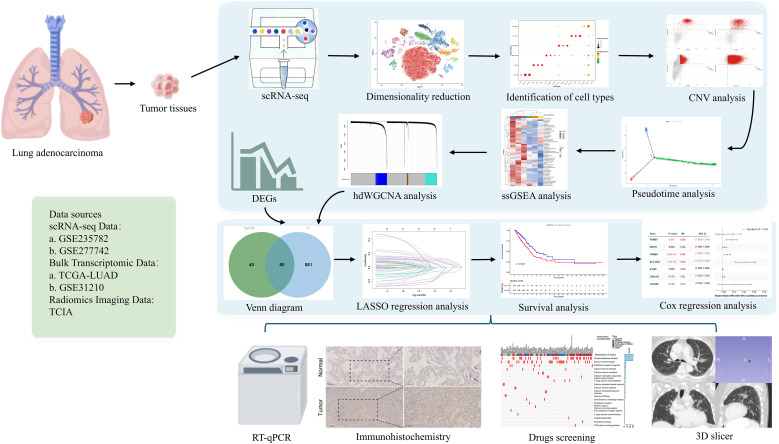 Figure 1
 Figure 2
 Figure 3
 Figure 4
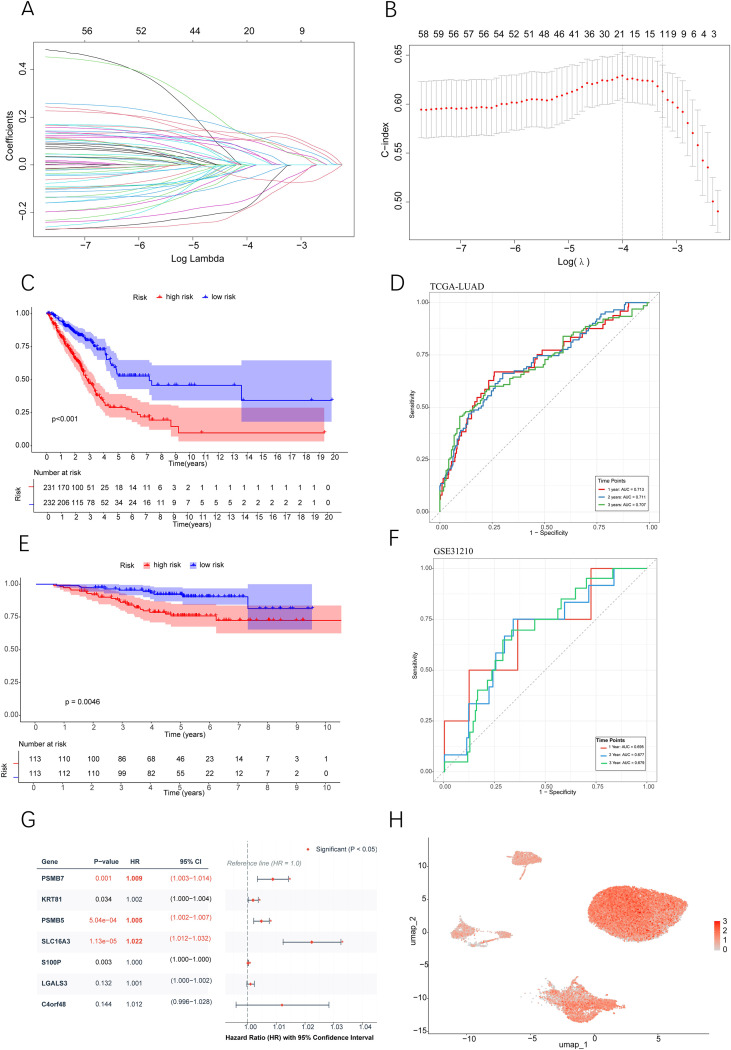 Figure 5
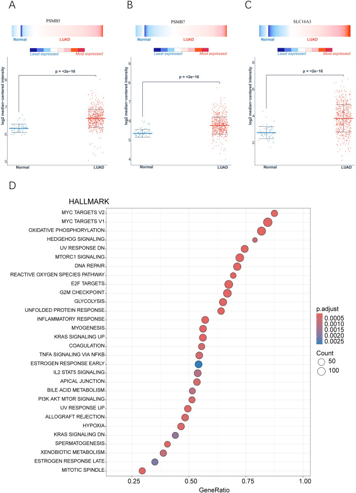 Figure 6
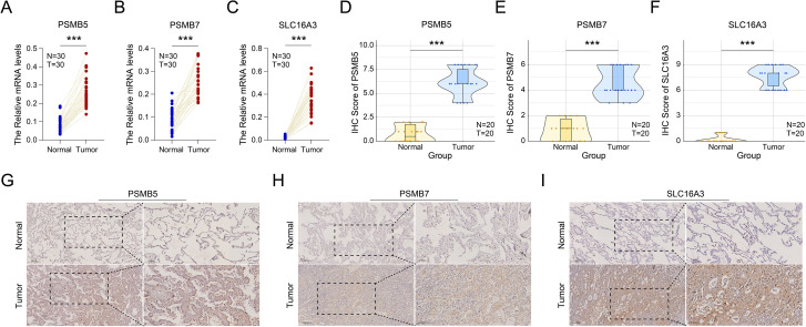 Figure 7
 Figure 8
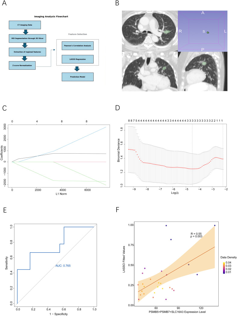 Figure 9
Peer Reviews
No public reviews on file for this paper yet. If you reviewed it on a platform where reviews are public (OpenReview, ICLR, NeurIPS, ICML), you can paste yours below so the community can read it here.
Videos
No videos yet. Explain this paper in a talk, walkthrough, or lecture? Add one.
Taxonomy
TopicsSingle-cell and spatial transcriptomics · Radiomics and Machine Learning in Medical Imaging · Ferroptosis and cancer prognosis