A Massive Cavernous Mediastinal Haemangioma Causing Superior Vena Cava Obstruction and Extending to the Supraclavicular Space: A Case Report
Antonios Charokopos, Andreas Granitsas, Georgios T Stathopoulos, Irene Zarvou, Stella Petrou, Pinelopi Anagnostopoulou, Nikos Chondros, Elena Theofanous, Konstantinos Markakis, Tonia Adamides

TL;DR
This case report describes a rare, large mediastinal haemangioma causing severe vein blockage and extending to the neck area in a young man.
Contribution
The paper presents a unique case of a massive cavernous haemangioma with extensive vascular involvement and management challenges.
Findings
The haemangioma caused aneurysmal dilatation of the superior vena cava and a large collateral venous network.
MRI and CT confirmed the mass as a cavernous haemangioma, with histology providing definitive diagnosis.
Embolisation was recommended due to venous-phase extravasation and worsening superior vena cava diameter.
Abstract
Mediastinal haemangiomas are exceptionally rare, benign vascular tumours and account for a very small proportion of all mediastinal masses. Their symptomatology ranges from an incidental finding to significant mass-mediated compression of vital thoracic structures. We report a unique case of a massive mediastinal cavernous haemangioma with supraclavicular and axillary extension, which led to central venous obstruction. A 24-year-old man, with a childhood history of a resected supraclavicular cyst, was found to have a symptomatic right-sided heterogeneous mediastinal mass. Computed tomography (CT) angiography identified the hypervascular mass extending from the anterior mediastinum to the supraclavicular fossa, which caused aneurysmal dilatation of the superior vena cava (SVC), with an extensive collateral venous network. Magnetic resonance imaging (MRI) appearance was highly suggestive…
Genes, proteins, chemicals, diseases, species, mutations and cell lines named across the full text — each resolved to its canonical identifier and authoritative record.
Click any figure to enlarge with its caption.
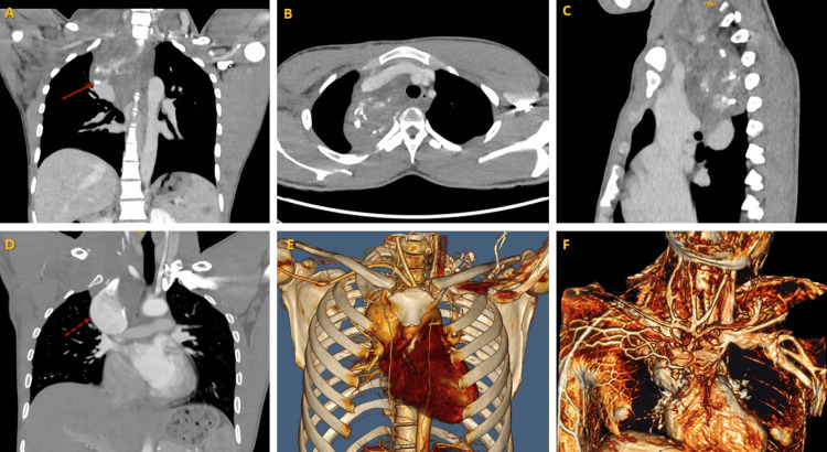 Figure 1
 Figure 2
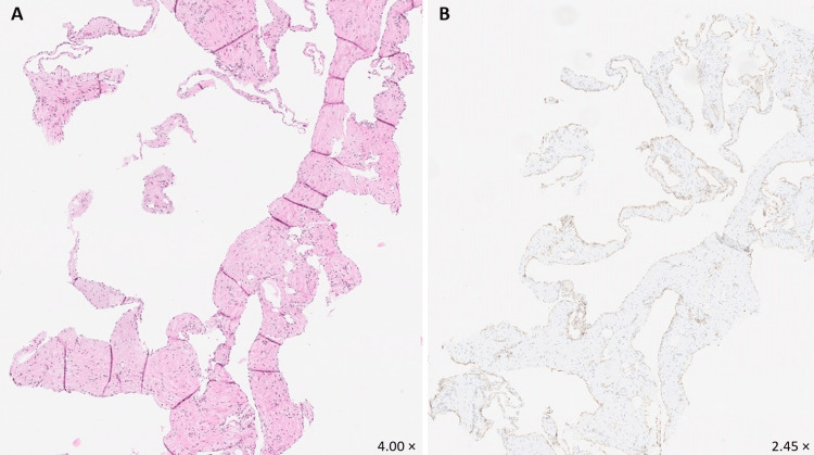 Figure 3
Peer Reviews
No public reviews on file for this paper yet. If you reviewed it on a platform where reviews are public (OpenReview, ICLR, NeurIPS, ICML), you can paste yours below so the community can read it here.
Videos
No videos yet. Explain this paper in a talk, walkthrough, or lecture? Add one.
Taxonomy
TopicsVascular Malformations and Hemangiomas · Vascular Tumors and Angiosarcomas · Cardiac tumors and thrombi