Spatial protein expression patterns across pathologically-associated fibers revealed molecular specialization in inclusion body myositis
T. I. Nijssen, S. Davis, R. A. O’Shaughnessy, E. Bos, A. J. van der Kooi, J. Raaphorst, E. Aronica, R. Fischer, B. M. Kessler, Vered Raz

TL;DR
The study reveals how different types of muscle fibers in inclusion body myositis have unique protein patterns linked to disease mechanisms.
Contribution
A novel myofiber-level proteomic workflow was developed to uncover subtype-specific molecular signatures in pathologically-associated fibers of IBM.
Findings
Regenerating fibers with embryonic myosin show molecular similarity to centrally nucleated fibers, not inflammation-adjacent fibers.
PAF subtypes exhibit distinct proteomic profiles linked to impaired proteostasis and aggregation-prone proteins.
HNRNPA1 localization changes suggest a role in protein aggregation and inflammation in IBM.
Abstract
In Inclusion Body Myositis (IBM), myofibers undergo structural and functional changes, including increased regeneration, atrophy, and fibrosis. The molecular mechanisms driving pathologically -associated fibers (PAF) remain poorly understood. We developed a myofiber-level proteomic workflow to identify protein signatures of three PAF subtypes. Laser-capture microdissection mass spectrometry of immunolabeled cryosections was performed, complemented by immunofluorescence and electron microscopy validation. Regenerating fibers expressing embryonic myosin heavy chain showed greater molecular similarity to centrally nucleated fibers than to fibers adjacent to inflammation, which were enriched in aggregation-prone proteins. These distinct proteomic profiles revealed disruptions in protein homeostasis and proteasome composition, implicating impaired proteostasis in defective regeneration. In…
Genes, proteins, chemicals, diseases, species, mutations and cell lines named across the full text — each resolved to its canonical identifier and authoritative record.
Click any figure to enlarge with its caption.
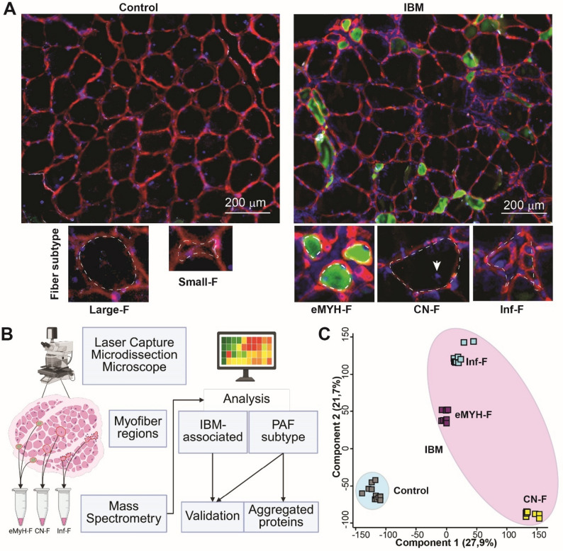 Figure 1
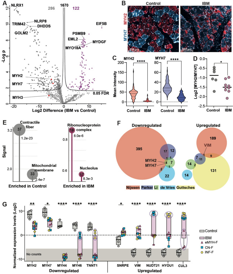 Figure 2
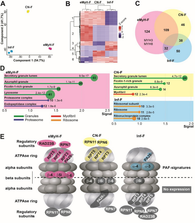 Figure 3
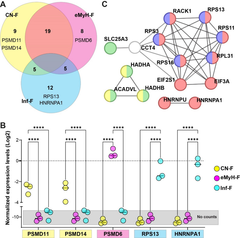 Figure 4
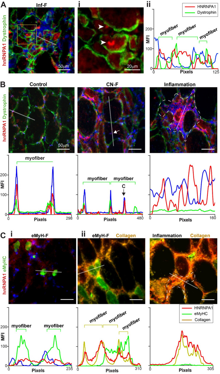 Figure 5
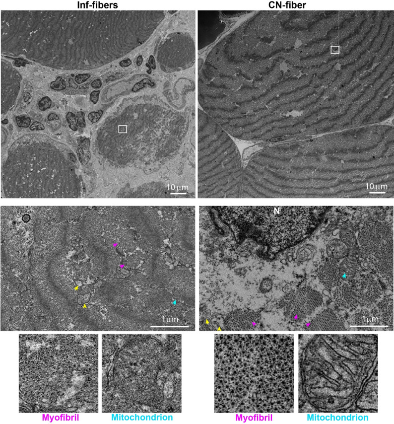 Figure 6
Peer Reviews
No public reviews on file for this paper yet. If you reviewed it on a platform where reviews are public (OpenReview, ICLR, NeurIPS, ICML), you can paste yours below so the community can read it here.
Videos
No videos yet. Explain this paper in a talk, walkthrough, or lecture? Add one.
Taxonomy
TopicsInflammatory Myopathies and Dermatomyositis · Muscle Physiology and Disorders · Systemic Sclerosis and Related Diseases