Association between coronary artery calcium scores and retinal ischemic perivascular lesions
Jay Bharatsingh Bisen, Hayden Sikora, Michael Drakopoulos, John Bryan, Maxwell Shramuk, Adin-Cristian Andrei, Richard L. Weinberg, Rukhsana G. Mirza

TL;DR
This study finds a link between high coronary artery calcium scores and retinal ischemic lesions, suggesting retinal changes may indicate cardiovascular disease risk.
Contribution
The study establishes a novel association between retinal ischemic perivascular lesions and elevated coronary artery calcium scores as a marker of cardiovascular risk.
Findings
High-risk patients (CAC ≥ 300) had significantly more retinal ischemic perivascular lesions than low-risk patients.
High-risk patients were more likely to have elevated RIPL counts (≥ 2) compared to low-risk patients.
Known cardiovascular risk factors partially mediated the relationship between CAC scores and RIPL counts.
Abstract
Coronary artery calcium (CAC) scores measure atherosclerosis and are associated with increased cardiovascular disease (CVD) risk. Retinal ischemic perivascular lesions (RIPLs) represent focal retinal infarcts and have been linked to systemic CVD. Given their association with retinal microcirculation dysfunction, RIPLs might serve as early indicators of systemic vascular dysfunction. This study aims to assess the relationship between CAC scores and RIPLs. Retrospective, single-institution, cross-sectional study. Patients consenting for research who had both high-quality, bilateral, macular optical coherence tomography (OCT) imaging and a CAC score of either 0 (low risk) or ≥ 300 (high risk) within one year after their macular OCT imaging. RIPLs were identified as regions of focal inner nuclear layer (INL) thinning, outer plexiform layer (OPL) inward deviation, and outer nuclear layer…
Genes, proteins, chemicals, diseases, species, mutations and cell lines named across the full text — each resolved to its canonical identifier and authoritative record.
Click any figure to enlarge with its caption.
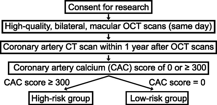 Figure 1
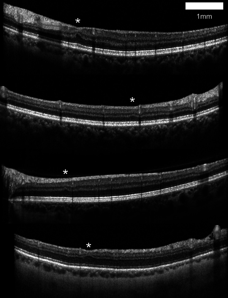 Figure 2
Peer Reviews
No public reviews on file for this paper yet. If you reviewed it on a platform where reviews are public (OpenReview, ICLR, NeurIPS, ICML), you can paste yours below so the community can read it here.
Videos
No videos yet. Explain this paper in a talk, walkthrough, or lecture? Add one.
Taxonomy
TopicsRetinal and Optic Conditions · Retinal Imaging and Analysis · Cerebrovascular and Carotid Artery Diseases