Dysferlin stabilizes membrane nanodomains of cardiomyocytes after myocardial infarction
Justus B. Wegener, Yannik Zühlke, Carolin Fleischhacker, Justus Marks, Brian Foo, Niklas Bader, Gabriel C. Riedemann, Jasper Wedemeyer, Kim-Chi Vu, Ana M. Vergel Leon, Nora Josefine Paulke, Tobias Kohl, Henning Urlaub, Constanze Schmidt, Gerd Hasenfuß, Tobias Moser

TL;DR
This study shows that the protein dysferlin helps protect heart cells after a heart attack by stabilizing key membrane structures, which may reduce heart function loss.
Contribution
The study identifies dysferlin as a novel molecular target for preserving sarcolemmal nanodomains in the heart after myocardial infarction.
Findings
Dysferlin expression increases by 230% in the MI border zone of wild-type mice.
Dysferlin-knockout mice show larger infarct sizes and reduced heart function after MI.
Dysferlin stabilizes TAT and ICD nanodomains, as shown by proteomic and imaging analyses.
Abstract
Despite advances in acute care medicine, myocardial infarction (MI) remains a predominant cause of premature death and heart failure. In the MI border zone, cardiomyocytes are exposed to high biomechanical stress that impairs the integrity of the sarcolemmal membrane. Hence, we hypothesized that the Ca2+-sensitive membrane repair protein dysferlin is crucial for preserving sarcolemmal nanodomains in the MI border zone, like the transverse-axial tubule (TAT) network and the intercalated disc (ICD) membrane folds, and thereby limits the post-MI loss of myocardial function. We employed left anterior descending artery ligation to induce MI in wild-type (WT) versus dysferlin-knockout (KO) mice. While immunohistology identified an upregulated dysferlin expression of 230% in cardiomyocytes of the WT MI border zone, KO mice presented larger infarct sizes and reduced left-ventricular systolic…
Genes, proteins, chemicals, diseases, species, mutations and cell lines named across the full text — each resolved to its canonical identifier and authoritative record.
Click any figure to enlarge with its caption.
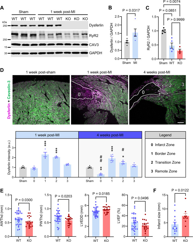 Figure 1
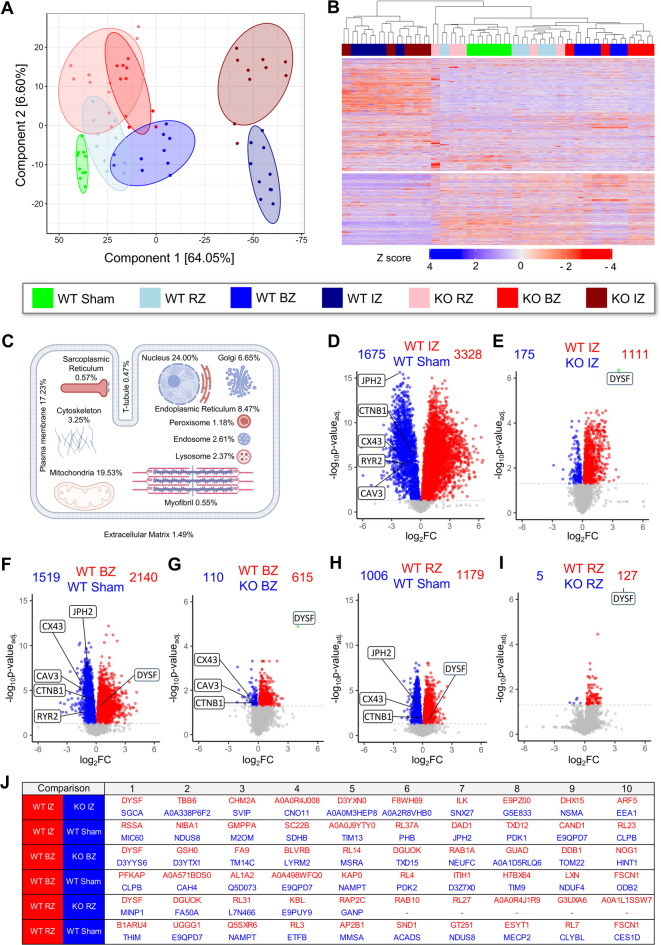 Figure 2
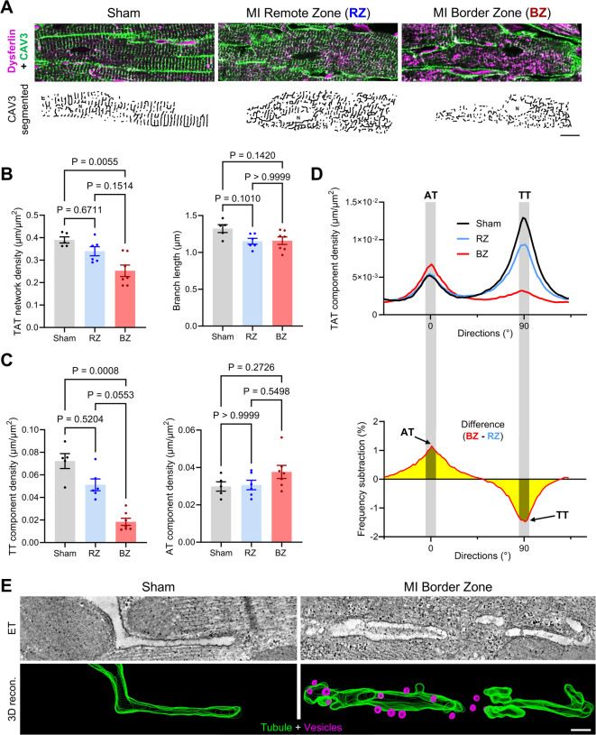 Figure 3
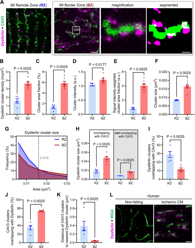 Figure 4
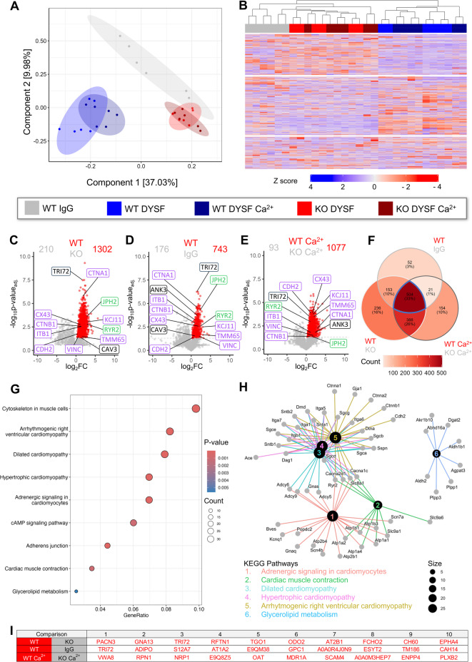 Figure 5
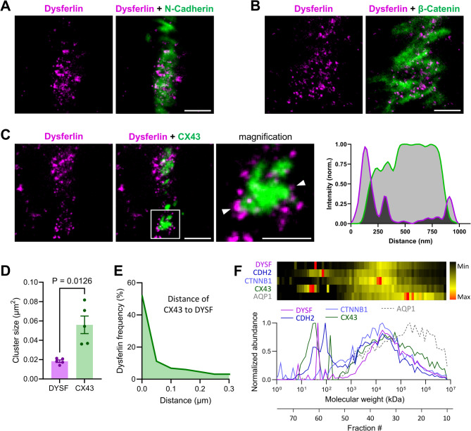 Figure 6
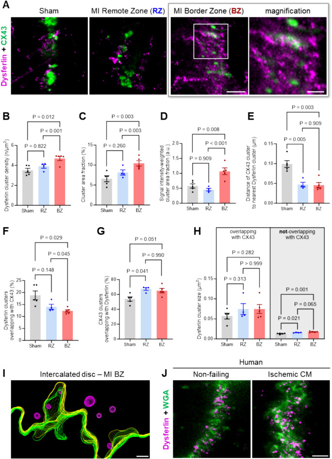 Figure 7
Peer Reviews
No public reviews on file for this paper yet. If you reviewed it on a platform where reviews are public (OpenReview, ICLR, NeurIPS, ICML), you can paste yours below so the community can read it here.
Videos
No videos yet. Explain this paper in a talk, walkthrough, or lecture? Add one.
Taxonomy
TopicsCardiac Fibrosis and Remodeling · Congenital heart defects research · Signaling Pathways in Disease