Imaging improvements reveal guttae development and posterior fibrillar layer formation in fuchs endothelial corneal dystrophy
Daniel B. Zander, Anne-Marie S. Kladny, Judith-Lisa Lieberum, Philip Maier, Thabo Lapp, Stefan J. Lang, Sonja Heinzelmann-Mink, Claudia Auw-Hädrich, Günther Schlunck, Katrin Wacker, Gottfried Martin

TL;DR
This study uses advanced imaging to better understand guttae and a related layer in a corneal disease, which could improve diagnosis and treatment.
Contribution
The study introduces a novel imaging method combining DIC, autofluorescence, and polarized light to analyze guttae and the posterior fibrillar layer in Fuchs endothelial corneal dystrophy.
Findings
Guttae were classified into peripheral, knob-like and central, flat types with gradual transition.
A posterior fibrillar layer containing COL1 was found to cover central guttae and was visualized using polarized light microscopy.
The presence of the posterior fibrillar layer correlated with clinical parameters like anterior scatter and post-operative corneal edema resolution.
Abstract
Guttae are a hallmark of Fuchs endothelial corneal dystrophy (FECD) and the disease’s progression. A posterior fibrillar layer (PFL) that covers central guttae has been previously described, but its formation and progression remain unclear. This study aims to further investigate the characteristics of guttae and the PFL in FECD. In a well-characterized prospective FECD patient cohort, a total of 43 DMEK (Descemet membrane endothelial keratoplasty) specimens were immunostained for ZO1 or COL1, flat-mounted, and analyzed using differential interference contrast (DIC), autofluorescence (excited at 480 nm), and polarized light microscopy. Guttae and PFL were quantified and correlated with clinical data. Guttae were visualized by DIC and autofluorescence imaging and classified into two types with gradual transition: peripheral, knob-like guttae and central, flat guttae. Guttae with low…
Genes, proteins, chemicals, diseases, species, mutations and cell lines named across the full text — each resolved to its canonical identifier and authoritative record.
Click any figure to enlarge with its caption.
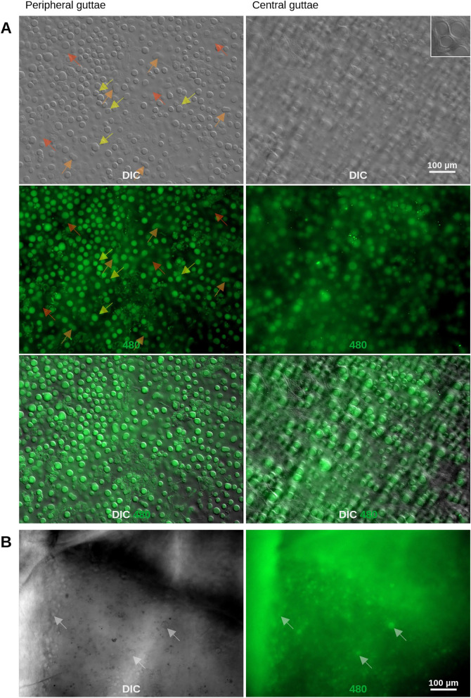 Figure 1
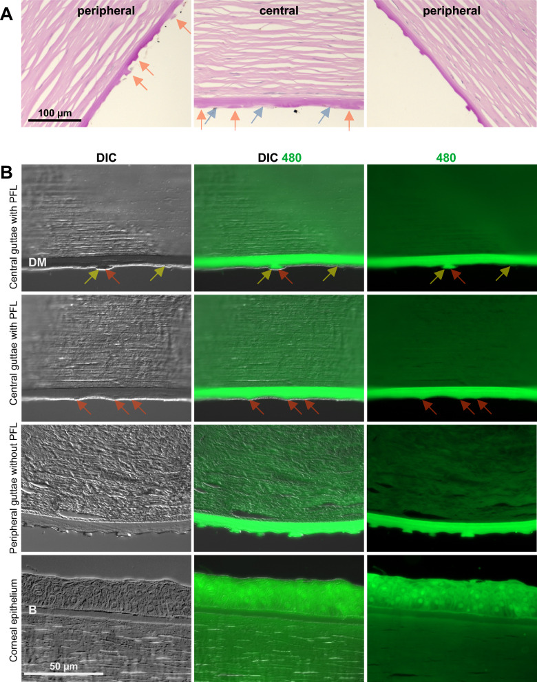 Figure 2
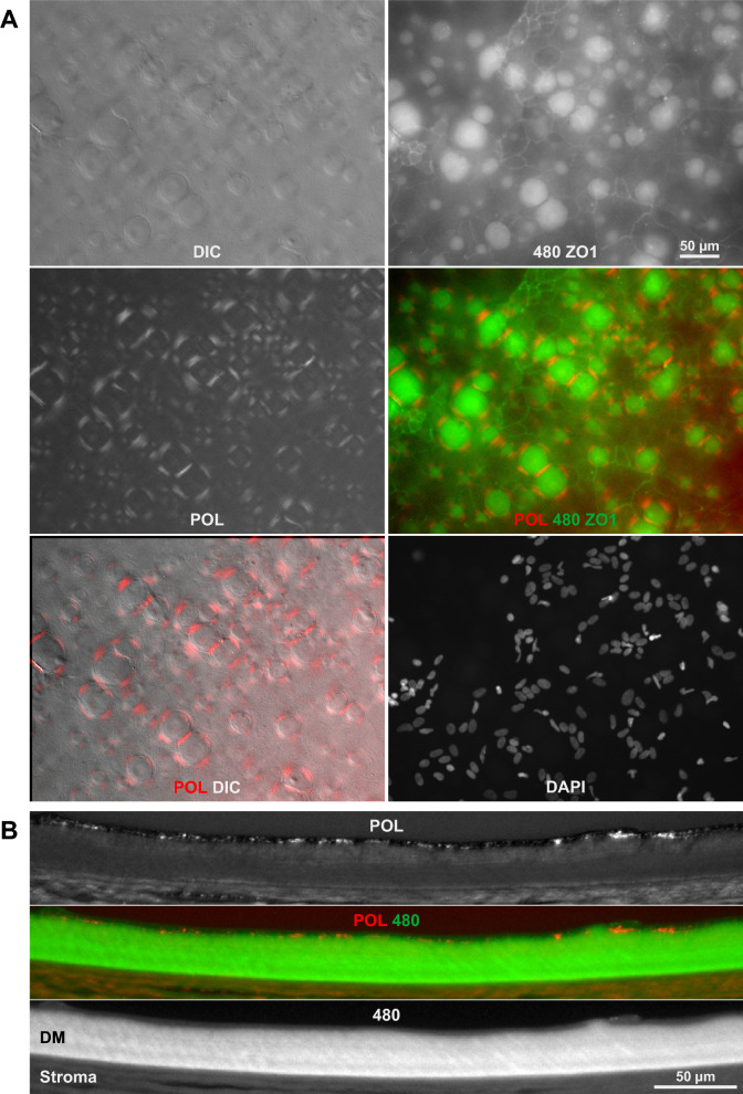 Figure 3
 Figure 4
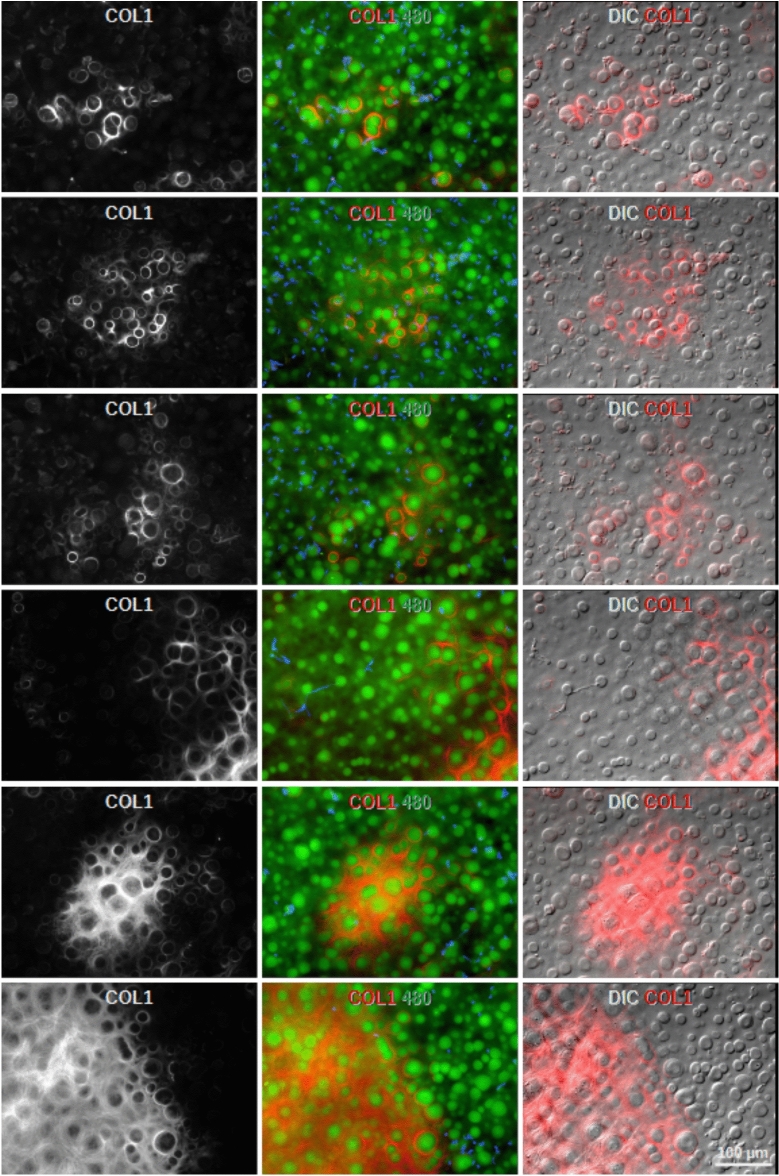 Figure 5
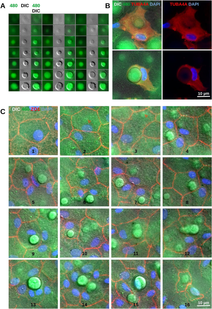 Figure 6
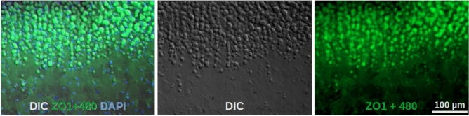 Figure 7
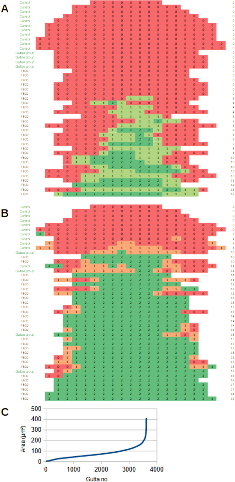 Figure 8
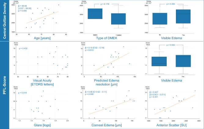 Figure 9
Peer Reviews
No public reviews on file for this paper yet. If you reviewed it on a platform where reviews are public (OpenReview, ICLR, NeurIPS, ICML), you can paste yours below so the community can read it here.
Videos
No videos yet. Explain this paper in a talk, walkthrough, or lecture? Add one.
Taxonomy
TopicsCorneal surgery and disorders · Corneal Surgery and Treatments · Ophthalmology and Visual Impairment Studies