Contextual anatomy-guided deep learning for accurate fovea segmentation in diabetic retinopathy fundus images
Sakon Chankhachon, Supaporn Kansomkeat, Patama Bhurayanontachai, Sathit Intajag

TL;DR
This paper introduces a new deep learning method for accurately identifying the fovea in diabetic retinopathy images by using anatomical context, improving segmentation and treatment decisions.
Contribution
The paper introduces a data-centric approach using anatomical landmarks and a new dataset and framework for robust fovea segmentation.
Findings
Incorporating anatomical landmarks like the optic disc and blood vessels improves fovea detection performance.
The proposed framework achieves high segmentation accuracy (fovea IoU = 0.812, F1 = 0.894).
The GEV-based augmentation technique outperforms baseline methods in detection rates.
Abstract
Accurate fovea segmentation in fundus images is a critical step in diabetic retinopathy screening; however, it remains a challenging task due to the indistinct boundaries of the fovea. Beyond simple localization, precise segmentation offers essential clinical value for Diabetic Macular Edema (DME) management, as treatment decisions–specifically the choice between intravitreal anti-VEGF injection for center-involved DME and laser therapy for extrafoveal edema–depend on the accurate delineation of the foveal region. While existing methods often rely on increasing model architecture complexity, the potential of anatomical context within the training process remains under-explored. This paper presents a data-centric approach that leverages contextual information to robustly identify the fovea. We demonstrate that progressively incorporating key anatomical landmarks–the optic disc, retina,…
Genes, proteins, chemicals, diseases, species, mutations and cell lines named across the full text — each resolved to its canonical identifier and authoritative record.
Click any figure to enlarge with its caption.
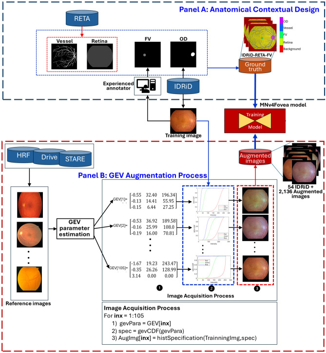 Figure 1
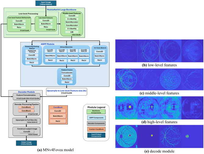 Figure 2
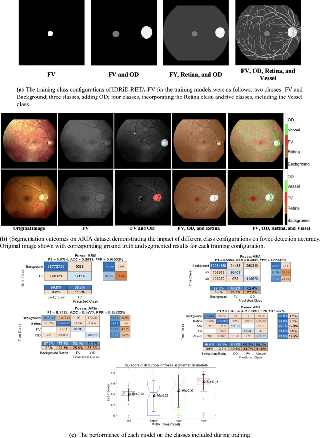 Figure 3
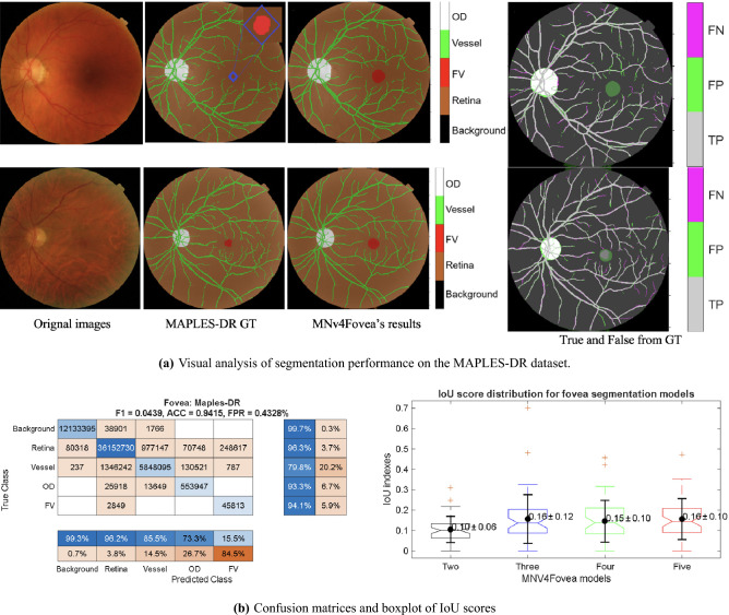 Figure 4
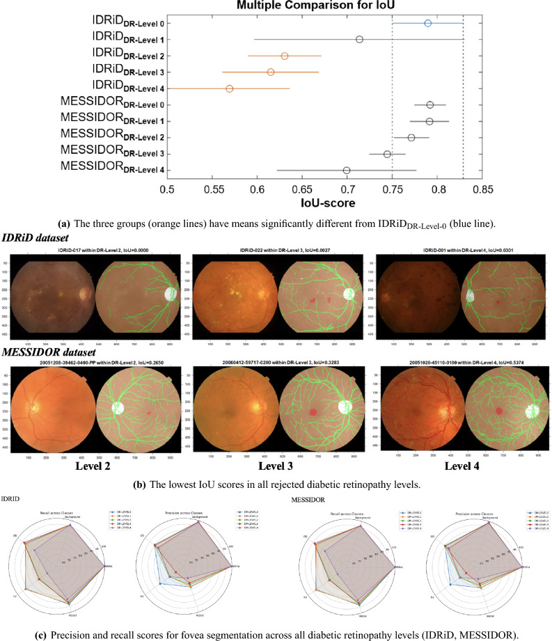 Figure 5
 Figure 6
Peer Reviews
No public reviews on file for this paper yet. If you reviewed it on a platform where reviews are public (OpenReview, ICLR, NeurIPS, ICML), you can paste yours below so the community can read it here.
Videos
No videos yet. Explain this paper in a talk, walkthrough, or lecture? Add one.
Taxonomy
TopicsRetinal Imaging and Analysis · Retinal Diseases and Treatments · Ocular Diseases and Behçet’s Syndrome