Matching accuracy between CT images and intraoral surface scans using glass-ceramic markers compared to gutta-percha markers
Yoshiyuki Nakano, Takuya Mino, Yoko Kurosaki, Hiroaki Shimizu, Mariko Nishizaki, Kenji Maekawa

TL;DR
This study compares the accuracy of using glass-ceramic versus gutta-percha markers for aligning CT scans with intraoral scans, finding that glass-ceramic markers offer better precision.
Contribution
The study introduces glass-ceramic markers as a more accurate alternative to gutta-percha for CT-surface scan matching in dental implant planning.
Findings
Glass-ceramic markers with three or six markers showed significantly lower matching errors than gutta-percha markers under air conditions.
Glass-ceramic markers also outperformed gutta-percha markers in water conditions, which simulate intraoral scattering.
The study demonstrates that fewer glass-ceramic markers can achieve better accuracy than more gutta-percha markers.
Abstract
This study investigated the accuracy of matching computed tomography (CT) images and intraoral surface scans using glass-ceramic (GC) and conventional gutta-percha (GP) markers. A mandibular master model of the right posterior edentulous region (teeth 45–47) was prepared with three training implants, each with a scan body fixed at the anterior central, left posterior, and right posterior sites. The model was scanned 10 times using an intraoral scanner, and 10 CT matching templates with six GP markers (GPCTMTs) and 10 with six GC markers (GCCTMTs) were fabricated. Each template was mounted on the model for CT imaging and intraoral scanning under air conditions. CT imaging was also performed with the model immersed in water to simulate intraoral scattering. A dentist blinded to the study purpose used implant simulation software for matching, which was performed with GP with six markers…
Genes, proteins, chemicals, diseases, species, mutations and cell lines named across the full text — each resolved to its canonical identifier and authoritative record.
Click any figure to enlarge with its caption.
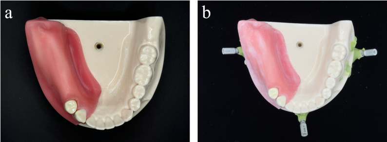 Figure 1
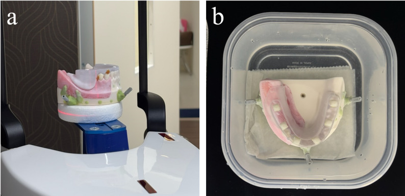 Figure 2
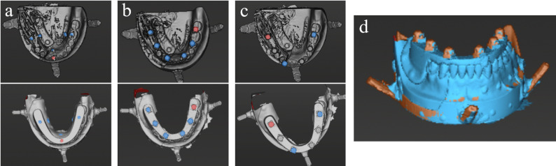 Figure 3
 Figure 4
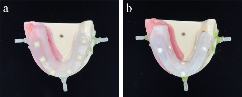 Figure 5
Peer Reviews
No public reviews on file for this paper yet. If you reviewed it on a platform where reviews are public (OpenReview, ICLR, NeurIPS, ICML), you can paste yours below so the community can read it here.
Videos
No videos yet. Explain this paper in a talk, walkthrough, or lecture? Add one.
Taxonomy
TopicsDental Radiography and Imaging · Dental materials and restorations · Dental Implant Techniques and Outcomes