Design, Implementation, and Advances in Indirect SERS Sensors for Biomedical and Human-Health-Related Analyte Detection
North Pinkley, Uchhwas Banik, Nayeem Anam, Aastha Oza, Kevin J. Ledford, Bhavya Sharma

TL;DR
This review explains how SERS nanotags are designed and used to detect health-related molecules with high sensitivity and specificity, offering insights into their structure, function, and future applications.
Contribution
The paper provides a comprehensive overview of indirect SERS nanotag design and applications, emphasizing recent trends and integration into diagnostic systems.
Findings
SERS nanotags enhance Raman signals for ultrasensitive detection of health-related analytes.
Recent advances in AI and machine learning are accelerating the design and optimization of SERS nanotags.
SERS nanotags are being integrated into point-of-care diagnostic formats like lateral flow assays and microfluidic devices.
Abstract
What are the main findings? This review provides an overview of SERS nanotags, explaining their structural design, functional components, and underlying mechanisms for Raman signal enhancement.This work summarizes the applications of SERS nanotags for the detection and diagnosis of human-health-related analyte detection, highlighting recent trends in assay design and application. This review provides an overview of SERS nanotags, explaining their structural design, functional components, and underlying mechanisms for Raman signal enhancement. This work summarizes the applications of SERS nanotags for the detection and diagnosis of human-health-related analyte detection, highlighting recent trends in assay design and application. What are the implications of the main findings? By unifying the fundamental principles and current biosensing applications of SERS nanotags, this review…
Genes, proteins, chemicals, diseases, species, mutations and cell lines named across the full text — each resolved to its canonical identifier and authoritative record.
Click any figure to enlarge with its caption.
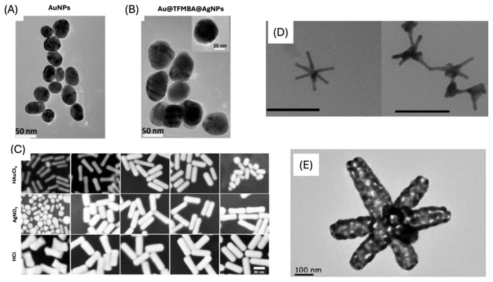 Figure 3
Peer Reviews
No public reviews on file for this paper yet. If you reviewed it on a platform where reviews are public (OpenReview, ICLR, NeurIPS, ICML), you can paste yours below so the community can read it here.
Videos
No videos yet. Explain this paper in a talk, walkthrough, or lecture? Add one.
Taxonomy
TopicsGold and Silver Nanoparticles Synthesis and Applications · Spectroscopy Techniques in Biomedical and Chemical Research · Innovative Microfluidic and Catalytic Techniques Innovation
1. Introduction
Advances in medicine are being driven by the development of new molecular-based technologies for disease diagnostics, imaging, and therapeutics [1,2]. These interrelated fields are aiding in improving the understanding of the mechanisms of disease, which leads to early and enhanced detection, as well as the discovery of new therapeutic targets. Additionally, advances in these fields, combined with artificial intelligence and machine learning approaches, are revolutionizing healthcare and the development of precision medicine [3,4,5]. Molecular diagnostics involves the monitoring of biomarkers, including proteins, nucleic acids, small molecules, and metabolites. Conventionally, biomarkers are monitored through techniques such as PCR, ELISAs, HPLC, mass spectrometry, and other lab-based testing methods. Conventional tests often require multi-step sample processing, long analysis times, and non-portable equipment. Wearable sensors, i.e., smart watches, fitness devices, continuous glucose monitors, etc., have limited selectivity and specificity, and issues with reusability and handling of large datasets [6,7,8,9,10]. There are also limited wearable chemical sensors for monitoring neurological health [11]. There is an unmet need for sensors that are highly sensitive and specific, rapid, low-cost, quantitative, and that require little to no sample preparation.
Raman spectroscopy is an inelastic light scattering technique that measures the frequency shift between incident laser photons and those scattered by a sample. These frequency shifts correspond to molecular vibrations that are highly specific to individual chemical bonds and molecular structures, enabling precise molecular fingerprinting [12]. Compared with other analytical techniques, Raman spectroscopy offers several advantages: it enables rapid, specific, and non-destructive analysis; requires minimal sample preparation; and can be implemented using portable instrumentation for on-site or in-field measurements [13]. Despite these advantages, conventional Raman spectroscopy suffers from inherently low sensitivity because Raman scattering is an intrinsically weak process; only about one in every 10^8^ photons is Raman scattered. This limitation renders normal Raman spectroscopy unsuitable for detecting low-abundance analytes, such as trace biomarkers in complex biological matrices, where weak target signals are easily masked by fluorescence or background contributions [14]. To overcome this sensitivity barrier while retaining many of the inherent advantages of regular Raman, surface-enhanced Raman spectroscopy (SERS) has emerged as a powerful alternative.
SERS enhances Raman signals by exploiting localized surface plasmon resonance (LSPR), a phenomenon that occurs when conduction electrons in a noble metal nanostructure collectively oscillate in response to incident light [15]. This oscillation generates intense, localized electromagnetic fields near the metal surface that can amplify the Raman scattering of molecules in close proximity by several orders of magnitude. When two or more plasmonic nanostructures are close enough for their electric fields to overlap, “hotspots” can form (Figure 1), which can generate signal enhancements of up to 10^10^-fold [16].
SERS-based sensing can be broadly classified into direct and indirect detection modes (Figure 2). In direct SERS, the analyte of interest is directly brought to the metal surface, producing an enhanced Raman spectrum that reflects the intrinsic molecular vibrations of the target analyte. However, many biologically relevant molecules have a low affinity for metal surfaces or exhibit overlapping vibrational signatures, complicating spectral interpretation in complex biological samples using direct sensing [14]. Consequently, indirect SERS approaches are commonly employed in biosensing applications, where signal transduction is achieved via a well-defined Raman reporter molecule rather than the analyte itself [18].
This review will focus on the design of SERS nanotags and the application of the nanotags for indirect SERS sensing. SERS nanotags are primarily nanoparticle (NP)-based sensors used to detect analytes with high specificity. SERS-based sensors can be applied for imaging [19], precision drug delivery [20], and photothermal therapy [21]; however, this review will focus on the applications of SERS sensors for diagnostic and analyte monitoring published within the past decade. SERS nanotags appear in the literature under different names, such as “SERS tags,” “bioconjugate probes,” and “nanosensors,” [22,23,24]. In this review, the term “nanotags” will be used for clarity and consistency. A SERS nanotag is typically comprised of three essential components: (1) a plasmonic nanostructure (such as a gold or silver nanoparticle) that provides the source of electromagnetic enhancement, (2) a Raman reporter molecule that generates a strong, distinct, and quantifiable vibrational signal, and (3) an analyte-specific targeting ligand (i.e., antibody, aptamer) that enables selective binding or recognition of the target analyte. The design of an effective SERS nanotag requires careful consideration of each component to optimize signal enhancement, stability, and analytical performance.
When an SERS nanotag binds to its target, nanoparticle aggregation or clustering can occur, creating additional electromagnetic hotspots that further amplify the Raman signal. The resulting increase in reporter signal intensity correlates directly with analyte concentration, enabling quantitative detection [25]. However, for alternative sensing methods such as competitive or displacement-based assays, higher analyte concentrations can lead to a decrease in reporter signal intensity as the analyte displaces reporter molecules or inhibits nanoparticle aggregation. In both cases, quantitative analysis is achieved by constructing calibration curves that relate analyte concentration to the integrated intensity of the reporter peak, allowing unknown sample concentrations to be determined from the fitted response [25]. This quantitative capability, arising from the precise interplay of nanostructure design, reporter selection, and surface chemistry, forms the foundation for reliable SERS-based sensing.
Through their high sensitivity, molecular specificity, and versatility in functionalization, SERS nanotag assays have emerged as powerful and adaptable sensing platforms for biological detection. Their ability to convert molecular recognition events into quantifiable optical signals has positioned SERS-based sensors as a promising tool for various applications, including point-of-care diagnostics, biomedical imaging, and drug monitoring [20,26,27]. This review will focus on the design, implementation, and advances of SERS sensors used for small molecule sensing and disease diagnostics.
2. Plasmonic Nanostructure
The first component of the SERS nanotag is the plasmonic nanostructure, which provides the electromagnetic enhancement necessary to generate a strong Raman signal from the reporter molecule. Since the inception of SERS in 1977, researchers have explored a wide variety of metals and morphologies to optimize plasmonic performance. Gold (Au) and silver (Ag) are the most frequently used metals for SERS nanostructures. Among these, gold and silver have become the dominant materials because their localized surface plasmon resonance (LSPR) bands can be tuned within the visible to near-infrared (NIR) range. These are the spectral regions where Raman scattering is least affected by fluorescence background and where biological samples are relatively transparent.
2.1. Spherical Nanoparticles
Colloidal gold nanoparticles (AuNPs) are a widely used plasmonic substrate for SERS nanotags (Figure 3A). Their popularity is due to the simple and reproducible synthesis, the chemical stability/inertness of gold, and the well-established methods available to control particle size and surface chemistry [28]. Typically, spherical AuNPs are synthesized by reducing tetrachloroaurate with trisodium citrate dihydrate, a method that allows for fine control over particle size through the stoichiometry of the reducing agent, reaction times, and temperature. Because of these favorable characteristics, AuNPs have been applied extensively in a variety of biophotonic applications, including SERS biosensing [29]. While gold is the most employed material, silver nanoparticles (AgNPs) also offer exceptionally strong plasmonic activity and can produce higher electromagnetic enhancement factors due to their sharper plasmon resonances. AgNPs also have quick, reliable, and tunable syntheses, making them preferable SERS substrates [30]. However, AgNPs are generally less chemically stable than AuNPs, tending to oxidize under ambient or biological conditions, which can alter their optical properties and limit their long-term use.
2.2. Anisotropic Nanoparticles
The chemical, optical, and electrical characteristics of NPs can change depending on their form. Beyond simple spheres, researchers have designed nanostructures with diverse morphologies, such as nanostars, nanorods, nanocubes, and nanoprisms [31]. Non-spherical NPs are considered anisotropic nanostructures (Figure 3). The properties of anisotropic NPs vary depending on the shape of the NPs, in contrast to spherical NPs, which have homogeneous qualities in all directions [31]. Among these anisotropic structures, nanostars (NSs) are a widely used substrate due to the fact that they have been shown to possess higher SERS enhancement capabilities compared to spherical NPs [32]. This increased enhancement is due to the strong electromagnetic field around the tips of the star, indicating more intense “hot spot” areas. Structures featuring sharp edges, tips, or interparticle gaps can generate stronger localized electromagnetic fields and create highly efficient SERS hotspots [33]. Another common type of anisotropic structure used is gold nanorods (AuNRs). These structures are unique because they have dual-mode LSPR maxima, arising from the transverse (perpendicular to the long axis) and longitudinal (along the long axis) plasmon modes, which are tunable through changing the aspect ratio (ratio of length to diameter) of the nanorods [34].
TEM images of (A) Au NPs and (B) Au@TFMBA@Ag NPs [35]. (C) SEM images of AuNRs [36]. (D) TEM images of the AuNS [32]. (E) TEM image of a typical Au nanocage [37]. (A,B) were adapted from Ref. [35]. (C) adapted with permission from Ref. [36]. (D) adapted from Ref. [32]. (E) adapted from Ref. [37].
2.3. Core-Shell Nanoparticles
Synthesis of core-shell nanoparticles is a common element in the development of SERS nanotags. Core-shell morphologies allow for RRMs bound to the core nanoparticle to be enhanced through the exploitation of the dual plasmonic effect, which arises when the LSPR from two metals overlap and hybridize to form an intense electric field between the two metal surfaces [38]. Additionally, embedding RRMs between the core and the shell can minimize desorption from the surface and help maintain the SERS signal for nanotags over extended periods of time [39]. Another common core-shell nanoparticle geometry is the combination of a metal shell with iron oxide core nanoparticles, which allows for the magnetic properties of iron oxide to be combined with the plasmonic properties of the metal shell [37]. Analytes of interest are bound to the surface of the magnetic nanoparticles, and then magnets are used to pull the analyte-bound nanotags from solution. Through this separation and isolation of the analytes, the limit of detection (LOD) can be lowered. To further lower LODs, nanoparticles are coated in silica, which protects adsorbed RRMs and prevents their dissociation from the metal surface, stabilizing the nanoparticles and resulting in increased SERS signal [25].
2.4. Supported Nanoparticles
Supported nanoparticles form a major class of SERS substrates in which plasmonic metals are immobilized on higher-dimensional materials to create stable, high-density hotspots for sensitive molecular detection. Graphene oxide (GO) is a 2D carbon structure to which AuNPs are easily attached (GO@AuNP) and are a common example of a type of supported morphology. GO provides a stable surface that reduces nanoparticle aggregation and increases SERS enhancement due to the electronic interaction between the surface plasmons and pi-conjugated domains of GO [40]. There are many other 2D and 3D structures conjugated with NPs that have been used to increase stability, optimize electrochemical enhancement, or facilitate analyte detection, such as metal-organic frameworks (MOFs), carbon-dots, polymers, paper, etc. [41].
Here, we summarize the major classes of plasmonic nanostructures commonly employed for SERS sensing. Rational control over nanoparticle shape, size, and interparticle spacing is essential for optimizing electromagnetic enhancement and overall sensing performance. Although precise experimental tuning of these parameters remains challenging, recent progress in computational modeling, high-throughput synthesis, and data-driven analysis has enabled more predictive design of plasmonic architectures with tailored optical properties. Machine-learning approaches trained on simulated and experimental datasets are now being used to uncover structure–property relationships, optimize synthetic conditions, and guide the fabrication of nanostructures with user-defined plasmonic responses [42]. Together, these advances offer a powerful route toward intelligently tunable, application-specific SERS substrates.
3. Raman Reporter Molecules
In SERS nanotags, signals from Raman reporter molecules (RRMs) are critical for the indirect quantification of target analytes. RRMs are chosen based on several key characteristics: strong and unique Raman peaks, a large Raman scattering cross-section, chemical stability, and compatibility with nanoparticle surface chemistry. When multiple nanotags bind to a target, the proximity of their plasmonic nanostructures generates SERS “hotspots,” dramatically enhancing the Raman signal of the attached reporter molecules. The intensity of the RRM correlates directly with the amount of target present, enabling quantitative analysis through calibration curves that relate RRM intensity or peak area to analyte concentration [25]. Selecting an appropriate RRM is therefore essential, as it determines signal strength, spectral reproducibility, and assay sensitivity. RRMs used in nanotags generally fall into two major categories: dye-based and non-dye-based molecules. A full table of common RRMs along with their structures is provided in Table 1.
Optimal SERS enhancement occurs when RRMs form a uniform monolayer or sub-monolayer on the nanoparticle surface. This means that during nanotag fabrication, parameters influencing surface monolayer formation are particularly critical. These include reporter concentration, incubation time, solvent composition, pH, ionic strength, and the presence of competing ligands or stabilizers (e.g., citrate, CTAB, PVP), all of which affect surface coverage, molecular orientation, and packing density [43,44,45]. Reporter molecules containing thiol (–SH) and amine (–NH_2_) groups are often favored due to their strong binding affinity for gold and silver nanoparticles, enabling stable attachment and controlled monolayer formation [43]. Incomplete ligand exchange, multilayer formation, and desorption under physiological conditions can all introduce spectral variability and batch-to-batch inconsistencies in SERS sensors [46,47]. It is essential to optimize co-absorption strategies with the selected targeting ligand to ensure optimal signal intensity is balanced with colloidal stability and ligand accessibility [19].
3.1. Dye RRMs
Dye molecules and aromatic chromophores are commonly used as RRMs because they possess extended conjugated π-electron systems, which produce strong Raman scattering due to increased molecular polarizability. These dyes exhibit intense, well-defined vibrational modes and often are in resonance with the excitation wavelength, further enhancing the SERS signals. Dye RRMs are valued for their high sensitivity, distinct spectral features, and robust signal output, making them especially suitable for indirect detection formats in which the RRM serves as the quantifiable element of the assay. Common dye RRMs include Cyanine 5 (Cy5), malachite green (MG), Rhodamine 6G (R6G), and Nile Blue A (NBA). Dye RRMs can operate bifunctionally, as both reporter molecules and targeting ligands. For example, Li et al. employed a modified Rhodamine B structure to bind and detect Hg^2+^ ions in contaminated milk samples [48].
3.2. Non-Dye RRMs
Non-dye RRMs derive their Raman activity from specific chemical moieties, such as thiols, amines, nitro groups, nitriles, and aromatic rings. These structures generate strong, characteristic Raman peaks. These RRMs provide highly reproducible spectral signatures and can be chemically tailored to serve additional roles. In many designs, non-dye reporters act as bifunctional linkers: one functional group binds the nanoparticle surface, while another reacts with targeting ligands such as antibodies, peptides, or aptamers. This dual functionality simplifies fabrication and ensures a well-defined geometry between the plasmonic core, the reporter, and the biorecognition element. For example, the Porter group at the University of Utah has popularized the use of the DSNB molecule (Figure 4) as a bifunctional linker where the thiol group binds to the metal surface, the nitro group provides a strong and distinct Raman signal, and an antibody is covalently attached using the succinimide group [49].
3.3. Multiplexing RRMs
Multiplex SERS employs two or more RRMs with non-overlapping vibrational signatures to enable the simultaneous detection of multiple analytes. Each RRM is incorporated into a distinct nanotag population, and their combined spectra allow for parallel identification/quantification from a single measurement. Effective multiplexing requires RRMs that provide strong, narrow, and well-separated peaks that remain distinguishable even in complex samples. Sometimes chemometric approaches such as PCA combined with partial least squares regression (PLS-R) can be used to separate overlapping features and enhance the reliability of multiplex detection. Kearns et al. developed a multiplex approach with three unique RRMs on SERS tags to detect three different pathogenic bacteria with PCA (Figure 5) [24]. Other authors also used this multiplexed RRMs technique to detect multiple analytes for cancer [50,51,52,53,54,55], cardiovascular diseases [56,57,58,59], and traumatic brain injury [60,61]. Multiplexed RRMs significantly expand the capabilities of SERS nanotags, offering higher information density, improved diagnostic throughput, and the ability to analyze multiple biomarkers or chemical targets simultaneously.
4. Targeting Ligand
Choosing an effective target ligand while designing an SERS nanotag is another important step in the nanotag synthesis process. The type of targeting ligand (antibody, aptamer, or other) is key in determining the specificity and sensitivity of the nanotag. Other considerations include selecting the proper conjugation method, stabilization method, and assay design to match the desired targeting ligand. All these considerations must be combined and optimized to create a nanotag that is specific and effective for detecting the target analyte.
4.1. Antibody
Antibodies are widely used ligands for SERS nanotags. Antibodies are y-shaped proteins that have high affinity and specificity for antigens. Antibodies are produced by many organisms as part of their adaptive immune system against pathogens, such as viruses or bacteria. However, advances in microbiology and molecular biology have allowed scientists to generate antibodies against almost any target of interest [62]. Antibodies have distinct binding pockets that utilize non-covalent interactions to form a very specific interaction with an antigen. Due to this specificity, multiple antibodies can bind to the same antigen at different sites, allowing for an antigen to be “sandwiched” between two antibodies. For SERS nanotags, IgG-type antibodies are typically used; however, some groups have explored using antibody variants, such as single-chain variable fragments (scFv) or nanobodies, as targeting ligands [63,64].
4.2. Aptamer
Aptamers are short, single-stranded DNA or RNA sequences that fold into unique three-dimensional structures capable of binding molecular targets with high affinity and specificity. Unlike antibodies, which possess fixed tertiary structures, aptamers adopt multiple conformations depending on their local environment, and this dynamic folding enables selective recognition through a combination of hydrogen bonding, electrostatic interactions, and van der Waals forces [65]. Aptamers are relatively easy to generate using the Systematic Evolution of Ligands by Exponential Enrichment (SELEX) process, an iterative in vitro selection method in which large randomized nucleic-acid libraries are enriched toward sequences that bind a desired target. Compared with antibodies, aptamers offer several advantages, including low-cost chemical synthesis, excellent batch-to-batch reproducibility, long-term stability, and the ability to regenerate their binding activity after denaturation [65]. For SERS nanotag construction, aptamers are commonly conjugated to plasmonic nanostructures through the incorporation of thiol-modified oligonucleotides into the aptamer sequence for conjugation via gold-sulfur bonding. Other conjugation approaches are very similar to antibody conjugation strategies, including covalent binding through EDC (1-ethyl-3-(-3-dimethylaminopropyl) carbodiimide hydrochloride) and NHS (N-hydroxysuccinimide) coupling chemistry. Despite their benefits, aptamers can be limited by susceptibility to nuclease degradation and by the sensitivity of their binding performance to environmental factors that alter their tertiary structure. Aptamer design and generation have been enhanced by advances in computational approaches. For example, Quarin et al. utilized an automated computational program to systemically improve their aptamer-based catalytic sensor for SERS-based detection of SARS-CoV-2 and malaria genetic biomarkers [66].
4.3. Other Targeting Ligands
While antibody and aptamer-based nanotags are extremely common and most prevalent for the scope of this work, there are other types of targeting ligands that can be used to bind and indirectly detect target analytes. One emerging targeting ligand is the molecularly imprinted polymer (MIP), which is a highly stable synthetic polymer engineered with a specific binding pocket to mimic an antibody [67]. Another major class includes non-antibody proteins like enzymes, receptor proteins, and binding proteins that can bind selectively to cell receptors, metal ions, and other biologically relevant targets. Finally, host–guest ligands are supramolecular molecules (e.g., cyclodextrins, cucurbiturils, crown ethers) that selectively bind target analytes through size- and shape-complementary noncovalent inclusion interactions, enabling stable and reversible recognition for indirect SERS detection. These alternative targeting ligands have been mentioned here to provide a broad overview of possible ligands; however, they are not commonly applied to the indirect sensing applications detailed in this work.
4.4. Conjugation Strategies
The conjugation of targeting ligands to plasmonic nanostructures is a fundamental step in SERS nanotag fabrication, as it determines biological specificity, as well as influencing colloidal stability and signal reliability. The selected ligand must preserve both the optical enhancement properties of the plasmonic core and the functional integrity of the Raman reporter molecules. Inadequate or poorly controlled ligand conjugation can result in reduced binding efficiency, substrate aggregation, or attenuation of electromagnetic enhancement due to excessive interfacial spacing. Accordingly, multiple strategies have been developed to balance stability, control over ligand orientation, and reproducibility. The following sections summarize and compare the primary conjugation approaches used in SERS nanotag synthesis for both antibodies and aptamer-type ligands.
4.4.1. Direct Adsorption
The simplest way to conjugate a plasmonic substrate with antibodies is through direct adsorption via electrostatic interactions [68]. In direct adsorption, the positively charged region of the antibodies interacts with the negatively charged surface of a nanoparticle, which arises from the reducing agent used during nanoparticle synthesis (such as trisodium citrate) [28]. Additionally, thiol bonds between the cysteine groups within antibodies and the Au or Ag nanoparticle surface can react, thereby impacting adsorption. To aid in the absorption of antibodies to the nanoparticle surface, pH can be adjusted (Figure 6) to maintain the negative charge of the nanoparticle surface and facilitate electrostatic interactions [69].
4.4.2. Linker Aided Direct Adsorption
Another method for conjugation of antibodies to the surface of nanostructures is through a linker molecule. Linker molecules can be homobifunctional or heterobifunctional. Homobifunctional linkers have the same reactive moiety on each side of the molecule, while heterobifunctional linkers have different reactive moieties on each side of the molecule. DSP [3,3′-dithiobis(succinimidylproprionate)] (Figure 7), sometimes also denoted as DTSP, has been used as a homobifunctional linker to conjugate antibodies to reporter-labeled nanoparticles. The NHS ester moieties react with amine groups of lysines within antibodies to form stable amide bonds. The thiol groups adsorb to the surface of nanoparticles via Au/Ag-S bonds. Bashir et al. adsorbed DSP to mesoporous AuNPs to conjugate anti-PLAP for ovarian cancer [70]. DSP has similarly been used to conjugate different antibodies to AuNPs [71] and Au@AgNPs [35]. DTSSP [3,3′-dithiobis(sulfosuccinimidylproprionate)] (Figure 7) is a water-soluble homobifunctional linker alternative to DSP. The sulfo-succinimidyl esters react with amines in the lysine groups in antibodies to create a stable amide bond. Antibodies are first incubated with DTSSP to form the amide bond, and then the di-thiol groups adsorb to the nanoparticle surface. Wang et al. activated multiple antibodies with DTSSP, which were then adsorbed to AuNPs [50]. Other groups have similarly utilized DTSSP with antibodies to conjugate reporter labeled AuNPs [72].
4.4.3. Covalent Conjugation Strategy
Linker molecules can be modified via an EDC/NHS crosslinking coupling reaction; the mechanism is shown in Figure 8. EDC [1-ethyl-3-(-3-dimethylaminopropyl) carbodiimide hydrochloride] reacts with carboxylic acid moieties to form an unstable o-acylisourea intermediate. NHS (N-hydroxysuccinimide) and its water-soluble alternative sulfo-NHS react with the unstable intermediate to form an amine-reactive NHS (or sulfo-NHS) ester that readily reacts with amines to form stable amide bonds. When preparing EDC and sulfo-NHS solutions, allow the powders to equilibrate to room temperature; they also hydrolize rapidly in water, so solutions should be used within five minutes of preparation. The linker molecule must contain a carboxylic acid moiety and a moiety that adsorbs to gold/silver, such as sulfhydryl groups. The most common linker used to conjugate antibodies to nanostructures is HS-PEG-COOH [73,74,75].
Wang et al. adsorbed 11-mercaptoundecanoic acid (MUA) on AuNPs, activated the carboxyl groups with EDC/sulfo-NHS, and covalently coupled detection aptamers for different exosomes [52]. Others similarly used MUA or mixed MUA/PEG-COOH layers on AuNPs, followed by EDC/NHS activation to attach multiple antibodies to reporter encoded tags [51,77]. Additionally, silica-encapsulated Au nanostars were functionalized with 3-(triethoxysilyl) propyl succinic anhydride (TEPSA) to introduce surface carboxyl groups, which were then activated with EDC/sulfo-NHS to immobilize two aptamers [78]. Other strategies included coating Au@Ag-Au nanogap particles with a PEG-COOH thiol, activating the termini with EDC/NHS, and conjugating detection antibodies for digital SERS quantification [79] or first modifying carboxylated magnetic beads or Ag-Au nanoflowers with EDC/NHS before grafting capture and detection antibodies [54,80]. In core-shell systems, an ultrathin silica shell was grown on Au@Ag nanoparticles, aminated with APTES buffer, carboxylated with succinic anhydride, and finally incorporated EDC/sulfo-NHS chemistry to bind six different antibodies on a seven-channel ovarian cancer platform [81].
Covalent conjugation of bio-recognition elements of SERS nanotags has largely replaced non-covalent physisorption in the diagnostics platforms owing to its superior stability, orientation control and reproducibility. Although simple adsorption is still used for rapid proof-of-concept studies [82,83], it is prone to ligand desorption in serum, random antibody orientation, and high batch-to-batch variability. In contrast, covalent strategies based on thiol-Au bonds [68,74,84], EDC/NHS amide coupling [50,51,54,77,78,79,80] or DSP/DTSSP crosslinkers [50,51,81] provide robust linkage that retains >90–95% activity after weeks of storage and exposure to undiluted biofluids [53,68,81], while heterobifunctional spacers enable controlled Fc-region orientation that routinely improves functional affinity two-to four-fold. These features yield inter-batch variation typically below 8–10%, a prerequisite for the quantitative clinical cohorts [35,70,72,81,84,85]. Additional polydopamine or silica encapsulation after covalent attachment further enhances antifouling properties [68,73]. Consequently, covalent conjugation has become the standard for transforming SERS nanotags into reliable, clinically translatable probes for biomarker detection.
4.5. Stabilization
Uncoated nanotags have multiple limitations, including aggregation, toxicity, oxidation, instability, and non-specific binding of biomolecules to the surface. Any of these can impact the in vitro or in vivo applications. Protective surface coatings, such as silica, polymers, and biomolecules, have been utilized to minimize the limitations created using plasmonic nanostructures. In this review, we focus on two stabilizing agents: bovine serum albumin (BSA) and polyethylene glycol (PEG). These are the two most widely adopted and representative stabilization strategies in SERS nanotag design. BSA and PEG offer distinct advantages depending on the intended application of the SERS nanotag.
4.5.1. BSA
Bovine serum albumin (BSA) is one of the most highly used molecules to prevent non-specific binding and aggregation of nanoparticles, while also increasing biocompatibility for SERS nanotags. BSA is a plasma protein, similar in structure to human serum albumin, that maintains extracellular fluid volume in the human body.
BSA serves as a universal stabilizing and blocking agent in virtually all SERS nanotag syntheses for diagnostic purposes. Added at 1–10% after reporter labeling and antibody/aptamer conjugation, BSA rapidly adsorbs onto any remaining bare Au/AgNPs surfaces, forming a protective protein corona that prevents aggregation in high-ionic-strength media and undiluted blood-based samples through combined electrostatic and steric repulsion. This simple step, explicitly used by many researchers, dramatically reduces non-specific binding (>90% background suppression), maintains colloidal stability for weeks to months, and ensures reproducible SERS performance in clinical samples, making BSA passivation an essential final protocol for translating nanotags into robust blood-based assays [51,61,81,86,87,88]. The volume and concentration are optimized based on the nanotag application. Zheng et al. compared AuNPs-RRM with and without a BSA surface coating over a range of pH values [89]. In Figure 9, AuNPs-RRM with BSA coating from pH 4–9 showed the red-wine color that is indicative of AuNP stability.
4.5.2. PEG
Another common stabilizing agent is polyethylene glycol (PEG). PEG is a polymer that consists of a large chain of repeating ether subunits. Most commonly, PEG groups with thiol groups attached are used to promote ease of conjugation. Similarly, PEG derivatives with amine or carboxyl groups increase covalent surface modification in nanoparticles. The hydrophilic polyether backbone creates electrostatic protection, which prevents protein adsorption that is common in uncoated nanoparticles in physiological environments [90]. Additionally, the ether oxygen atoms form hydrogen bonds with surrounding water molecules, creating a hydration shell [91]. This shell helps prevent nanoparticles from interacting with other biological components, ultimately decreasing protein adsorption on the surface of the nanoparticle. This protection helps prevent aggregation of the nanotags, as well as increasing specificity towards the target molecule by limiting off-target binding. Thus, PEG is a vital stabilizing agent that provides many benefits in vitro and in vivo diagnostic applications.
Between these two stabilizing agents, BSA is widely favored for in-vitro diagnostic platforms due to its simplicity, low cost, and rapid adsorption-based blocking, which effectively suppresses aggregation and non-specific binding under physiological buffer conditions. In contrast, PEG provides covalently anchored, chemically defined surface modification with superior antifouling properties and long-term colloidal stability, making it more suitable for highly complex matrices and potential in vivo applications.
5. Integrating Nanotags into Assays
After nanotags have been carefully designed and synthesized, the final task is to implement them into a suitable assay format. Nanotags have been implemented in a wide variety of creative and useful assay formats, depending on the application and sample type. Implementing the correct assay format is key to reducing background signal and improving the interaction between SERS nanotags and the target analyte, with the goal of improving the limit of detection (LOD) of the nanotags. LOD is the standard measure of performance for a biosensor, referring to the lowest concentration of an analyte that can be distinguished from the blank (Equation (1)):
where sigma (σ) is the standard deviation of blank, and m is the slope of the calibration curve. Here, we will focus on summarizing the most common assay formats for detecting biomarkers and diseases using SERS nanotags.
5.1. Solution-Based Assay
The simplest format of the SERS nanotag assay is the solution-based assay. In a typical workflow for this type of assay, SERS nanotags are combined directly with the liquid sample of interest to detect the target analyte. The nanotags bind to the analyte in solution, forming aggregates or complexes that create strong electromagnetic “hotspots”, or the SERS signal is turned “on” or “off” after the targeting ligand binds to the analyte [18]. However, because biological and environmental samples often contain interfering substances that can dampen or obscure the SERS signal, many solution-based assays incorporate separation or washing steps to remove excess reagents and matrix components. Magnetic nanoparticles are particularly useful in this context, as they allow rapid and efficient magnetic separation of the target-bound complexes from the bulk solution, improving reproducibility and signal.
5.2. Immunoassays
Immunoassays, like lateral flow assays (LFA), have been implemented since the 1970s. However, the Porter group first integrated SERS and immunoassays together in 1999 to create an extremely versatile and powerful assay [92]. Today, immunoassays are a very common assay format used to implement SERS nanotags. Immunoassays primarily rely on antibody–antigen interactions; however, aptamers can be implemented as part of the SERS nanotag as the recognition element. There are multiple types and variations in immunoassays; here, we will focus on understanding the mechanism of the SERS-lateral flow assay (SERS-LFA) since this is the most common assay format for this type of sensing.
SERS-LFA works in a very similar manner to an Enzyme-Linked Immunosorbent Assay (ELISA). First, a sample flows across a nitrocellulose test strip, and if present, the target analyte will bind with a capture antibody on the strip and subsequently a SERS nanotag to create a “sandwich” complex (Figure 10). The capture antibodies are deposited in a line across the test strip, and thus, when the target analyte is present, there will be an identifiable test line that can be scanned with a laser to determine the SERS signal intensity. Advantages of this assay include a rapid colorimetric result to determine if the target is present, combined with quantification of the analyte concentration through analysis of the SERS signal intensity. The SERS-LFA design (Figure 11) has been implemented for a variety of applications, including cancer and brain injury diagnostics. Current advances include using portable Raman spectrometers in conjunction with SERS-LFAs for point-of-care applications in clinical settings [93].
5.3. Microfluidic Assays
Microfluidic devices are powerful assay platforms for surface-enhanced Raman scattering (SERS)-based diagnostics. They offer precise fluid control and integration of sample preparation, mixing, and detection within a compact “lab-on-a-chip” system. By confining analytes in microscale channels, these devices enhance molecular interactions with SERS-nanotags, improving detection sensitivity and reproducibility [95]. Microfluidic systems can also be integrated with other assay platforms, including immunoassays and magnetic nanoparticle-based assays, to enhance nanotag interactions with the target and analyte separation, respectively. Together, these features make microfluidic SERS devices highly efficient, rapid, and versatile tools for sensitive and label-free detection in biomedical diagnostics.
6. Applications of SERS Nanotags
Having now discussed the composition of nanotags and various assay platforms, we now summarize recent and notable applications of SERS nanotags for the detection of biomarkers and diseases. We discuss applications of SERS nanotags to detect bacteria, viruses, cancer, cardiovascular diseases, traumatic brain injury, and food contaminants. We also provide a table summarizing the nanotag components, assay type, and LOD for each listed application.
6.1. Bacteria
Rapid detection and identification of bacteria in food and patient samples are integral to human health and safety. SERS-based nanotags offer a compelling alternative to conventional culture-based or genetic tests for bacterial detection and identification, which are relatively slow and require specialized personnel [96]. Many recent SERS-based bacterial assays focus on rapid detection in clinical environments (Figure 12), including an aptamer-based SERS sensor for the detection of E. coli in urine samples for diagnosis of UTIs, where a 95% accuracy was achieved [97]. Other novel assays include SERS-LFAs for: rapid diagnosis of Group A Streptococcus pyogenes (GAS) from a throat swab [98], monitoring the concentration of P. gingivalis as a biomarker of oral health from saliva using a handheld Raman spectrometer for point-of-care applications [99], multiplexed identification and quantification of three different bacterial pathogens that are common sources of food-borne illness [100], and quantification of E. coli O157:H7 concentration from spiked Romaine lettuce [99]. These studies (summarized in Table 2) highlight the applications of SERS nanotags for rapid and multiplexed bacterial detection, for both human disease diagnosis and food contamination.
6.2. Virus
Detection of viruses is challenging due to their small size (~20–400 nm) and environmental resilience; SERS-based methods, however, offer a powerful approach to detect low viral loads. Reports on SERS viral detection have increased rapidly since the COVID-19 pandemic, with SERS sensing proposed as an alternative method for detection of the SARS-CoV-2 virus [17], including development of a magnetic microdroplet-based SERS assay to detect SARS-CoV-2 nucleocapsid protein at levels as low as 0.22 PFU/mL [105]. In addition to the SARS-CoV-2 virus, SERS-LFAs incorporating Ag@Au nanostars were used to quantify levels of anti-Influenza A antibody from both the colorimetric and SERS response of the assay [106], and a unique CRISPR-cas9-based assay (Figure 13) was developed to detect DNA from human papillomavirus (HPV) utilizing silica-coated nanostars as the plasmonic substrate [107]. SERS nanotags have also been employed for the detection of prevalent endemic tropical diseases, such as Zika, Dengue, and Monkeypox viruses, which are often difficult to diagnose in resource-limited tropical regions due to their overlapping symptoms, the need for rapid detection, and limited access to advanced diagnostic tools [63,108,109]. Viral detection using SERS nanotags has predominantly been applied to human viruses; however, there are now applications in development for plant and animal viruses. Kissell et al. implemented a hydrogel assay system in combination with SERS nanotags that rapidly uptakes viral RNA and detects the presence of tobacco mosaic virus (TMV) [110]. These distinct SERS-based viral assays highlight the versatility of SERS-based nanotags in accurately detecting and quantifying viral species across a variety of sample types, demonstrating their potential for widespread diagnostic applications (Table 3).
6.3. Cancer
SERS nanotag assays have emerged as a versatile platform for non-invasive cancer diagnosis via liquid biopsy, targeting diverse malignancies, such as breast cancer [55,72,87] melanoma [50], lung cancer [35,84], ovarian cancer [70], prostate cancer [80,113,114], pancreatic cancer [68] osteosarcoma [51], gastric cancer [115], hepatocellular carcinoma [53], and multiple myeloma [71], while enabling accurate staging and monitoring of therapeutic response. Various research groups have developed innovative nanotag designs to achieve ultrasensitive multiplex detection of exosomal proteins, tumor antigens, and nucleic acids directly in clinical biofluids. The innovative assays include a signal-off magnetic assay using DTNB/2NAT/MMC-encoded AuNP tags targeting HER2, CEA, and PSMA exosomes with LODs ranging from 32 to 203 exosomes µL^−1^ [52] (Figure 14). Other examples are embedded-reporter gold–silver–silver heterostructure nanotags for displacement-based profiling of PSMA, HER2, and AFP exosomes with LODs of 26–72 exosomes µL^−1^ in serum [53] (Figure 15); polydopamine-wrapped PEARL tags for pancreatic markers, including MIF, GPC1, and EGFR, with an ultralow LOD of approximately one exosome in 2 µL serum [68]; MBA/DTNB/TFMBA-encoded AuNP tags on CD63-magnetic beads targeting EpCAM, GPC1, and CD44v6 pancreatic exosomes [54];.an electro-osmotic microarray with quadruple-reporter (MBA/TFMBA/DTNB/MPY) barcodes for tetraspanin and tumor-specific markers (MCSP/MCAM/ErbB3) for melanoma, lung, and breast cancer EVs (LOD 10^3^ EVs mL^−1^) [50].; MGITC-loaded Au@Ag core-shell tags in a seven-channel microfluidic system targeting CD81, CD9, EpCAM, EGFR, CD24, and CA125 on ovarian cancer EVs (LOD 10 particles mL^−1^ and AUC >0.94 for early-stage detection) [81]; and mesoporous Au nanotags combined with MOF-derived magnetic nanorods for MUC16, CLDN3, and FOLR1 in ovarian cancer (LOD 2.13 EVs µL^−1^) and demonstrated performance superior to CA-125 [85]. These diverse advancements in plasmonic nanostructures and encoding strategies highlight the robust potential of SERS nanotags for precise multiplexed molecular profiling in early cancer detection (Table 4).
6.4. Cardiovascular Diseases
Acute myocardial infarction (AMI) [117] heart failure/congestive heart failure (CHF) [58], coronary artery disease (CAD)/myocardial infarction (MI) vs. CRD stratification [118], acute coronary syndrome (ACS)-sudden cardiac death (ACS-SCD) [119], and stroke/cerebrovascular injury [120] have all been studied via SERS nanotags/substrates. General strategies include nanotag-based SERS-LFIA or multiplexed assays that allow for low limits of detection (pg to fg mL^−1^), high sensitivity and high specificity (>80%) for cardiac markers, such as cardiac troponin I (cTnI); lactate dehydrogenase B (LDHB), an enzyme that converts lactate into pyruvate in the presence of oxygen in heart tissue (Figure 16); creatine kinase MB (CK-MB), a cardiac enzyme that is released with heart damage; 3-hydroxybutyrate dehydrogenase 1 (BDH1), a mitochondrial enzyme that is critical in energy metabolism and heart disease outcomes; cardiac troponin T (cTnT), a protein released in the blood due to heart damage; and aspartate aminotransferase (AST), an enzyme that is elevated with heart tissue injury (although it is also present in other organs; Table 5) [57]. Beyond cardiac markers, multiplex SERS-LFIAs and core–shell–satellite tags were used to detect matrix metalloproteinase-9 (MMP-9), an enzyme involved with breakdown of extracellular matrix, neuron-specific enolase (NSE), an enzyme found in neurons and neuroendocrine cells, N-terminal pro-B-type natriuretic peptide (NT-proBNP), a peptide fragment that is a strong biomarker in assessing ischemic and hemorrhagic stroke, and S100B, a protein found in astrocytes that is elevated in stroke, with fg–pg mL^−1^ sensitivities and serum validation [121].
6.5. Traumatic Brain Injury
Traumatic brain injury (TBI) biomarkers, including glial fibrillary acidic protein (GFAP), a structural protein in astrocytes that is released with TBI; UCH-L1 (ubiquitin carboxyl-terminal hydrolase L1), an enzyme that is abundant in the brain and plays a role in managing protein breakdown; S-100B; myelin basic protein (MBP), a protein associated with breakdown of myelin on nerve fibers; NSE; and cytokines related to regulation of inflammation, such as interleukin-10 (IL-10) and tumor necrosis factor- α (TNF-α), have all been assayed using SERS-based nanotags. Multiple assay formats have been employed for TBI biomarker detection: paper lateral-flow SERS (SERS-PLFS; Figure 17), sandwich assays on engineered plasmonic substrates, hierarchical “tag + substrate” hot-spot architectures, and nanozyme-assisted turn-on SERS [86]. Nanotag designs span Au nanostars, Au nanorods, Au@Ag core–shells, and silica-encapsulated tags, most commonly encoded with 4-MBA/DTNB/NBA/THI and coupled via EDC/NHS or amine-reactive linkers; silica shells or polymer layers stabilize tags in plasma/whole blood [86]. Across these reports, LODs range from fg mL^−1^ (substrate-enhanced or nanozyme-amplified formats) to low ng mL^−1^ (PLFS), with SERS spectra frequently excited at 785 nm using handheld Raman systems and cross-validated against ELISA or clinical samples (Table 6).
6.6. Food and Drink Contaminants
SERS nanotags have matured from proof-of-concept into practical, field-ready assays that can be applied for screening of illegal additives (β_2_-agonists, heavy metals, mycotoxins) with pg–fg·mL^−1^ sensitivities in food supplies. These assays can be implemented using multiplexing LFAs or vertical-flow chips and offer robust performance in real food samples (Table 7). Wu et al., 2022 [130] built a magnetic dual-readout ICA with rough Fe_3_O_4_@Au DTNB-encoded tags for the detection of clenbuterol and ractopamine, β-agonists used to increase lean muscle in meats, using a portable 785 nm Raman system and achieving LODs in the pg·mL^−1^ range. This idea was extended to a dual-channel clenbuterol + Cd^2+^ assay using raspberry-like Fe@RAu tags with embedded DTNB with pg·mL^−1^ LODs and tested in milk and pork [131]. For mycotoxins, Chen et al., 2023 [132] achieved single-T-line multiplexing detection of Aflatoxin B1 (AFB1) and Ochratoxin A (OTA), highly toxic carcinogenic molds, at sub-pg·mL^−1^ LODs in approximately 15 min in cereals with Au@SiO_2_ tags carrying orthogonal 4-MBA/DTNB reporters. Sun et al., 2021 [75] used silica photonic-crystal microspheres with AuNP@NBA immunotags to competitively screen for mycotoxins AFB1, OTA, and zearalenone (ZEN) with improved repeatability. Advancing substrates, Chen et al., 2025 [133] paired Au@Ag tags (NBA/4-MBA/DNTB) with photonic nitrocellulose inverse-opal vertical-flow chips to reach fg·mL^−1^ for OTA/AFB1/ZEN (Figure 18), and Chen et al., 2025 [134] embedded reporters in Fe_3_O_4_@Au shells for a magnetic three-plex VFA detection of mycotoxins Fumonisin B1 (FB1), AFB1, and deoxynivalenol (DON), enabling single-spot fg·mL^−1^ quantitation without preprocessing. Zhang et al., 2025 [135] introduced a membrane-like GO@PEI–AuNP tag (<3 nm gaps) plus a Fe_3_O_4_@Ag magnetic aptaprobe for a signal-off AFB1 aptasensor with pg·mL^−1^ LODs and strong HPLC concordance. Targeting another major area of contamination, other groups have produced nanotags to detect metal ions in beverage samples. Utilizing dual-reporter/targeting ligands that bind metal ions, Li et al. and Dugandžić et al. were able to detect low concentrations of mercury (II) ions in milk and copper (II) ions in white wine, respectively [48,136].
6.7. Alzheimer’s Disease
Alzheimer’s disease (AD) is a neurodegenerative disorder that leads to progressive dementia, memory loss, and cognitive decline [137,138]. It severely affects the physical and mental well-being of older adults and places a significant burden on families and society. Worldwide, more than 50 million people currently live with dementia, and this number is projected to rise to about 152 million by 2050 [139], with AD accounting for 60–70% of cases. As global populations continue to age, AD has become an increasingly serious public health and social challenge. AD is defined by two main pathological features: the buildup of amyloid-beta (Aβ) in extracellular plaques and the accumulation of abnormal tau protein [140,141]. Aβ, produced from the cleavage of amyloid precursor protein (APP), exists mainly in two peptide fragment lengths, Aβ40 and Aβ42, and becomes harmful when it aggregates, causing oxidative stress, inflammation, and synaptic dysfunction. Tau, a protein that stabilizes microtubules, becomes dysfunctional when excessively modified, contributing to neuronal degeneration. In recent years, blood-based biomarkers, such as Aβ, tau, and neurofilament light chain (NFL) [142], have shown potential for predicting AD progression, although their levels in blood are very low. Since AD is complex and variable, no single biomarker can provide a reliable early diagnosis, making sensitive multi-biomarker detection essential for accurate clinical assessment. SERS nanotags can serve as an effective tool for the specific identification and quantitative analysis of Alzheimer’s disease biomarkers (Table 8). Zhan et al. [143] developed a multiplex SERS-based lateral flow assay (SERS-LFA) using Au@SiO_2_ nanotags to simultaneously detect four major Alzheimer’s disease biomarkers—Aβ42, Aβ40, tau, and NFL. The nanotags were prepared by encoding gold core–silver shell nanoparticles with 4-MBA or DNTB as the Raman reporter molecules and coating them with a silica shell to improve stability. Antibodies were attached using epoxy-silane chemistry, allowing the formation of two sandwich immunoassays on a single test strip. The system showed high specificity, wide linear ranges (0.001–1000 ng/mL), and very low detection limits: 138.1 fg/mL for Aβ42, 191.2 fg/mL for Aβ40, 257.1 fg/mL for tau, and 309.1 fg/mL for NFL (Figure 19). These values are two orders of magnitude below typical blood concentrations, demonstrating strong potential for early AD screening in portable, low-cost formats. Similarly, Yang et al. [144] reported an ultrasensitive SERS immunosensor for tau protein detection using Cy3-labeled gold nanotags and a plasmonic head-flocked gold nanopillar substrate. By converting full IgG antibodies into half-antibody fragments with TCEP, they reduced the binding distance between nanotags and the nanopillar surface, greatly enhancing plasmonic coupling and SERS signal strength. This approach improved sensitivity by 135-fold compared to whole-antibody sensors and achieved a detection limit of 3.21 fM over a broad working range (10 fM–1 μM). The sensor also successfully measured tau levels in clinical plasma samples and clearly distinguished AD patients from healthy individuals.
6.8. Pregnancy and Infertility
SERS nanotags have emerged as highly sensitive analytical platforms for detecting pregnancy and infertility-related biomarkers, due to their strong signal enhancement, molecular specificity, and compatibility with rapid point-of-care formats, such as lateral flow assays and microfluidic immunosensors (Table 9). Song et al. [148] reported an SERS-colorimetric dual-signal LFA for the simultaneous and ultrasensitive detection of PI3K and CRAF, two vital biomarkers [149,150] associated with fetal growth restriction (FGR). In this work, AgNPs embedded with the Raman reporter 4-MPA were conjugated with anti-PI3K and anti-CRAF antibodies to form SERS nanotags capable of direct analysis in unprocessed blood. Notably, the assay reached detection limits of 0.76 fg/mL (PI3K) and 0.61 fg/mL (CRAF), outperforming conventional ELISA and enabling reliable quantification in clinical samples. Here, this ultrasensitive and straightforward SERS assay provides a tool for the detection of double targets in pregnancy. Male infertility diagnosis increasingly relies on the accurate assessment of sperm-specific biomarkers, among which the intra-acrosomal protein SP10 [151] is recognized as a highly reliable indicator of sperm concentration and overall reproductive potential. Liu et al. [152] developed an ultrasensitive SERS-based lateral flow immunoassay (SERS-LFA) for quantitative detection of SP10. In this platform, gold–silver core–shell nanorod SERS nanotags embedded with DTNB Raman reporters were conjugated with anti-SP10 antibodies and integrated into a paper-based LFA device. The assay exhibited an impressive linear range from 100 fg/mL to 10 ng/mL and achieved a detection limit of 25.12 fg/mL, corresponding to only a few hundred sperm per milliliter—substantially lower than commercial colorimetric fertility strips (Figure 20). As SP10 is a key acrosomal protein released during sperm maturation, its sensitive quantification offers valuable insight into male fertility status. Overall, this SERS-nanotag-enabled LFA represents a promising, low-cost, and rapid point-of-care device for quantitative male infertility diagnostics.
6.9. Veterinary Drugs
Veterinary antibiotics are extensively used in livestock and aquaculture to manage animal diseases, reduce infection rates, and prevent the spread of pathogens within herds and flocks [155]. While these medications play an important role in maintaining animal health and supporting agricultural productivity, their excessive or inappropriate use can have serious unintended consequences. One major concern is that these veterinary drugs can move through the food chain and ultimately reach humans. This not only reduces the effectiveness of essential antibiotics but also poses significant risks to public health and environmental stability. SERS nanotags have emerged as powerful analytical tools for rapid, ultrasensitive detection of veterinary drug residues in food and animal-derived samples (Table 10). Liu et al. [156] developed an ultrasensitive multiplex magnetic–SERS immunoassay for simultaneous quantification of the veterinary antibiotics chloramphenicol (CAP) and tetracycline (TTC). The system employs gold–silver core–shell nanostar nanotags (AuNS@Ag) in which Raman reporters (4-MBA or DTNB) are embedded within the nanogap region created during Ag shell overgrowth, producing strong plasmonic hotspots and highly stable spectral signatures. These nanotags are paired with monoclonal anti-antibiotic antibodies immobilized on Fe_3_O_4_ magnetic nanoparticles, allowing rapid target extraction. The assay achieves remarkably low detection limits 159.49 fg/mL for CAP and 294.12 fg/mL for TTC—with broad linear ranges from 10 pg/mL to 1 μg/mL (Figure 21). Application to whole-milk samples demonstrates excellent accuracy (92–110% recovery) and strong linear correlations, underscoring the robustness of this platform for real-world food-safety surveillance.
6.10. Water Contamination Monitoring
There is increasing concern regarding contamination of the water supply and environment by pesticides, heavy metals, and plastics. These threats make large-scale and efficient water monitoring methods essential [161,162]. Pesticides are widely used to maintain agricultural productivity by controlling pests, weeds, and disease vectors. However, excessive or improper application has led to residue accumulation in the water supply that poses serious environmental and public health risks [163]. Similarly, heavy metal ions and microplastics entering the water supply from various sources pose similar threats to public health and safety. To address these contaminants, SERS nanotags have emerged as powerful analytical tools, offering high sensitivity, strong specificity, and rapid on-site detection capabilities for water and runoff monitoring (Table 11). To detect pesticides, Sheng et al. [164] developed a multiplex SERS-LFA test strip for the simultaneous detection of three widely used pesticides—chlorothalonil (CHL), imidacloprid (IMI), and oxyfluorfen (OXY). For this application, a nanotag was created using 4-nitrothiophenol (4-NTP), which was embedded between a silver core and gold shell, as the Raman reporter. A complete SERS-LFA setup was created by utilizing ssDNA–streptavidin to create a dual-binding interaction with nanotags, effectively boosting the overall detection signal to achieve ultralow detection limits with high sensitivity in real samples. Santhoshkumar et al. developed a AgNP/GO/g-CN nanohybrid to detect potentially dangerous levels of mercury (II) (Hg^2+^) ions at ppm levels in aqueous solutions, demonstrating that rationally designed SERS substrates can be used to monitor wastewater for metal ion contamination [165]. Kumar et al. developed a toluene sample dispersion combined with SERS on filter paper to detect various types of microplastics from lake and salt water samples [166].
7. Conclusions
This review seeks to summarize SERS nanotags and their use as sensitive, versatile, and customizable diagnostic sensors. The three main components of the nanotag, the plasmonic core, the Raman reporter molecule, and the targeting ligand, must be carefully integrated to achieve optimal performance in biomedical sensing applications. Successful nanotag development requires thorough consideration of multiple interdependent factors, including the selection of plasmonic materials and geometries for maximum enhancement, the choice of reporter molecules with distinct spectral signatures for multiplexing capabilities, and the functionalization strategies that ensure stability and sensitivity in complex biological environments.
Recent advances in SERS nanotags include using machine learning and computational modeling to create nanostructures with controlled optical properties and to optimize assay conditions with unprecedented precision. In addition, the integration of SERS nanotags into practical diagnostic platforms, particularly lateral flow assays and microfluidic devices, demonstrates their potential to bridge the gap between laboratory research and clinical point-of-care testing. Despite significant progress, several challenges remain. Standardization of nanotag synthesis and characterization protocols is needed to improve the reproducibility of SERS nanotags across laboratories. Also, long-term stability of SERS nanotags and the scalability of production are hurdles still to be fully addressed.
Looking forward, SERS nanotags are poised to play an increasingly important role in a wide range of biomedical fields. Bringing together spectroscopy and nanotechnology has created opportunities for developing next-generation diagnostic tools that are not only highly sensitive and specific but also accessible and affordable. As this field continues to mature, collaborative efforts between chemists, biologists, clinicians, and engineers will be essential to fully realize the potential of SERS nanotags and to address the growing demand for rapid, multiplexed diagnostic solutions in healthcare and many other fields.
The reference list from the paper itself. Each links out to its DOI / PubMed record.
- 1Becatti M. Cho W.C. Editorial: Insights in Molecular Diagnostics and Therapeutics: 2022 Front. Mol. Biosci.202310120984410.3389/fmolb.2023.120984437214341 PMC 10196617 · doi ↗ · pubmed ↗
- 2Bustin S.A. Jellinger K.A. Advances in Molecular Medicine: Unravelling Disease Complexity and Pioneering Precision Healthcare Int. J. Mol. Sci.2023241416810.3390/ijms 24181416837762471 PMC 10531697 · doi ↗ · pubmed ↗
- 3Alowais S.A. Alghamdi S.S. Alsuhebany N. Alqahtani T. Alshaya A.I. Almohareb S.N. Aldairem A. Alrashed M. Bin Saleh K. Badreldin H.A. Revolutionizing Healthcare: The Role of Artificial Intelligence in Clinical Practice BMC Med. Educ.20232368910.1186/S 12909-023-04698-Z 37740191 PMC 10517477 · doi ↗ · pubmed ↗
- 4Esteva A. Robicquet A. Ramsundar B. Kuleshov V. De Pristo M. Chou K. Cui C. Corrado G. Thrun S. Dean J. A Guide to Deep Learning in Healthcare Nat. Med.201925242910.1038/s 41591-018-0316-z 30617335 · doi ↗ · pubmed ↗
- 5Johnson K.B. Wei W.Q. Weeraratne D. Frisse M.E. Misulis K. Rhee K. Zhao J. Snowdon J.L. Precision Medicine, AI, and the Future of Personalized Health Care Clin. Transl. Sci.202114869310.1111/cts.1288432961010 PMC 7877825 · doi ↗ · pubmed ↗
- 6Hu J. Dun G. Geng X. Chen J. Wu X. Ren T.L. Recent Progress in Flexible Micro-Pressure Sensors for Wearable Health Monitoring Nanoscale Adv.202353131314510.1039/D 2NA 00866 A 37325539 PMC 10262959 · doi ↗ · pubmed ↗
- 7Kalasin S. Surareungchai W. Challenges of Emerging Wearable Sensors for Remote Monitoring toward Telemedicine Healthcare Anal. Chem.2023951773178410.1021/acs.analchem.2c 0264236629753 · doi ↗ · pubmed ↗
- 8Kaur B. Kumar S. Kaushik B.K. Novel Wearable Optical Sensors for Vital Health Monitoring Systems—A Review Biosensors 20231318110.3390/bios 1302018136831947 PMC 9954035 · doi ↗ · pubmed ↗