Measuring cerebral glucose metabolism by chemical exchange-sensitive spin-lock (CESL) MRI of 2-deoxy-D-glucose in rodents
Philipp Boehm-Sturm, Patrick Schuenke, Marco Foddis, Susanne Mueller, Stefan P. Koch, Daniel J. Beard, Paul Holloway, Amin Mottahedin, Leif Schröder, Alastair M. Buchan, Philipp Mergenthaler

TL;DR
This paper introduces a new MRI method using 2-deoxy-D-glucose to measure brain glucose metabolism in rodents, which could help study neurological diseases and treatment responses.
Contribution
The protocol introduces a novel application of CESL MRI with 2DG for metabolic imaging in rodent brains.
Findings
The CESL MRI protocol successfully characterized metabolic changes in ischemic tissue in a rat stroke model.
The method is adaptable to other disease models with minimal modifications.
CESL MRI is sensitive to glucose analogs suitable for both animal and human studies.
Abstract
Magnetic resonance imaging (MRI) of glucose metabolism shows significant potential for identifying disease biomarkers and monitoring therapeutic responses in neurological conditions. Here, we present a protocol utilizing chemical exchange-sensitive spin-lock (CESL) MRI with the glucose analogue 2-deoxy-D-glucose (2DG) in the rat brain. We employed this method to characterize metabolic changes in ischemic tissue in a rat model of stroke. However, the utility of the technique is not limited to stroke and may be adapted to other disease models with minimal modifications. Previous research has demonstrated that CESL MRI is sensitive to various glucose analogs, including regular D-glucose, which is suitable for human application. Consequently, our protocol provides a foundation for a wide range of future applications in both basic and translational research, with potential utility in animal…
Genes, proteins, chemicals, diseases, species, mutations and cell lines named across the full text — each resolved to its canonical identifier and authoritative record.
Click any figure to enlarge with its caption.
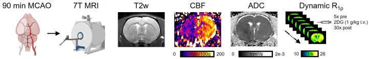 Fig 1
Fig 1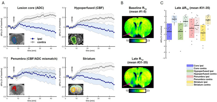 Fig 2
Fig 2- —http://dx.doi.org/10.13039/501100006188Einstein Stiftung Berlin
- —http://dx.doi.org/10.13039/501100006188Einstein Stiftung Berlin
- —http://dx.doi.org/10.13039/501100006188Einstein Stiftung Berlin
- —http://dx.doi.org/10.13039/501100001674Fondation Leducq
- —http://dx.doi.org/10.13039/501100002347Bundesministerium für Bildung und Forschung
- —http://dx.doi.org/10.13039/501100001659Deutsche Forschungsgemeinschaft
- —http://dx.doi.org/10.13039/501100001659Deutsche Forschungsgemeinschaft
- —http://dx.doi.org/10.13039/501100001659Deutsche Forschungsgemeinschaft
Peer Reviews
No public reviews on file for this paper yet. If you reviewed it on a platform where reviews are public (OpenReview, ICLR, NeurIPS, ICML), you can paste yours below so the community can read it here.
Videos
No videos yet. Explain this paper in a talk, walkthrough, or lecture? Add one.
Taxonomy
TopicsLanthanide and Transition Metal Complexes · Advanced MRI Techniques and Applications · Electron Spin Resonance Studies
Introduction
Cerebral glucose metabolism is critical to sustain brain function, as emphasized by the fact that the brain is the main consumer of glucose-derived energy in mammals [1,2]. Disturbance or even breakdown of cerebral glucose metabolism and subsequent energy deficit is associated with several brain disorders, including acute stroke [1,3,4]. We recently reported the application and utility of Chemical Exchange Sensitive Spin Lock (CESL) magnetic resonance imaging (MRI) of the glucose analogue 2-deoxy-d-glucose (2DG) as a novel imaging biomarker to quantify glucose uptake and metabolism in the middle cerebral artery occlusion (MCAO) model of transient focal ischemic stroke in rats [5]. 2DG CESL MRI was compared to standard MRI imaging sequences measuring reduced cerebral blood flow (CBF) using perfusion MRI, and diffusion MRI of the apparent diffusion coefficient (ADC). ADC is a surrogate of the extent of the ischemic lesion core and the mismatch with perfusion MRI is a clinically established marker of the penumbra which is defined as potentially salvageable tissue when restoring perfusion [5]. In our study, we demonstrated that 2DG CESL MRI allowed measuring the cellular uptake and metabolism of the glucose analogue in ischemic tissue and allowed precise mapping of the hypometabolic ischemic core [5]. Importantly, 2DG CESL is not specific to brain ischemia. We believe that it can provide biomarkers of metabolism in many other animal models of brain disorders, such as brain tumors or neurodegenerative disease, or other models entirely.
The underlying principle of 2DG CESL MRI is based on measuring chemical exchange between exchangeable protons on the molecule and the bulk water pool, which can be performed for D-glucose and other glucose analogues as well [6–11]. In CESL, the relaxation rate in the rotating frame (R_1ρ_) is measured, which increases linearly with increasing concentration of 2DG protons in the non-water pool. Thus, the change ΔR_1ρ_ after 2DG injection compared to baseline is a marker of local 2DG concentration. The principle is very similar to measuring glucose via Chemical Exchange Saturation Transfer (gluco-CEST), but the sensitivity of CESL was shown to be higher [12].
2DG behaves chemically almost identical to 2-Deoxy-2-[^18^F]fluoroglucose (FDG), a well established tracer of metabolism in positron emission tomography (PET). Thus, 2DG CESL MRI may present an alternative to FDG-PET without the need of expensive radiochemistry and the high demands on logistics and legal administration of a radionuclide facility. Depending on the intended application, next to using 2DG CESL MRI, it might be useful considering replacing 2DG with the glucose analogue 3-O-methyl-D-glucose (3OMG) which is also detectable using CESL and contrary to 2DG is not metabolized by hexokinase [13].
Here, we provide an experimentally validated protocol [5] to measure cerebral uptake and metabolism of 2DG using CESL MRI in the context of stroke in rodents (Fig 1).
Overview of the protocol detailing experimental and analytical steps.A) Rats undergo 90 min transient MCAO. After surgery, animals are directly transferred to a 7 T MRI system for T2-weighted (T2w) MRI, perfusion MRI of cerebral blood flow (CBF), diffusion MRI of apparent diffusion coefficient (ADC) followed by dynamic R1ρ mapping with CESL MRI before and after injection of 2DG. Figure and legend were previously published and are reproduced from [5] under a CC-BY-NC license.
Materials and methods
The protocol described in this peer-reviewed article is published on protocols.io, https://dx.doi.org/10.17504/protocols.io.n92ldnoz8v5b/v1 and is included for printing as supporting information S1 File with this article.
All animal procedures underlying the reporting of this protocol were performed after approval by the regulating authority (Landesamt für Gesundheit und Soziales Berlin). Studies were performed in accordance with the German Animal Welfare Act and EU regulations.
Expected results
Using the protocol described herein, it will be possible to measure cerebral uptake and metabolism of the glucose analogue 2DG after MCAO as recently described [5]. Even though we expect that this protocol can be readily implemented, we suggest performing initial CESL measurements using phantoms of different 2DG concentrations similarly to outlined herein (supporting information S1 File).
When performing 2DG CESL MRI in conjunction with standard measurements of blood flow and diffusion in the rat MCAO stroke model, it is expected that measurements follow previous results [5] showing an increase of R_1ρ_ over the period of CESL imaging in metabolically active tissue (e.g., contralateral to the stroke as shown here, Fig 2). However, R_1ρ_ is expected to be stable or to decrease in metabolically compromised tissue (e.g., in the stroke territory, Fig 2). Quantifications should focus on late ΔR_1ρ_ measurements, e.g., representing the mean of the last five scans (Fig 2C).
Example of in vivo metabolic MRI in the context of stroke.A) Quantification of mean R1ρ in the lesion core, hypo-perfused areas, penumbra, ipsilateral striatum (blue) and in corresponding mirrored ROIs (gray) which showed strong differences continuously increasing over time. Shaded areas correspond to 95% confidence intervals (CI). Syringes indicate injection of 2DG at 8 minutes after start of CESL imaging. B) Representative baseline and late (mean of first and last 5 maps) R1ρ maps show an increase in contralateral tissue and slight decrease in the lesion territory. C) Quantification of late R1ρ showed strongest effects of ipsi- vs. contralateral values in striatum and lesion core. Here, the contrast was most pronounced in striatum (ipsi: −1.54 ± 2.62%, contra: 2.80 ± 1.45%, t = −5.27, p = 0.00026, significant after Bonferroni correction, Cohen’s d = 1.52) and lesion core (ipsi: −1.11 ± 2.70%, contra: 3.09 ± 1.10%, t = −5.08, p = 0.00036, significant after Bonferroni correction, Cohen’s d = 1.47) but smaller in hypoperfused tissue (ipsi: −1.04 ± 2.76%, contra: 2.33 ± 1.73%, t = −3.84, p = 0.0027, significant after Bonferroni correction, Cohen’s d = 1.11) and not significant in the penumbra (ipsi: 0.04 ± 3.19%, contra: 0.99 ± 2.55%, t = −1.16, p = 0.27, Cohen’s d = 0.33). Figure and legend were previously published and are reproduced with modifications from [5] under a CC-BY-NC license.
Our previous study focused on cerebral 2DG uptake and metabolism in stroke at the time of reperfusion [5]. However, with minimal adjustments, measurements can be made at any timepoint after reperfusion. Given the relatively high concentrations of 2DG required for this protocol, it might be advantageous to replace 2DG with other glucose analogues such as 3OMG to avoid 2DG toxicity from blocking hexokinase [14], which should be possible with minor modifications as performed in other studies [13]. Minor adjustments should allow the protocol to be adapted for use in other disease models and other organs.
In summary, here we have described a protocol for noninvasive metabolic imaging of the brain.
Associated content
Experimental study describing the utility of 2DG-CESL-MRI to measure cerebral glucose metabolism in a rat model of transient ischemic stroke: https://doi.org/10.1177/0271678X251355049Protocol on Protocols.io: https://doi.org/10.17504/protocols.io.n92ldnoz8v5b/v1Data and code associated with this manuscript are available on Zenodo (https://zenodo.org/records/14526092; DOI: 10.5281/zenodo.14526091), including MRI image data to reproduce the steps described in this protocol.
Supporting information
S1 FileStep-by-step protocol, also available on protocols.io (DOI: 10.17504/protocols.io.n92ldnoz8v5b/v1).(PDF)
The reference list from the paper itself. Each links out to its DOI / PubMed record.
- 1Mergenthaler P, Lindauer U, Dienel GA, Meisel A. Sugar for the brain: the role of glucose in physiological and pathological brain function. Trends Neurosci. 2013;36(10):587–97. doi: 10.1016/j.tins.2013.07.001 23968694 PMC 3900881 · doi ↗ · pubmed ↗
- 2Yellen G. Fueling thought: Management of glycolysis and oxidative phosphorylation in neuronal metabolism. J Cell Biol. 2018;217(7):2235–46. doi: 10.1083/jcb.201803152 29752396 PMC 6028533 · doi ↗ · pubmed ↗
- 3Mergenthaler P, Balami JS, Neuhaus AA, Mottahedin A, Albers GW, Rothwell PM, et al. Stroke in the time of circadian medicine. Circ Res. 2024;134(6):770–90. doi: 10.1161/CIRCRESAHA.124.323508 38484031 · doi ↗ · pubmed ↗
- 4Walther J, Kirsch EM, Hellwig L, Schmerbeck SS, Holloway PM, Buchan AM, et al. Reinventing the penumbra - the emerging clockwork of a multi-modal mechanistic paradigm. Transl Stroke Res. 2023;14(5):643–66. doi: 10.1007/s 12975-022-01090-9 36219377 PMC 10444697 · doi ↗ · pubmed ↗
- 5Boehm-Sturm P, Schuenke P, Foddis M, Mueller S, Koch SP, Beard DJ, et al. 2-deoxy-D-glucose chemical exchange-sensitive spin-lock MRI of cerebral glucose metabolism after transient focal stroke in the rat. J Cereb Blood Flow Metab. 2025;45(12):2370–80. doi: 10.1177/0271678 X 251355049 40626496 PMC 12237932 · doi ↗ · pubmed ↗
- 6Jin T, Iordanova B, Hitchens TK, Modo M, Wang P, Mehrens H, et al. Chemical exchange-sensitive spin-lock (CESL) MRI of glucose and analogs in brain tumors. Magn Reson Med. 2018;80(2):488–95. doi: 10.1002/mrm.27183 29569739 PMC 5910214 · doi ↗ · pubmed ↗
- 7Jin T, Mehrens H, Hendrich KS, Kim S-G. Mapping brain glucose uptake with chemical exchange-sensitive spin-lock magnetic resonance imaging. J Cereb Blood Flow Metab. 2014;34(8):1402–10. doi: 10.1038/jcbfm.2014.97 24865996 PMC 4126103 · doi ↗ · pubmed ↗
- 8Paech D, Schuenke P, Koehler C, Windschuh J, Mundiyanapurath S, Bickelhaupt S, et al. T 1ρ-weighted dynamic glucose-enhanced MR imaging in the human brain. Radiology. 2017;285(3):914–22. doi: 10.1148/radiol.2017162351 28628422 · doi ↗ · pubmed ↗