Lensless Quantitative Phase Imaging with Bayer-Filtered Color Sensors Under Sequential RGB-LED Illumination
Jiajia Wu, Yining Li, Yuheng Luo, Leiting Pan, Pengming Song, Qiang Xu

TL;DR
This paper introduces a new method for high-resolution, lensless imaging using color sensors and RGB-LEDs, enabling detailed biological imaging without labels.
Contribution
A novel Wirtinger Poly-Gradient Solver (WPGS) that improves phase imaging with Bayer-filtered sensors under RGB-LED illumination.
Findings
The method achieves pixel super-resolution beyond the Nyquist limit using a 1.85 μm pixel-pitch sensor.
Label-free imaging of HeLa cells and unstained tissue sections was successfully demonstrated.
The approach suppresses CFA-induced artifacts and enables high-throughput digital pathology.
Abstract
Lensless on-chip microscopy enables high-throughput, wide-FOV imaging; however, the Bayer color filter array (CFA) in standard color sensors spatially multiplexes spectral channels, introducing sub-sampling and spectral crosstalk that degrade phase retrieval. We propose a Wirtinger Poly-Gradient Solver (WPGS) for quantitative phase reconstruction with Bayer-filtered color sensors under sequential Red–Green–Blue Light-Emitting Diode (RGB-LED) illumination. The method combines Transport of Intensity Equation (TIE)-based initialization with polychromatic Wirtinger optimization to suppress CFA-induced artifacts and enable pixel super-resolution (PSR). Experiments resolve a 2.76 μm linewidth using a 1.85 μm pixel-pitch sensor, exceeding the nominal Nyquist limit imposed by pixel sampling. We further demonstrate label-free imaging of HeLa cells and unstained tissue sections, supporting…
Genes, proteins, chemicals, diseases, species, mutations and cell lines named across the full text — each resolved to its canonical identifier and authoritative record.
Click any figure to enlarge with its caption.
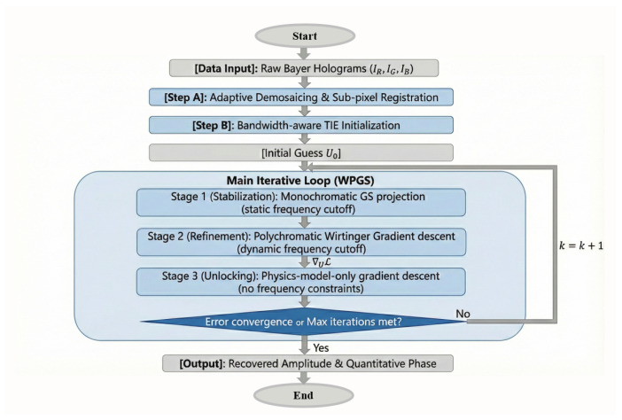 Figure 1
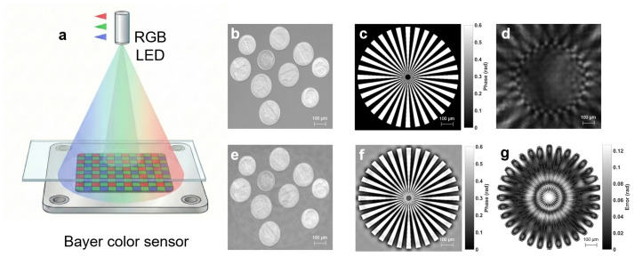 Figure 2
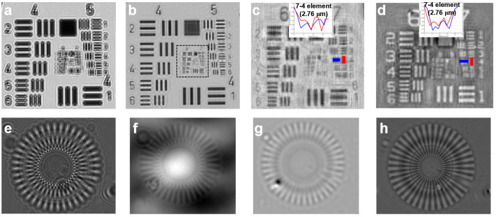 Figure 3
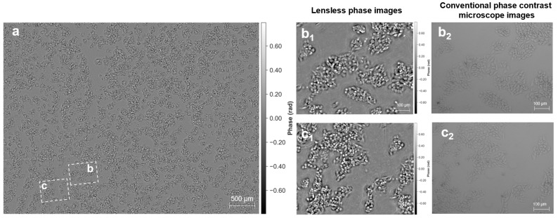 Figure 4
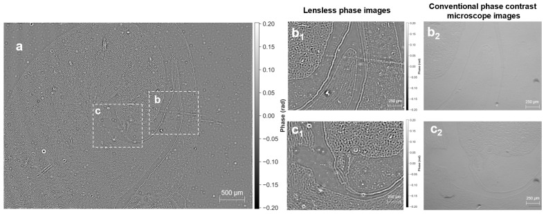 Figure 5
- —Guangzhou National Laboratory
- —National Natural Science Foundation of China
Peer Reviews
No public reviews on file for this paper yet. If you reviewed it on a platform where reviews are public (OpenReview, ICLR, NeurIPS, ICML), you can paste yours below so the community can read it here.
Videos
No videos yet. Explain this paper in a talk, walkthrough, or lecture? Add one.
Taxonomy
TopicsDigital Holography and Microscopy · Advanced X-ray Imaging Techniques · Random lasers and scattering media
1. Introduction
While indispensable for life sciences [1,2], conventional microscopy is fundamentally constrained by the field of view (FOV) versus resolution trade-off, dictated by physical invariants such as optical étendue and the space-bandwidth product (SBP) [3,4,5]. In practice, achieving high resolution typically requires high-numerical-aperture objectives, which inevitably restrict the FOV and limit throughput in applications such as large-scale cell monitoring and comprehensive histological analysis [6,7,8].
Lensless on-chip microscopy addresses this limitation by placing the specimen in close proximity to the image sensor and reconstructing the complex field computationally from recorded diffraction patterns [9,10]. Despite its compact form factor and large FOV, many high-performance lensless systems still rely on monochromatic detection [11,12]. Standard color CMOS sensors are attractive due to their accessibility and low cost, yet the Bayer color filter array (CFA) spatially multiplexes spectral channels, introducing sub-sampling and spectral crosstalk [13,14]. These effects reduce the effective spatial-frequency support and induce model-mismatch errors in phase retrieval [15,16], resulting in chromatic artifacts and degraded reconstruction fidelity [17,18].
Existing mitigation strategies typically trade hardware simplicity for spectral efficiency, for example, by physically removing the CFA or operating on a single color channel [19,20]. Moreover, conventional demosaicing methods—largely optimized for human visual perception—tend to attenuate the high-frequency oscillatory fringes that are essential for coherent diffraction imaging. To overcome these limitations without modifying standard sensors [15,16,17], we introduce the Wirtinger Poly-Gradient Solver (WPGS), a multi-spectral computational framework for quantitative phase recovery. The key contributions of this work are summarized as follows: (i) We propose a physics-based polychromatic model to numerically decouple the periodic CFA modulation; (ii) We develop a three-stage iterative engine that progressively unlocks sub-pixel structural details; (iii) We demonstrate that by leveraging spectral diversity as a strong physical constraint, the space-bandwidth product (SBP) can be effectively extended on commercial Bayer-filtered sensors.
By combining sequential Red–Green–Blue Light-Emitting Diode (RGB-LED) illumination with this iterative framework, WPGS suppresses crosstalk [21] and enables pixel super-resolution (PSR). Experiments successfully resolve a linewidth using a sensor with a pixel pitch, exceeding the nominal Nyquist limit. We further demonstrate label-free imaging of HeLa cell populations and unstained tissue sections, offering significant potential for longitudinal live-cell monitoring and digital pathology.
2. Materials and Methods
In this section, we present the hardware and algorithmic components of our lensless imaging system. The process involves sequential RGB illumination, adaptive demosaicing, and a multi-spectral iterative reconstruction to achieve quantitative phase recovery from Bayer-filtered holograms.
2.1. Experimental Configuration and Sequential Illumination
The imaging system is a compact lensless on-chip microscope designed for scan-free quantitative phase acquisition. Diffraction patterns are recorded using a Sony IMX226 CMOS sensor (Sony Corporation, Tokyo, Japan) ( , pixel pitch). The sample is placed at a sensor distance of approximately to balance space-bandwidth product and fringe contrast. Illumination is provided by an integrated RGB-LED module with center wavelengths of , , and (Full Width at Half Maximum, FWHM ). The three LEDs are triggered sequentially to acquire a multi-wavelength hologram stack within seconds, providing spectral diversity for the subsequent reconstruction.
2.2. Biological Specimen Preparation and Comparative Imaging
To assess performance on biologically relevant specimens, we imaged fixed HeLa cells and unstained mouse kidney tissue sections. HeLa cells were cultured in Dulbecco’s Modified Eagle Medium (DMEM) supplemented with 10% fetal bovine serum and 1% penicillin–streptomycin (Gibco, Carlsbad, CA, USA) at 37 °C in 5% CO_2_. Cells were seeded on glass-bottom dishes (NEST, Wuxi, China) and fixed at 70–80% confluency using 4% paraformaldehyde (Biosharp, Hefei, China), followed by rinsing with phosphate-buffered saline. Unstained mouse kidney tissue slices (Tianjian, Xinxiang, China) were deparaffinized and rehydrated prior to imaging. For lensless acquisition and quantitative analysis, the sample was maintained in phosphate-buffered saline (PBS), which was treated as the surrounding medium in subsequent dry-mass estimation. For reference, corresponding fields of view were acquired using a commercial inverted microscope (Axio Observer A1, Carl Zeiss AG, Oberkochen, Germany) with phase-contrast objectives ( and , 0.4 NA). These images were used as a reference to evaluate the reconstruction fidelity of WPGS.
2.3. Forward Modeling of Polychromatic Bayer-Filtered Holography
In practical on-chip microscopy, the monochromatic assumption is violated by the finite spectral bandwidth of LED sources, leading to polychromatic averaging and wavelength-dependent propagation. For the Sony IMX226 sensor, the recorded intensity for channel is modeled as an integral over wavelength, weighted by the illumination spectral power distribution (SPD) and the combined CFA–sensor spectral response:
Here, represents the approximately Gaussian SPD of the c-th LED, and denotes the combined spectral response of the CFA–sensor system (incorporating both CFA transmittance and sensor quantum efficiency) for channel c. The operator defines free-space propagation based on the angular spectrum method.
2.4. Adaptive Demosaicing and Multi-Spectral Registration
Because the Bayer CFA spatially sub-samples each color channel, conventional demosaicing can introduce interpolation artifacts. We adopt an edge-aware strategy where horizontal and vertical gradients ( ) determine the interpolation direction:
Ties are resolved by averaging both directions. Following demosaicing, wavelength-dependent lateral shifts are compensated using Discrete Fourier Transform (DFT)-based registration to enforce spatial correspondence across multi-wavelength channels.
2.5. The WPGS Reconstruction Pipeline
The WPGS framework addresses the inherent low-frequency sensitivity of Transport of Intensity Equation (TIE) and the high-frequency convergence challenges of iterative phase retrieval. The logical flow of the algorithm is detailed in Figure 1.
2.5.1. Phase Initialization via Bandwidth-Aware TIE
The reconstruction pipeline is initiated with a deterministic phase estimate derived from a Fast Fourier transform (FFT)-based TIE solver [22]. To circumvent the requirement for physical axial scanning, we adopt a heuristic spectral-to-axial mapping that approximates the axial intensity derivative by leveraging the intensity contrast between disparate spectral channels. To account for polychromatic decoherence and compensate for the inherent spectral responsivity mismatch between color channels, we introduce a bandwidth-aware correction factor :
where and denote the holograms captured under two distinct center wavelengths. The parameter is a bandwidth-aware correction factor that also serves as a dynamic step-size during the optimization (decaying linearly from 0.5 to 0.1), and represents the equivalent axial defocus separation facilitated by wavelength diversity via Fresnel scaling. Here, is the sample-to-sensor distance, and is the center wavelength of the primary imaging channel. To compensate for the global piston ambiguity and systematic gain attenuation inherent in TIE-based solvers, we apply a linear calibration only in numerical simulations where a ground-truth phase is available, prior to quantitative evaluation and visualization. For experimental results, we do not rescale the phase values; instead, we only perform median-background piston removal and sign normalization as described in Section 2.6.
2.5.2. Non-Linear Refinement via Wirtinger Poly-Gradient Optimization
The initial estimate is refined through Wirtinger optimization [23]. The objective function compares measured amplitude with band-integrated predictions:
In Equation (4), U represents the complex amplitude of the specimen to be recovered, and is the free-space propagation operator at wavelength . The nonnegative weights satisfy and are computed as , where and denote the source emission spectrum and the Bayer filter’s spectral response, respectively. Additionally, S denotes the number of discrete spectral sampling points (set to to balance reconstruction fidelity and efficiency).
To ensure convergence and sub-pixel accuracy, the optimization is executed for 100 total iterations, divided into three structured stages corresponding to the flowchart in Figure 1: Stage 1 (Iterations 1–20): Focuses on monochromatic stabilization. By using monochromatic projections with a static frequency cutoff, we stabilize the initial phase estimate and suppress coarse noise. Stage 2 (Iterations 21–70): Introduces the polychromatic Wirtinger gradient with a dynamic frequency cutoff. To ensure robust convergence, the step size is linearly decayed from 0.5 to 0.1, effectively decoupling the CFA-induced crosstalk. Stage 3 (Iterations 71–100): Performs final resolution unlocking. By removing all frequency constraints, the physics-model-only descent recovers high-frequency diffraction details and achieves final pixel super-resolution. The computational efficiency of the WPGS framework is optimized for high-throughput applications. In our implementation using graphics processing unit (GPU) acceleration (NVIDIA RTX 3090), a typical pixel reconstruction with 100 iterations requires approximately 3.4 s, making it suitable for rapid biological screening and live-cell monitoring.
2.6. Quantitative Dry-Mass Estimation
For lensless phase retrieval, the recovered phase contains a global piston ambiguity. Therefore, a baseline correction was applied by subtracting the median phase measured in a non-cell background region. A sign convention was subsequently enforced by flipping the phase map when the mean phase within the segmented cell region was negative, ensuring that cellular regions exhibit positive optical path delay. For dry-mass quantification, a whole-mask integration strategy was adopted. To suppress residual negative contributions caused by noise or slight model mismatch, we set negative phase values inside the cellular mask to zero before integration, i.e., . Note that this non-negative truncation is applied only for dry-mass integration; phase maps are visualized using the piston-corrected and sign-normalized phase without truncation. The total dry mass within the segmented cellular region is estimated as [24,25]:
where denotes the piston-corrected and sign-normalized phase (in radians) after non-negative truncation, is the effective wavelength (520 nm, Plan A), is the refractive index increment (0.18–0.20 mL/g), and is the pixel area. The estimated mean dry mass per cell is reported as , where N denotes the number of segmented cells within the ROI. For reproducible visualization of the experimental phase maps, all phase maps are displayed in radians using a symmetric dynamic range , where m is determined by the 99th percentile of within the displayed ROI.
3. Results
In this section, we evaluate the performance of WPGS by first validating the forward model and noise robustness in numerical simulations, and then assessing practical imaging performance in experiments.
3.1. Numerical Simulation and Quantitative Evaluation
Numerical simulations using a complex phantom validated the reconstruction fidelity under configurations matched to Section 2.1, with a noise surrogate introduced for experimental perturbations. Specifically, the simulation employed a complex phantom with a pixel size of . We modeled the polychromatic illumination using the measured spectral power distributions (SPD) of RGB LEDs and introduced a Gaussian noise surrogate (std = 0.01) to simulate experimental perturbations. Compared to the blurred raw hologram (Figure 2d), the WPGS framework significantly improved structural preservation, achieving an amplitude Root Mean Square Error (RMSE) of 0.0229 and a phase RMSE of within the strict object ROI after linear calibration (corresponding to a relative error of approximately with respect to the phase peak). The phase Structural Similarity Index (SSIM) increased from (the TIE-only baseline) to post-optimization. To compensate for visualization consistency and systematic gain attenuation inherent in TIE-based solvers, a linear calibration was applied within the effective object ROI prior to visualization and quantitative evaluation. Specifically, the phase residual map (Figure 2g) confirms that absolute errors remain below for of pixels within the strict object ROI (approximately of the full field, calculated by counting pixels with an absolute error less than relative to the total pixel count), with minor deviations localized at high-frequency boundaries where recovery is ill-conditioned due to Bayer sub-sampling limits. The residual in Figure 2g is visualized as in radians (no normalization); therefore, its dynamic range is intentionally much smaller than that of the phase maps in Figure 2c,f. The absence of periodic checkerboard artifacts validates effective CFA decoupling and robust fine-scale phase recovery. Although some minor grid-like artifacts are visible in the reconstructed amplitude (Figure 2e), they are primarily attributed to the inherent spatial sub-sampling of the Bayer CFA. Crucially, these artifacts have a negligible impact on the quantitative phase accuracy, as the phase residual remains extremely low (Figure 2g). This spatial error distribution confirms the physical consistency of our model: the framework excels in stabilizing bulk phase retrieval, while the localized boundary deviations highlight the inherent trade-off between sub-pixel resolution and the discrete sampling limits of the Bayer CFA. It is important to acknowledge the inherent limitations of the proposed framework at this stage. While WPGS significantly enhances phase fidelity, its performance is subject to a dual constraint: first, the physical information loss caused by the Bayer CFA occlusion limits the recovery of extreme high-frequency components; and second, the iterative solver faces inherent challenges in maintaining convergence stability when reconstructing sharp, non-continuous phase transitions from sub-sampled data. Recognizing these boundaries is crucial for further optimization of the sampling and reconstruction engine.
3.2. Experimental Performance and Resolution Analysis
We evaluated the practical imaging performance of WPGS using a USAF 1951 target and a Siemens star target under the configuration described in Section 2.1. Figure 3a shows the raw diffraction pattern of the USAF target, which is affected by Bayer CFA modulation and exhibits mosaic artifacts and diffraction-induced blur.
The full FOV amplitude reconstruction is shown in Figure 3b, and a magnified region for resolution assessment is provided in Figure 3c. WPGS resolves Group 7 Element 4, corresponding to a linewidth of . To clearly substantiate this claim, we have enlarged the intensity profile insets in Figure 3c,d. The calculated peak-to-valley ratio is approximately 20%, which indicates resolvability of Group 7-4 under a commonly used bar-target modulation criterion. The consistent resolution of these features in both amplitude and phase reconstructions further confirms the reliability of our PSR performance. For a sensor pixel pitch of , the nominal Nyquist limit imposed by pixel sampling is ; therefore, resolving features indicates pixel super-resolution (PSR) by surpassing the nominal sampling limit of a single Bayer-subsampled channel. The corresponding phase reconstruction (Figure 3d) preserves contrast with a stable background across Groups 6 and 7.
Compared with numerical simulations, the experimentally achieved resolution is lower than the best-case prediction (e.g., access to Group 8 elements). This gap is mainly attributable to the Bayer CFA, which imposes periodic sub-sampling and wavelength-dependent crosstalk that attenuate high-frequency fringes at the measurement stage. Under experimental noise, the reduced fringe contrast limits the recovery of sub-pixel structures, thereby constraining the attainable resolution.
To further isolate the contribution of polychromatic optimization, we additionally evaluated phase recovery using a Siemens star target (Figure 3e–h). The TIE-only result (Figure 3f) provides a stable low-frequency estimate but remains strongly blurred, preventing reliable resolution of the inner spokes. The monochromatic iterative reconstruction (Figure 3g) improves apparent sharpness but exhibits pronounced ringing and spatially varying phase inconsistencies, consistent with insufficient constraints to decouple CFA-induced modulation. In contrast, WPGS (Figure 3h) reduces ringing artifacts and yields a more spatially consistent phase map by exploiting spectral diversity under sequential RGB illumination, enabling clearer visibility of spokes toward the center region. This performance leap is inherently rooted in the spectral redundancy provided by the multi-spectral model. Unlike monochromatic baselines that suffer from ill-posed sub-sampling, WPGS leverages the overlapping spectral responses as a physical constraint to effectively resolve high-frequency features beyond the nominal Nyquist limit.
3.3. Application to Biological Specimen Imaging
We demonstrate label-free quantitative phase imaging on fixed HeLa cells and unstained mouse kidney tissue sections. These specimens exhibit limited contrast in bright-field imaging, whereas the proposed system recovers quantitative phase maps without exogenous staining. Figure 4a shows a full-FOV reconstruction covering approximately , enabling visualization of cell distributions across the sensor area. Magnified regions (Figure 4(b1,c1)) reveal cellular morphology and subcellular density variations that are consistent with the corresponding reference phase-contrast images (Figure 4(b2,c2)). To provide a physically interpretable quantitative metric, we report a conservative whole-mask dry-mass estimate for the HeLa-cell ROI acquired in PBS (treated as the surrounding medium). Following median-background piston removal, sign normalization, and non-negative truncation as described in Section 2.6, the total dry mass within the segmented cellular region was estimated as – pg (using – mL/g and nm). With segmented cells, the corresponding mean dry mass is 142.68–158.53 pg per cell. These values are in line with the reported dry-mass range of mammalian cells measured by quantitative phase microscopy. We emphasize that this whole-mask integration avoids per-cell boundary ambiguity and provides a robust population-level quantitative metric. The phase maps shown in Figure 4 are visualized in radians using a symmetric 99th-percentile-based dynamic range (units: rad), following the same percentile rule described in Section 2.6 for reproducible display.
For unstained mouse kidney tissue (Figure 5), histological structures such as glomeruli and convoluted tubules are readily identified. In addition, the reconstructed phase maps show reduced halo artifacts compared with conventional phase-contrast microscopy, leading to a more uniform background across dense tissue regions.
4. Discussion
The numerical and experimental results in Section 3 demonstrate that WPGS enables robust lensless quantitative phase imaging with Bayer-filtered color sensors. By combining deterministic TIE-based initialization with polychromatic Wirtinger optimization under sequential RGB illumination, the framework improves reconstruction stability while preserving a scan-free and compact hardware configuration.
4.1. Physical Synergy of the Hybrid Framework
The robustness of WPGS stems from integrating complementary mechanisms. TIE provides a deterministic low-frequency phase estimate from the axial intensity derivative, which improves initialization and alleviates low-frequency stagnation and local-minimum trapping in non-linear phase retrieval. Building on this initialization, polychromatic optimization exploits spectral diversity to refine the solution and recover higher spatial-frequency components. The demonstrated sub-pixel enhancement is primarily facilitated by fusing complementary frequency supports across RGB wavelengths. While Section 3.2 presents the quantitative resolution gain, here we emphasize that the framework’s ability to exceed the Nyquist limit is a direct consequence of spectral diversity serving as a strong physical constraint, which numerically mitigates the spatial information loss inherent in Bayer CFA modulation.
4.2. Comparison with Alternative Lensless Methodologies
WPGS offers distinct advantages over scanning-based architectures like coded ptychography. While the latter requires extensive frame acquisition and is sensitive to mechanical drift [26,27], our framework achieves multi-wavelength constraints without translational scanning by sequentially switching static RGB LEDs. This configuration significantly enhances temporal efficiency and environmental robustness, rendering the system inherently amenable to prospective longitudinal biological observation without the artifacts associated with mechanical instability. Furthermore, in contrast to monochromatic iterative solvers, the multi-wavelength optimization framework provides intrinsic spectral redundancy across channels. This redundancy facilitates the suppression of background noise, mitigates ringing artifacts, and minimizes cross-channel model inconsistencies, ultimately yielding superior phase reconstruction fidelity.
4.3. Error Analysis and Bottlenecks of the Bayer Architecture
Despite the demonstrated resolution improvement, a gap remains between experimental reconstructions and idealized simulations, largely due to the structural constraints of the Bayer CFA. The CFA imposes periodic sub-sampling and wavelength-dependent crosstalk, which attenuate high-frequency diffraction fringes at the measurement stage. While adaptive demosaicing reduces interpolation artifacts, information physically lost due to CFA occlusion cannot be fully recovered computationally. Residual deviations are further exacerbated by partial source coherence and sensor noise, which reduce fringe contrast and increase model mismatch in practical measurements.
4.4. Clinical Potential and Future Trajectories
The reconstructions of HeLa cells and unstained tissue sections indicate the potential of WPGS for high-throughput, label-free quantitative pathology. The unit-magnification geometry provides a large SBP, enabling micrometer-scale resolution across the full active sensor area. In addition, the recovered quantitative phase maps reduce halo artifacts commonly observed in phase-contrast microscopy, improving interpretability in dense histological structures. Future work will extend the framework to multi-angle illumination for three-dimensional diffraction tomography and investigate hardware-accelerated implementations for real-time biomedical imaging.
5. Conclusions
We presented WPGS for lensless quantitative phase imaging with Bayer-filtered color sensors. By combining bandwidth-aware TIE initialization with three-stage polychromatic Wirtinger optimization, WPGS numerically mitigates CFA-induced crosstalk and enables PSR. Experiments resolved features over an approximately field of view and demonstrated label-free imaging of cells and unstained tissue sections, supporting high-throughput digital pathology. While the current study validates morphological phase contrast on fixed specimens and further demonstrates dry-mass estimation in PBS, future work will extend this framework to time-lapse live-cell imaging to track dynamic dry-mass changes and growth rates, and to further benchmark quantitative accuracy under physiologically relevant conditions.
The reference list from the paper itself. Each links out to its DOI / PubMed record.
- 1Amos B. Lessons from the history of light microscopy Nat. Cell Biol.20002 E 151E 15210.1038/3501963910934490 · doi ↗ · pubmed ↗
- 2Stephens D.J. Allan V.J. Light microscopy techniques for live cell imaging Science 2003300828610.1126/science.108216012677057 · doi ↗ · pubmed ↗
- 3Ozcan A. Mc Leod E. Lensless imaging and sensing Annu. Rev. Biomed. Eng.2016187710210.1146/annurev-bioeng-092515-01084927420569 · doi ↗ · pubmed ↗
- 4Greenbaum A. Luo W. Su T.W. Göröcs Z. Xue L. Isikman S.O. Coskun A.F. Mudanyali O. Ozcan A. Imaging without lenses: Achievements and remaining challenges of wide-field on-chip microscopy Nat. Methods 2012988989510.1038/nmeth.211422936170 PMC 3477589 · doi ↗ · pubmed ↗
- 5Lukosz W. Optical systems with resolving powers exceeding the classical limit J. Opt. Soc. Am.1966561463147110.1364/JOSA.56.001463 · doi ↗
- 6Bishara W. Sikora U. Mudanyali O. Su T.W. Yaglidere O. Luckhart S. Ozcan A. Holographic pixel super-resolution in portable lensless on-chip microscopy using a fiber-optic array Lab Chip 2011111276127910.1039/c 0lc 00684 j 21365087 PMC 3151573 · doi ↗ · pubmed ↗
- 7Isikman S.O. Greenbaum A. Luo W. Coskun A.F. Ozcan A. Giga-Pixel Lensfree Holographic Microscopy and Tomography Using Color Image Sensors P Lo S ONE 20127 e 4504410.1371/journal.pone.004504422984606 PMC 3440383 · doi ↗ · pubmed ↗
- 8Mudanyali O. Tseng D. Oh C. Isikman S.O. Sencan I. Bishara W. Oztoprak C. Seo S. Khademhosseini B. Ozcan A. Compact, light-weight and cost-effective microscope based on lensless incoherent holography for telemedicine applications Lab Chip 2010101417142810.1039/c 000453 g 20401422 PMC 2902728 · doi ↗ · pubmed ↗