Double Staining Immunohistochemistry and Digital Pathology: Moving Towards Standardization of the Proliferative Index Evaluation in Meningiomas
Viscardo Paolo Fabbri, Giuseppe Broggi, Giovanna Attolini, Vincenzo L’Imperio, Fabio Pagni, Angela Guerriero, Stefano Marletta, Francesco Fiorentino, Stefania Caramaschi, Claudia Malagoli, Albino Eccher, Rosario Caltabiano

TL;DR
This study shows that double staining and digital pathology can improve the accuracy of evaluating the proliferation index in meningiomas, helping standardize tumor grading.
Contribution
The study introduces a standardized method using double staining immunohistochemistry and digital pathology for meningioma proliferation index evaluation.
Findings
Ki-67 expression varied by meningioma grade, with digital image analysis showing high agreement with manual assessments.
Double staining immunohistochemistry improved accuracy in evaluating diagnostic and proliferative markers in tumor samples.
Abstract
Background: Although Ki-67 is not included among the grading criteria in the current WHO Classification of Tumours of the Central Nervous System (CNS), it provides valuable, albeit limited, prognostic information. Immunohistochemistry for Ki-67 can reveal uneven proliferation patterns and assist in the assessment of mitotic counts. Several studies indicate that meningiomas with a proliferation index > 4% show recurrence rates comparable to CNS WHO grade 2 (atypical) tumors, while tumors with an index > 20% are associated with mortality rates similar to CNS WHO grade 3 (anaplastic) meningiomas. Issues related to Ki-67 assessment include interobserver variability, the use of different cut-off values among pathologists, and the presence of a complex inflammatory tumour microenvironment, which may lead to an overestimation of the proliferative index (PI). Methods: In this study, we describe…
Genes, proteins, chemicals, diseases, species, mutations and cell lines named across the full text — each resolved to its canonical identifier and authoritative record.
Click any figure to enlarge with its caption.
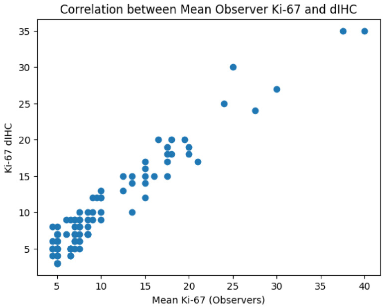 Figure 1
 Figure 2
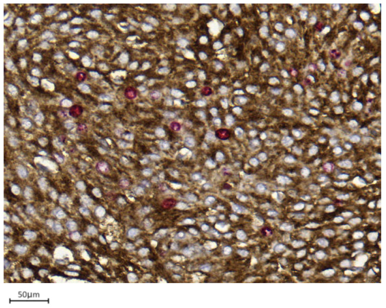 Figure 3
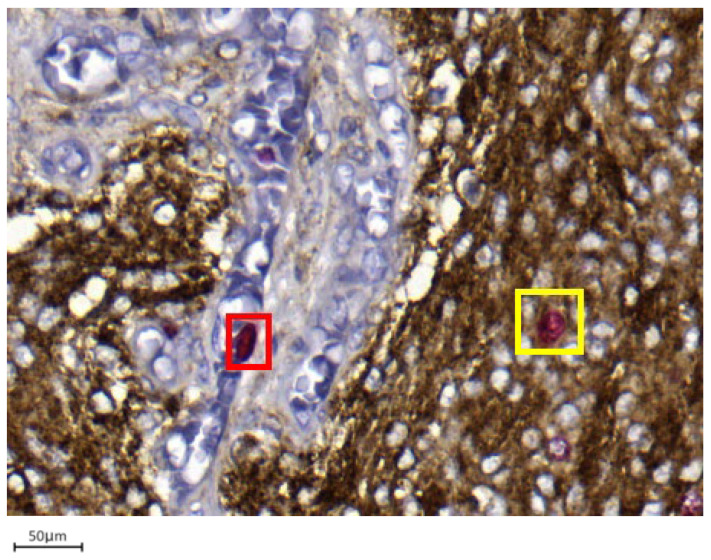 Figure 4
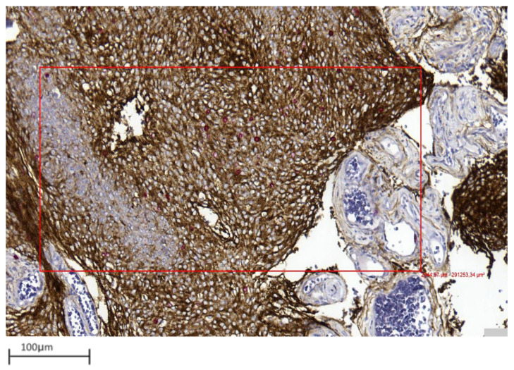 Figure 5
 Figure 6
 Figure 7
Peer Reviews
No public reviews on file for this paper yet. If you reviewed it on a platform where reviews are public (OpenReview, ICLR, NeurIPS, ICML), you can paste yours below so the community can read it here.
Videos
No videos yet. Explain this paper in a talk, walkthrough, or lecture? Add one.
Taxonomy
TopicsMeningioma and schwannoma management · Glioma Diagnosis and Treatment · Brain Metastases and Treatment