ClearScope: A Fully Integrated Light-Sheet Theta Microscope for Sub-Micron-Resolution Imaging Without Lateral Size Constraints
Matthew G. Fay, Peter J. Lang, David S. Denu, Nathan J. O’Connor, Benjamin Haydock, Jeffrey Blaisdell, Nicolas Roussel, Alissa Wilson, Sage R. Aronson, Veronica Pessino, Paul J. Angstman, Cheng Gong, Tanvi Butola, Orrin Devinsky, Jayeeta Basu, Raju Tomer, Jacob R. Glaser

TL;DR
ClearScope is a new microscope that can image large brain samples in high detail without size limits, helping study brain structure and disease.
Contribution
A fully integrated and user-friendly light-sheet theta microscope system that enables sub-micron-resolution imaging of large biological specimens without lateral size constraints.
Findings
ClearScope enables subcellular-resolution imaging of large cleared brain specimens.
The system supports seamless workflow from image acquisition to quantitative analysis.
Demonstrated performance includes high-resolution 3D imaging of mouse and human brain samples.
Abstract
Three-dimensional (3D) ex vivo imaging of cleared tissue from intact brains from animal models, human brain surgical specimens, and large postmortem human and non-human primate brain specimens is essential for understanding physiological neural connectivity and pathological alterations underlying neurological and neuropsychiatric disorders. Contemporary light-sheet microscopy enables rapid, high-resolution imaging of large, cleared samples but is limited by the orthogonal arrangement of illumination and detection optics, which constrains specimen size. Light-sheet theta microscopy (LSTM) overcomes this limitation by employing two oblique illumination paths while maintaining a perpendicular detection geometry. Here, we report the development of a next-generation, fully integrated and user-friendly LSTM system that enables uniform subcellular-resolution imaging (with subcellular…
Genes, proteins, chemicals, diseases, species, mutations and cell lines named across the full text — each resolved to its canonical identifier and authoritative record.
Click any figure to enlarge with its caption.
 Figure 1
 Figure 2
 Figure 3
 Figure 4
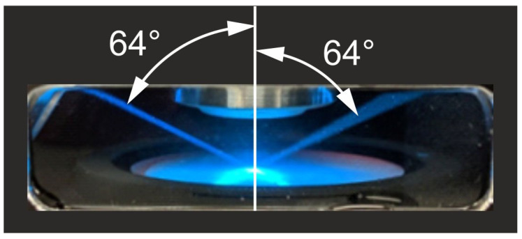 Figure 5
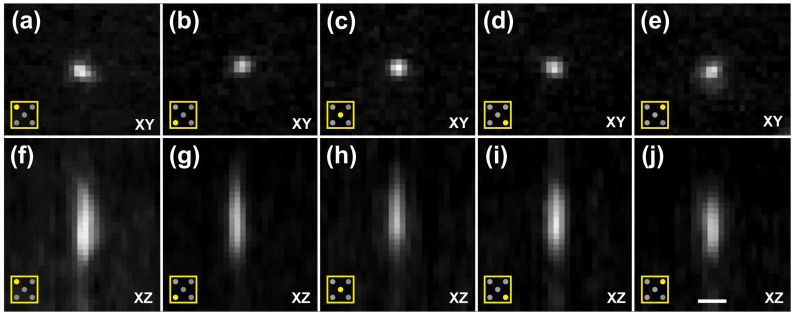 Figure 6
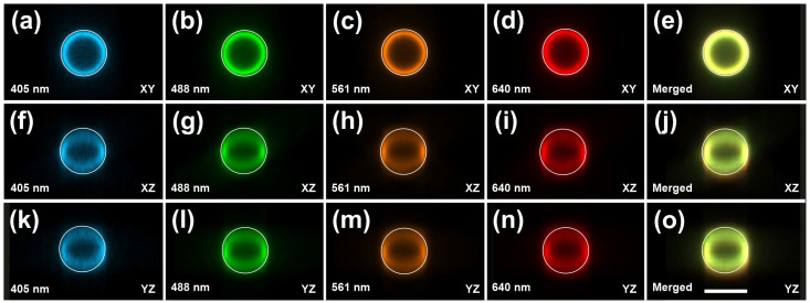 Figure 7
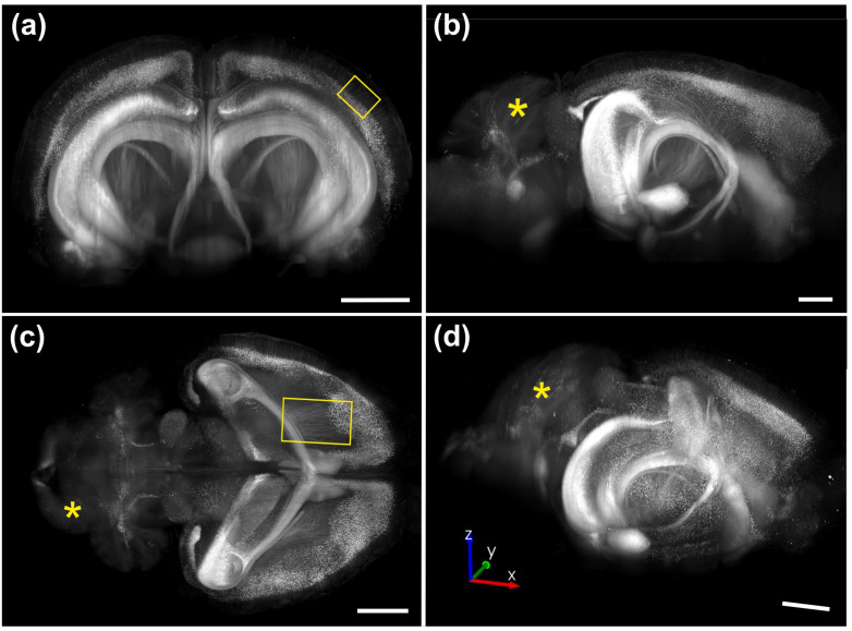 Figure 8
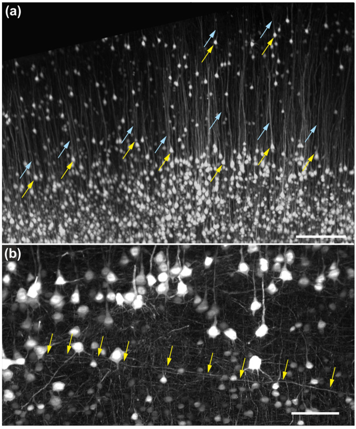 Figure 9
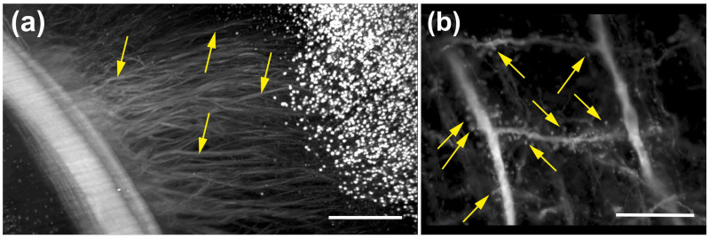 Figure 10
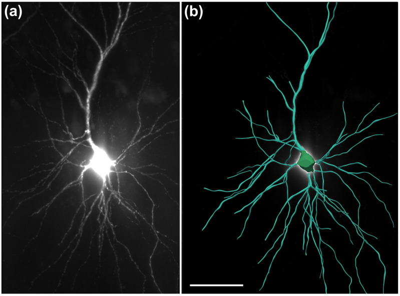 Figure 11
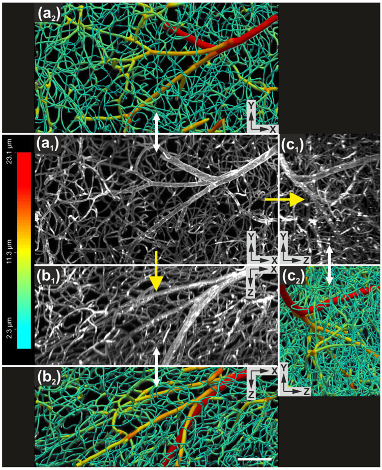 Figure 12
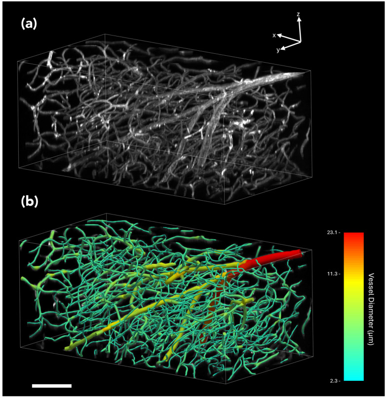 Figure 13
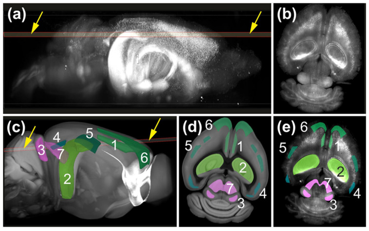 Figure 14
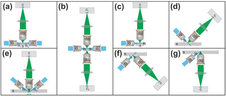 Figure 15
Peer Reviews
No public reviews on file for this paper yet. If you reviewed it on a platform where reviews are public (OpenReview, ICLR, NeurIPS, ICML), you can paste yours below so the community can read it here.
Videos
No videos yet. Explain this paper in a talk, walkthrough, or lecture? Add one.
Taxonomy
TopicsAdvanced Fluorescence Microscopy Techniques · Cell Image Analysis Techniques · Digital Holography and Microscopy