Automated Malaria Ring Form Classification in Blood Smear Images Using Ensemble Parallel Neural Networks
Pongphan Pongpanitanont, Naparat Suttidate, Manit Nuinoon, Natthida Khampeeramao, Sakhone Laymanivong, Penchom Janwan

TL;DR
This paper introduces an AI system that can detect malaria ring-form parasites in blood smear images with high accuracy, potentially improving malaria diagnosis efficiency.
Contribution
The novel contribution is a dual-branch neural network architecture combining convolutional and attention-based features for malaria ring-form classification.
Findings
The model achieved an ROC–AUC of approximately 0.99 and a macro F1-score of 0.97 on an independent test set.
Moderate-capacity feature fusion outperformed more complex architectures, which suffered from higher false positives.
The system shows promise for automated malaria screening but requires external validation before clinical use.
Abstract
Manual microscopy for malaria diagnosis is labor-intensive and prone to inter-observer variability. This study presents an automated binary classification approach for detecting malaria ring-form infections in thin blood smear single-cell images using a parallel neural network framework. Utilizing a balanced Kaggle dataset of 27,558 erythrocyte crops, images were standardized to 128 × 128 pixels and subjected to on-the-fly augmentation. The proposed architecture employs a dual-branch fusion strategy, integrating a convolutional neural network for local morphological feature extraction with a multi-head self-attention branch to capture global spatial relationships. Performance was rigorously evaluated using 10-fold stratified cross-validation and an independent 10% hold-out test set. Results demonstrated high-level discrimination, with all models achieving an ROC–AUC of approximately…
Genes, proteins, chemicals, diseases, species, mutations and cell lines named across the full text — each resolved to its canonical identifier and authoritative record.
Click any figure to enlarge with its caption.
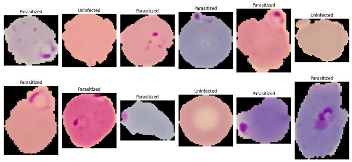 Figure 1
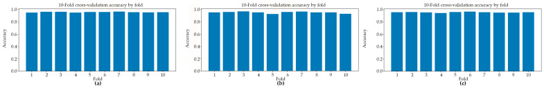 Figure 2
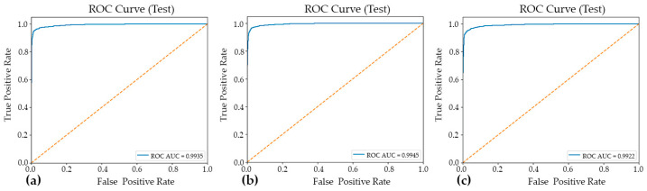 Figure 3
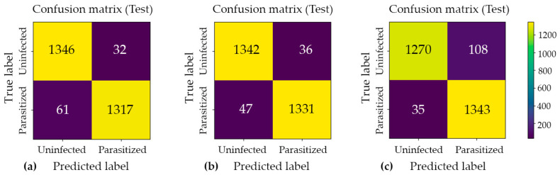 Figure 4
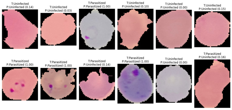 Figure 5
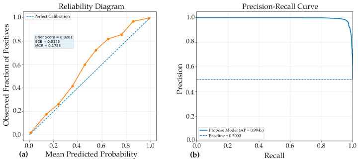 Figure 6
Peer Reviews
No public reviews on file for this paper yet. If you reviewed it on a platform where reviews are public (OpenReview, ICLR, NeurIPS, ICML), you can paste yours below so the community can read it here.
Videos
No videos yet. Explain this paper in a talk, walkthrough, or lecture? Add one.
Taxonomy
TopicsDigital Imaging for Blood Diseases · Cell Image Analysis Techniques · AI in cancer detection