Evaluation of Gingival Sulcus Width Gain After Nd: YAG Laser and Astringent Retraction Paste Using Intraoral and Laboratory STL Analysis: A Pilot Split-Mouth Study
Edwin Sever Bechir, Andrei-Mario Bădărău-Șuster, Mircea Suciu, Anca-Georgiana Zamfir, Zsuzsanna Bardocz-Veres, Farah Bechir

TL;DR
This study compares two methods for widening the gum sulcus to improve dental impressions, finding both effective but with differences in measurement depending on the scanner used.
Contribution
The study introduces a novel split-mouth design using intraoral and laboratory STL analysis to evaluate sulcus width gain from two soft tissue displacement methods.
Findings
Both Nd: YAG laser and astringent retraction paste achieved sulcus width gains above the clinical threshold of 0.20 mm.
The laboratory scanner recorded significantly higher sulcus width measurements compared to the intraoral scanner.
No statistically significant difference was found between the two displacement methods.
Abstract
Background/Objectives: Advancements in digital dentistry have led to new approaches for soft tissue management aimed at improving impression accuracy. This pilot split-mouth study included a single 39-year-old male patient with 19 abutment teeth (114 measurement points). Sulcus width gain was measured at six standardized points per abutment tooth (mesio-buccal, centro-buccal, disto-buccal, disto-oral, centro-oral, mesio-oral) using Exocad software. Methods: Nineteen abutment teeth (114 measurement sections) from one patient were included in a randomized split-mouth design. Gingival displacement was performed either with a Nd: YAG laser or astringent retraction paste. Sulcus width gain was measured at six standardized points per abutment using Exocad software version 3.1 on superimposed STL files obtained by intraoral (IOS) and laboratory (LABSCAN) scanners. Statistical analysis was…
Genes, proteins, chemicals, diseases, species, mutations and cell lines named across the full text — each resolved to its canonical identifier and authoritative record.
Click any figure to enlarge with its caption.
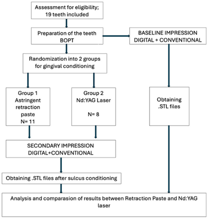 Figure 1
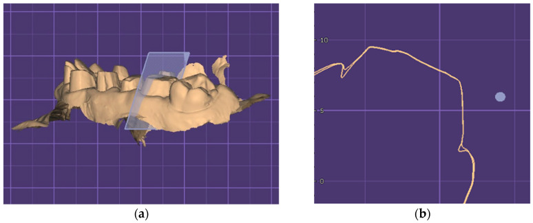 Figure 2
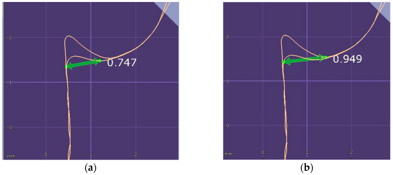 Figure 3
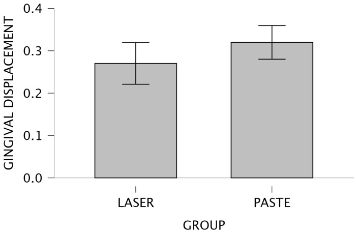 Figure 4
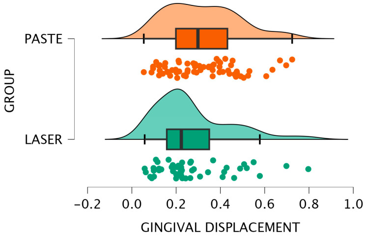 Figure 5
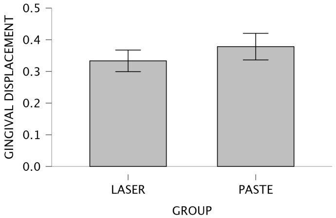 Figure 6
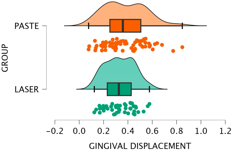 Figure 7
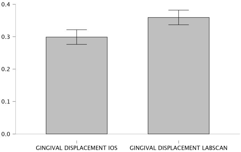 Figure 8
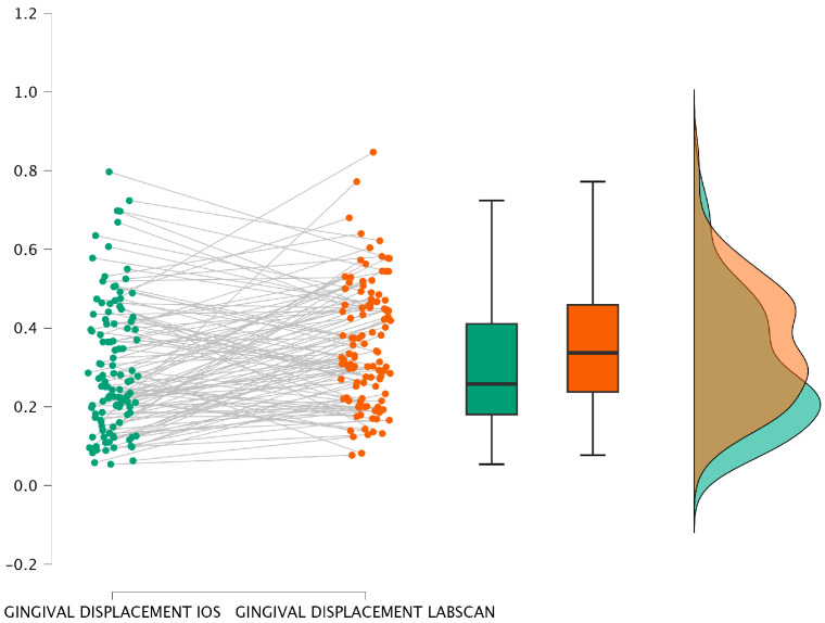 Figure 9
Peer Reviews
No public reviews on file for this paper yet. If you reviewed it on a platform where reviews are public (OpenReview, ICLR, NeurIPS, ICML), you can paste yours below so the community can read it here.
Videos
No videos yet. Explain this paper in a talk, walkthrough, or lecture? Add one.
Taxonomy
TopicsLaser Applications in Dentistry and Medicine · Dental materials and restorations · Periodontal Regeneration and Treatments