Sectoral Analysis of Corneal Thickness in Glaucoma and Healthy Eyes and Its Relationship with RNFL and Rim Area
Piotr Miklaszewski, Anna Maria Gadamer, Zuzanna Lelek, Dominika Janiszewska-Bil, Anita Lyssek-Boroń, Dariusz Dobrowolski, Edward Wylęgała, Beniamin Oskar Grabarek, Michael Janusz Koss, Katarzyna Krysik

TL;DR
This study finds that corneal thickness is reduced in glaucoma eyes but does not directly correlate with retinal nerve fiber layer thickness or optic disc rim area after adjusting for other factors.
Contribution
The study provides new insights into the relationship between corneal thickness and glaucoma, suggesting it reflects susceptibility rather than direct neuroretinal damage.
Findings
Eyes with glaucoma had significantly thinner corneas in all sectors compared to healthy eyes.
No independent associations were found between corneal thickness and retinal nerve fiber layer thickness or optic disc rim area after adjustment.
Glaucoma diagnosis and severity were consistently linked to reduced retinal nerve fiber layer thickness.
Abstract
Background/Objectives: To characterize sectoral corneal thickness (CT) profiles in eyes with primary open-angle glaucoma (POAG) compared with healthy eyes and to evaluate potential associations between CT, retinal nerve fiber layer (RNFL) thickness, and optic disc rim area (RA). Methods: In this cross-sectional study, 192 participants (91 with POAG and 101 controls) contributed 297 eyes (145 glaucoma eyes and 152 control eyes). All participants underwent comprehensive ophthalmological examination and spectral-domain optical coherence tomography (OCT; Optovue Solix, Fremont, CA, USA) to obtain peripapillary RNFL measurements, optic disc rim area, and corneal pachymetry maps across five sectors (central, superior, inferior, temporal, and nasal). Repeated-measures correlation analyses were used to assess within-subject associations between CT and RA, and generalized estimating equation…
Genes, proteins, chemicals, diseases, species, mutations and cell lines named across the full text — each resolved to its canonical identifier and authoritative record.
Click any figure to enlarge with its caption.
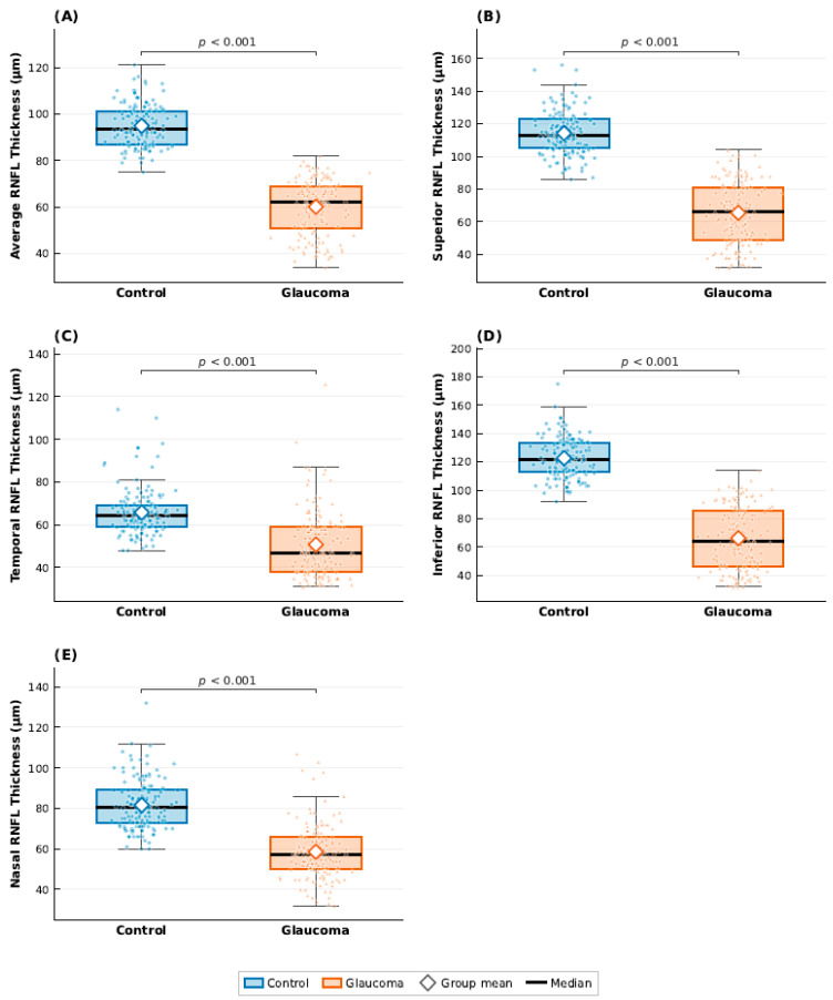 Figure 1
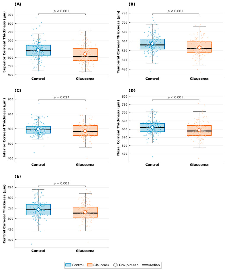 Figure 2
Peer Reviews
No public reviews on file for this paper yet. If you reviewed it on a platform where reviews are public (OpenReview, ICLR, NeurIPS, ICML), you can paste yours below so the community can read it here.
Videos
No videos yet. Explain this paper in a talk, walkthrough, or lecture? Add one.
Taxonomy
TopicsGlaucoma and retinal disorders · Corneal surgery and disorders · Ocular Surface and Contact Lens