Analysis of ATF6 and PLAT Expressions in Relation to hsa-miR-340-5p in Childhood Obesity
Yaşar Topal, Tuba Edgünlü, Dilek Akbaş, Çilem Özdemir, Hatice Topal, Habip Almiş, Ecenur Özdemir

TL;DR
This study explores how gene expressions related to vascular risk are altered in children with obesity, suggesting early molecular changes before clinical symptoms appear.
Contribution
The study identifies the ATF6/PLAT regulatory axis as a potential biomarker for early vascular risk in childhood obesity.
Findings
ATF6 expression was significantly downregulated in children with obesity compared to healthy controls.
PLAT expression was significantly upregulated in the obese group.
PPI analysis linked ATF6 to metabolic stress and PLAT to coagulation pathways.
Abstract
Childhood obesity is a complex pathology that triggers early vascular damage through endoplasmic reticulum (ER) stress and fibrinolytic imbalance; however, the role of the ATF6/PLAT regulatory axis in this process has not yet been fully elucidated. This study aims to investigate the molecular basis of vascular risk by determining the expression levels of these genes and the potential regulatory hsa-miR-340-5p in children with obesity. Gene expression analyses were performed using the RT-qPCR method on blood samples obtained from 55 children with obesity and 40 healthy controls, while in silico protein–protein interaction (PPI) networks were mapped using the STRING database. The findings revealed that ATF6 expression was significantly downregulated (p < 0.001) and PLAT expression was significantly upregulated (p = 0.005) in the obese group compared to controls. No significant difference…
Genes, proteins, chemicals, diseases, species, mutations and cell lines named across the full text — each resolved to its canonical identifier and authoritative record.
Click any figure to enlarge with its caption.
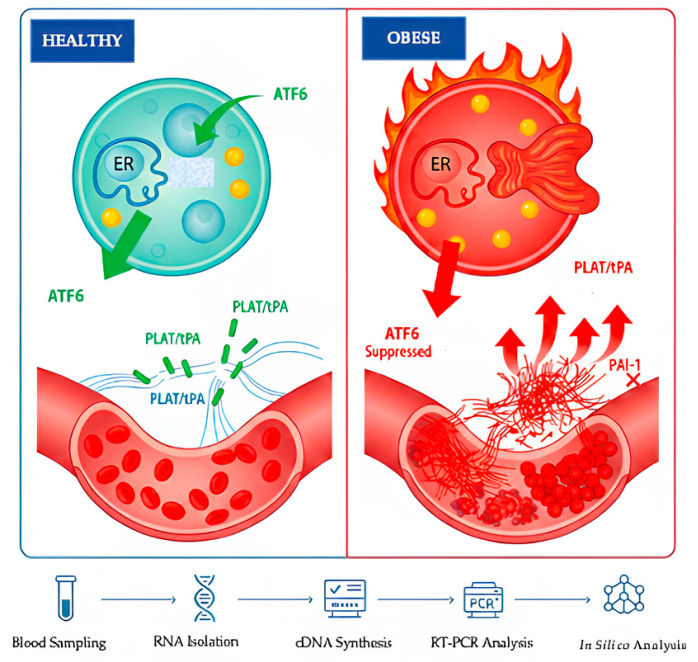 Figure 1
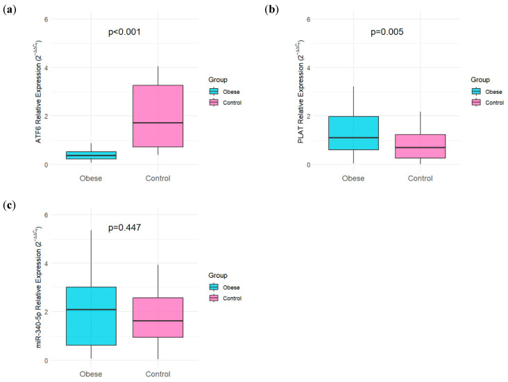 Figure 2
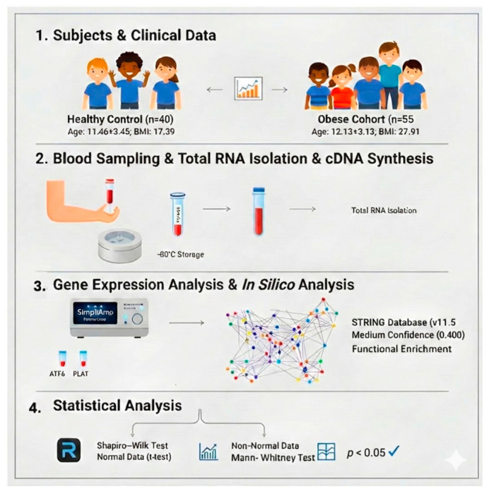 Figure 3
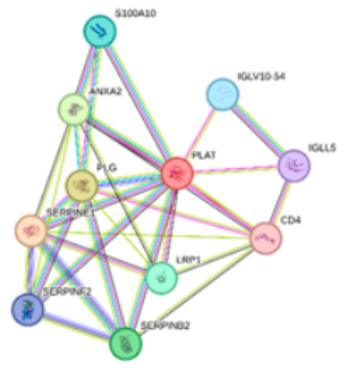 Figure 4
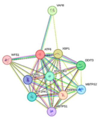 Figure 5
Peer Reviews
No public reviews on file for this paper yet. If you reviewed it on a platform where reviews are public (OpenReview, ICLR, NeurIPS, ICML), you can paste yours below so the community can read it here.
Videos
No videos yet. Explain this paper in a talk, walkthrough, or lecture? Add one.
Taxonomy
TopicsEndoplasmic Reticulum Stress and Disease · Genetic Associations and Epidemiology · Protein Kinase Regulation and GTPase Signaling
1. Introduction
Childhood obesity is increasingly recognized as a complex multifactorial pathology rather than a simple consequence of excess weight [1,2]. In addition to genetic predisposition, contemporary sedentary lifestyles and dysregulated dietary habits have transformed childhood obesity into a critical public health concern [1,2]. These environmental and behavioral factors culminate in a chronic state of positive energy balance, which not only drives adipose tissue expansion but also perturbs cellular homeostasis, thereby precipitating a spectrum of metabolic comorbidities [3]. Central to this cellular dysfunction is the impairment of the endoplasmic reticulum (ER), often referred to as the cell’s ‘quality control center’ [4]. Under conditions of chronic nutrient oversupply and consequent insulin resistance, the protein-folding capacity of the ER is overwhelmed, precipitating a state of cellular dysfunction known as Endoplasmic Reticulum (ER) stress [4,5]. In response to this proteotoxic stress, the cell activates an adaptive signaling pathway known as the Unfolded Protein Response (UPR) [3,6]. Among the canonical UPR transducers, Activating Transcription Factor 6 (ATF6) initially fulfills a protective function by upregulating chaperone genes to restore ER proteostasis (Figure 1) [7]. Conversely, under the persistent strain of chronic metabolic overload characteristic of obesity, this adaptive mechanism falters, and the sustained activation of ATF6 undergoes a deleterious functional shift [7]. While primarily acting as a transcriptional activator, its prolonged signaling has been implicated in the exacerbation of hepatic steatosis and the worsening of systemic insulin resistance [5,7].
Crucially, this disruption of lipid metabolism and ER function is not confined to tissue-specific stress but acts as a precursor to systemic vascular inflammation and endothelial dysfunction [8]. In this context, ATF6 functions not only as a stress sensor but also as a direct transcriptional regulator of the Plasminogen Activator, Tissue Type (PLAT) gene, which encodes the primary enzyme responsible for intravascular fibrinolysis [7]. Although these molecular mechanisms are broadly characterized in adult models, clinical evidence indicates that hemostatic disturbances, particularly impaired fibrinolysis and endothelial activation, manifest early in the clinical course of pediatric obesity [9]. Under physiological conditions, PLAT-derived tissue plasminogen activator (tPA) maintains vascular patency by converting plasminogen to plasmin; however, in the obese state, this protective pathway is frequently inhibited by elevated levels of Plasminogen Activator Inhibitor-1 (PAI-1), which is secreted abundantly by both dysfunctional adipose tissue and hepatocytes [10,11,12]. Mechanistically, the binding of PAI-1 to the Low-Density Lipoprotein Receptor-Related Protein 1 (LRP1) interferes with the downstream PKA/CREB1 signaling axis, specifically by reducing the phosphorylation of CREB1, which in turn diminishes the transcriptional activation of PLAT [7,12].
The regulation of this critical balance between ER stress and fibrinolysis extends beyond transcriptional control to include post-transcriptional modulation by microRNAs (miRNAs) [13,14]. Because these small non-coding RNAs can simultaneously target multiple components of the UPR and hemostatic systems, they represent key functional links between metabolic dysfunction and thrombotic risk [15]. Within this regulatory network, hsa-miR-340-5p has emerged as a significant candidate for investigation, particularly due to bioinformatic evidence suggesting its potential involvement in the modulation of UPR-related pathways and fibrinolytic gene expression. Given the complex interplay between cellular stress and hemostatic imbalance, this study aimed to evaluate the expression profiles of ATF6, PLAT, and hsa-miR-340-5p in children with obesity compared to a healthy control group.
2. Results
2.1. Demographic and Clinical Characteristics of Obese and Control Groups
A total of 95 participants were included in the study, comprising 55 obese and 40 control individuals. The demographic and clinical characteristics of the study population are summarized in Table 1.
There was no statistically significant difference in age between the obese and control groups (p = 0.341). Similarly, birth weight did not differ significantly between groups (p = 0.755). As expected, body mass index (BMI) was significantly higher in the obese group compared with controls (p < 0.001). The obese group also exhibited a significantly longer sleep duration (p = 0.042) and greater daily mobile screen time (p = 0.010). No significant difference was observed between the groups in terms of television viewing time (p = 0.140). Regarding gender distribution, the proportion of males and females was comparable between the obese and control groups (p = 0.548).
2.2. Expression Analysis of Target Genes and miRNA
The relative expression levels of the target genes and miRNA were normalized using the 2^−ΔΔCT^ method, and comparisons between groups were performed using the Mann–Whitney U test. A significant downregulation of ATF6 was observed in the obese group compared to the control group (p < 0.001). Conversely, PLAT expression levels were significantly higher in obese patients than in healthy controls (p = 0.005). However, no statistically significant difference was found between the groups regarding miR-340-5p expression levels (p = 0.447). These results indicate that while ATF6 and PLAT expressions are robustly associated with obesity status, miR-340-5p expression does not exhibit a similar clinical correlation in this specific study population (Table 2 and Figure 2).
2.3. Bioinformatic Assessment of Protein Interactions
To further investigate the functional landscape of PLAT and ATF6, we performed protein–protein interaction (PPI) and enrichment analyses using the STRING database. Details and enrichment results for Network 1 and Network 2 are presented in Table 3. The PLAT-centered network (Network 1) revealed a strong association with hemostatic processes. Specifically, PLAT was found to cluster with ANXA2, PLG, SERPINE1, SERPINF2, and SERPINB2 within the fibrinolysis. The regulation of plasminogen activation was further supported by the interactions between S100A10, ANXA2, PLG, SERPINE1, and SERPINF2. Additionally, the network showed significant involvement in Complement and coagulation cascades, represented by the core group of PLAT, PLG, SERPINE1, SERPINF2, and SERPINB2.
The ATF6-centered network (Network 2) demonstrated a robust profile related to cellular stress and metabolic regulation. A comprehensive set of proteins, including VAPB, XBP1, DDIT3, MBTPS2, HSPA5, MBTPS1, ERN1, and EIF2AK3, was identified as part of the Endoplasmic Reticulum (ER) unfolded protein response [17]. Within this network, the ER-nucleus signaling pathway was specifically mediated by the interaction of ATF6, XBP1, DDIT3, MBTPS2, HSPA5, MBTPS1, and EIF2AK3. Also, the analysis linked ERN1, XBP1, DDIT3, and EIF2AK3 to Non-alcoholic fatty liver disease (NAFLD), suggesting a metabolic dimension to the ATF6 interactome. These findings collectively indicate that PLAT and ATF6 serve as critical nodes in fibrinolytic activity and ER-stress management, respectively, through their interactions with these specialized protein clusters.
3. Discussion
This study provides the first clinical evidence regarding the interplay between ER stress markers and the fibrinolytic system in pediatric obesity. Our primary findings reveal a significant downregulation of ATF6 and a concurrent upregulation of PLAT in children with obesity compared to healthy controls. These results clinically validate the hypothesis that chronic metabolic stress disrupts the adaptive UPR capacity and alters fibrinolytic balance even at an early age, consistent with recent studies linking ER stress to metabolic dysregulation [8,11]. However, contrary to our in silico predictions, circulating miR-340-5p levels did not show a significant difference between groups, nor did they correlate with target gene expression. This discrepancy suggests that while the bioinformatic analysis correctly identified a potential regulatory network, the systemic reflection of this axis in pediatric circulation may be masked by tissue-specific mechanisms or the dominance of other regulatory miRNAs in the early stages of obesity [12].
3.1. The Paradox of ATF6 Decline in Chronic Obesity
One of the most striking findings of our study is the marked suppression of ATF6 expression in the obese group. Classically, ATF6 is recognized as a primary sensor of the UPR, typically upregulated to enhance chaperone capacity during acute ER stress [7]. However, our data align with the concept of “UPR exhaustion” or maladaptive remodeling under conditions of chronic metabolic stress. It is well-established that while acute lipotoxicity triggers a protective UPR surge, prolonged exposure to excess nutrients, as seen in childhood obesity, can lead to the attenuation of canonical UPR branches by exceeding the protein folding capacity [18]. The significant decrease in ATF6 observed in our cohort likely reflects a failure of cellular quality control mechanisms to sustain an adaptive response against persistent stress. This downregulation is critical because ATF6 deficiency has been linked to the deepening of insulin resistance and systemic inflammation, rather than resolving the proteotoxic load [8,19]. Furthermore, recent pediatric evidence suggests that such ER stress alterations are not merely cellular responses but key contributors to the pathogenesis of metabolic complications in children [20].
3.2. Fibrinolytic Imbalance and PLAT Increase
In contrast to the suppression of the UPR sensor, PLAT (tPA) expression levels were significantly elevated in the obese group. Under physiological homeostasis, ATF6 acts as a positive transcriptional regulator of PLAT, maintaining basal fibrinolytic capacity [12]. However, the inverse relationship observed in our study, downregulated ATF6 versus upregulated PLAT, suggests a “decoupled” regulatory mechanism specific to the obese state. We hypothesize that this elevation in PLAT represents a compensatory response to the chronic pro-thrombotic environment characterized by elevated PAI-1 levels. Recent studies in 2025 confirm that in obesity-induced endothelial dysfunction, PAI-1 is not merely an inhibitor but acts as a signaling molecule that can paradoxically induce PLAT expression via the PAI-1/LRP1/PKA/CREB1 axis to prevent thrombotic occlusion [11,21]. Furthermore, chronic low-grade inflammation (e.g., TNF-α and IL-6 signaling) has been shown to independently drive endothelial tPA release, bypassing the exhausted ER stress pathway [21,22]. This “fibrinolytic struggle” demonstrates that obese children are already exhibiting subclinical molecular signs of vascular stress and thrombosis risk.
3.3. The Effect of miRNAs and Lifestyle Factors
Although our in silico models identified hsa-miR-340-5p as a potential regulator, our experimental data did not show a significant correlation between this miRNA and its target genes. This discrepancy highlights the complexity of epigenetic regulation in childhood obesity, suggesting that tissue-specific interactions or other dominant metabolic miRNAs may override this axis in systemic circulation. More importantly, our demographic analysis revealed a significant increase in mobile screen time in the obese group. We propose that this sedentary behavior acts as a critical environmental trigger for chronic low-grade inflammation [21]. Sustained inflammatory signaling is known to deepen cellular ER stress, thereby contributing to the “exhaustion” of ATF6 and the disruption of fibrinolytic balance observed in our study [22].
3.4. Strengths and Limitations
This study provides crucial clinical evidence regarding the molecular fingerprints of early-stage childhood obesity, distinguishing itself from preclinical animal models. Validating our experimental findings with STRING database analysis has further clarified the functional clustering between ER stress failure and coagulation cascades. However, the cross-sectional design limits our ability to establish a definitive causal relationship between ATF6 downregulation and PLAT upregulation. Additionally, while transcriptional changes provide immediate insight into cellular reprogramming, future studies incorporating circulating protein levels (tPA and PAI-1) would offer a more comprehensive picture of the translational impact on fibrinolytic potential.
4. Materials and Methods
The overall experimental workflow, including subject recruitment, molecular procedures, and bioinformatics analysis, is summarized in Figure 3.
4.1. Ethics Committee Approval
Ethical approval for this study was obtained from the Scientific Research Ethics Committee of Sincan Training and Research Hospital (Approval Code: BAEK-2025-41).
4.2. Subjects
Following approval of the protocol by the Ethics Committee of Sincan Training and Research Hospital, the study was conducted in accordance with the Declaration of Helsinki. Informed consent was obtained from the parents of all participants. The study group consisted of 55 children and adolescents with obesity (OW/OB; Age: 12.13 ± 3.13 years; BMI: 27.91 [25.30–31.48] kg/m^2^) and 40 age- and gender-matched healthy controls (Age: 11.46 ± 3.45 years; BMI: 17.39 [16.20–21.16] kg/m^2^) (Figure 3). CDC growth curves were used to group participants. Exclusion criteria for both groups included a history of chronic metabolic/endocrine diseases (e.g., diabetes, hypothyroidism, Cushing’s syndrome), genetic obesity syndromes (e.g., Prader–Willi syndrome), active infection, and the use of medications affecting body weight within the last three months. Anthropometric data were collected following standard procedures.
4.3. Blood Sampling
Venous blood samples (2 mL) were collected in vacuum tubes containing K2-EDTA. The samples were immediately centrifuged at 3000 rpm for 10 min at 4 °C. The resulting plasma and cell pellets were stored at −80 °C until the analysis stage.
4.4. Gene Expression Analysis
Total RNA isolation, including the small RNA fraction, was performed using the TRIzol™ Reagent RNA isolation kit (Thermo Fisher Scientific, Carlsbad, CA, USA; Cat No: 15596026) according to the manufacturer’s instructions. Complementary DNA (cDNA) synthesis was performed from 1 µg of total RNA using the cDNA Reverse Transcription Kit (NucleoGene (Istanbul, Turkey; Cat. No: NGMM007)) according to the manufacturer’s protocol [23]. The expression levels of the ATF6 and PLAT genes and hsa-miR-340-5p were determined using the quantitative reverse transcription-polymerase chain reaction (RT-qPCR) method. The RT-qPCR analysis was performed using an automated thermal cycler (SimpliAmp Thermal Cycler, Thermo Fisher Scientific, Carlsbad, CA, USA; Cat No: 15596026) and SYBR Green reagent (NucleoGene, Istanbul, Turkey; Cat. No: NGMM007) in a final volume of 20 µL. GAPDH and U6 were used as internal controls for the normalization of mRNA and miRNA expression levels, respectively [24]. Relative expression levels were calculated using the 2^−ΔΔCt^ method. To ensure technical reproducibility, all measurements were performed in triplicate.
4.5. In Silico Analysis
To complement our experimental observations, we conducted in silico analyses. Potential miRNA targets regulating the ATF6 and PLAT genes were predicted using the miRDB online database (http://mirdb.org; accessed on 5 January 2026), utilizing an algorithm for miRNA target prediction and functional annotation [25]. Additionally, protein–protein interaction (PPI) networks for PLAT and ATF6 proteins were mapped using the STRING database (https://string-db.org/; accessed on 5 January 2026) [26]. The analysis was performed using default parameters with a medium confidence score (0.400) to identify functional enrichment of biological pathways.
4.6. Statistical Analysis
All statistical analyses were performed using R software (version 4.1.3). Continuous variables were first assessed for normality using the Shapiro–Wilk test [27]. Variables showing a normal distribution were presented as Mean ± Standard Deviation (SD) and compared using the Independent Samples t-test [28]. Non-normally distributed data were presented as Median and Interquartile Range (IQR), and group comparisons were conducted using the Mann–Whitney U test. Categorical variables were expressed as number and percentage (n, %) and compared using the Chi-square test. A p-value < 0.05 was considered statistically significant.
5. Conclusions
In summary, our study demonstrates that childhood obesity is characterized by the exhaustion of the ATF6-mediated ER stress response and a concurrent upregulation of the PLAT gene. This distinct molecular signature indicates that the cellular “quality control” system is compromised, triggering a compensatory fibrinolytic response aimed at protecting vascular health. Our findings emphasize that obesity-induced vascular stress begins at the transcriptomic level during childhood, preceding the onset of clinical symptoms. Consequently, the potential use of ATF6 and PLAT expression levels as biomarkers for early cardiovascular risk assessment in the pediatric population should be supported by future longitudinal studies.
The reference list from the paper itself. Each links out to its DOI / PubMed record.
- 1Di Pietro N. Marcovecchio M.L. Di Silvestre S. de Giorgis T. Cordone V.G.P. Lanuti P. Chiarelli F. Bologna G. Mohn A. Pandolfi A. Plasma from pre-pubertal obese children impairs insulin stimulated Nitric Oxide (NO) bioavailability in endothelial cells: Role of ER stress Mol. Cell. Endocrinol.2017443526210.1016/j.mce.2017.01.00128062198 PMC 5320395 · doi ↗ · pubmed ↗
- 2World Health Organization Obesity and Overweight [Fact Sheet]World Health Organization Geneva, Switzerland 2024 Available online: https://www.who.int/news-room/fact-sheets/detail/obesity-and-overweight(accessed on 25 February 2026)
- 3Ma K. Zhang Y. Zhao J. Zhou L. Li M. Endoplasmic reticulum stress: Bridging inflammation and obesity-associated adipose tissue Front. Immunol.202415138122710.3389/fimmu.2024.138122738638434 PMC 11024263 · doi ↗ · pubmed ↗
- 4Antoniotti V. Bellone S. Gonçalves Correia F.P. Peri C. Tini S. Ricotti R. Mancioppi V. Gagliardi M. Spadaccini D. Caputo M. Calreticulin and PDIA 3, two markers of endoplasmic reticulum stress, are associated with metabolic alterations and insulin resistance in pediatric obesity: A pilot study Front. Endocrinol.202213100391910.3389/fendo.2022.1003919 PMC 953738136213269 · doi ↗ · pubmed ↗
- 5Choo Y.N. Ravi R.N. Subramaniyan V. Insulin resistance induced by obesity: Mechanisms, metabolic implications and therapeutic approaches Mol. Biol. Rep.20265335710.1007/s 11033-026-11509-341636917 PMC 12872655 · doi ↗ · pubmed ↗
- 6Walter P. Ron D. The Unfolded Protein Response: From Stress Pathway to Homeostatic Regulation Science 20113341081108610.1126/science.120903822116877 · doi ↗ · pubmed ↗
- 7Zheng Z. Nayak L. Wang W. Yurdagul A.Jr. Wang X. Cai B. Lapping S. Ozcan L. Ramakrishnan R. Pestell R.G. An ATF 6-t PA pathway in hepatocytes contributes to systemic fibrinolysis and is repressed by DACH 1Blood J. Am. Soc. Hematol.201913374375310.1182/blood-2018-07-864843 PMC 637628330504459 · doi ↗ · pubmed ↗
- 8Zhang Z. Rodriguez M. Zheng Z. Clot or Not? Reviewing the Reciprocal Regulation Between Lipids and Blood Clotting Arterioscler. Thromb. Vasc. Biol.20244453354410.1161/ATVBAHA.123.31828638235555 PMC 10922732 · doi ↗ · pubmed ↗