Clinical Value of Optical Coherence Tomography in Craniopharyngioma
Klaudia Rakusiewicz-Krasnodębska, Agnieszka Bogusz-Wójcik, Anna Chmielarz-Czarnocińska, Elżbieta Moszczyńska, Wojciech Hautz

TL;DR
Optical coherence tomography (OCT) and OCT angiography (OCTA) help detect and monitor optic nerve damage in craniopharyngioma patients, improving visual outcome predictions.
Contribution
This review proposes OCT and OCTA as objective tools for assessing and predicting visual outcomes in craniopharyngioma.
Findings
OCT detects retinal nerve fiber layer and ganglion cell complex thinning linked to visual impairment.
OCTA identifies microvascular changes before structural damage occurs.
OCT metrics can predict postoperative visual recovery in craniopharyngioma patients.
Abstract
Craniopharyngioma is a rare benign brain tumor that often develops near the optic nerves and optic chiasm, which can lead to significant vision problems in both children and adults. Early detection of optic nerve damage is important to preserve visual function. Optical coherence tomography (OCT) and OCT angiography (OCTA) are noninvasive imaging techniques that allow detailed evaluation of the retina and its blood vessels. Changes such as thinning of the retinal nerve fiber layer and ganglion cell complex are associated with visual impairment and may help predict visual recovery after neurosurgery. OCTA can also detect microvascular alterations that may appear before structural damage. This review summarizes current knowledge about the role of OCT and OCTA in diagnosis, monitoring, and prognosis of visual outcomes in patients with craniopharyngioma. Craniopharyngioma (CP) is a rare…
Genes, proteins, chemicals, diseases, species, mutations and cell lines named across the full text — each resolved to its canonical identifier and authoritative record.
Click any figure to enlarge with its caption.
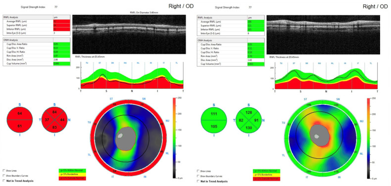 Figure 1
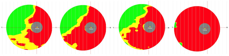 Figure 2
Peer Reviews
No public reviews on file for this paper yet. If you reviewed it on a platform where reviews are public (OpenReview, ICLR, NeurIPS, ICML), you can paste yours below so the community can read it here.
Videos
No videos yet. Explain this paper in a talk, walkthrough, or lecture? Add one.
Taxonomy
TopicsPituitary Gland Disorders and Treatments · Cerebral Venous Sinus Thrombosis · Glaucoma and retinal disorders