Artificial Intelligence in Prostate MRI: Comparison of an AI-Based Software and an Experienced Radiologist for Detecting Clinically Significant Prostate Cancer
Roberto Castellana, Simona Marzi, Andrea Russo, Maria Consiglia Ferriero, Irene Terrenato, Eugenia Papaleo, Giuseppe Navanteri, Davide Vitale, Giuseppe Pizzi, Antonello Vidiri, Luca Bertini

TL;DR
This study compares an AI-based software with an experienced radiologist for detecting significant prostate cancer on MRI and finds they perform similarly, with AI showing potential as a supportive tool.
Contribution
Demonstrates that AI-based software can match an expert radiologist in detecting clinically significant prostate cancer on MRI.
Findings
AI software and the radiologist showed comparable sensitivity and specificity in detecting clinically significant prostate cancer.
The AI detected more lesions, especially in the transition zone, but should be used as a support tool rather than a replacement for radiologists.
Both methods had extremely low negative likelihood ratios, indicating strong ability to rule out significant cancer.
Abstract
Prostate cancer is one of the most common cancers in men, and magnetic resonance imaging (MRI) plays a key role in identifying tumors that require treatment. However, interpreting prostate MRI requires experience, and results may vary between readers. Artificial intelligence tools have been developed to assist radiologists, but their real clinical value is still being evaluated. In this study, we compared an artificial intelligence–based software with an experienced radiologist in detecting clinically significant prostate cancer on MRI, using biopsy results as reference. We found that the software performed similarly to the expert reader, particularly in ruling out significant cancer when MRI findings were negative. Although the software detected more suspicious areas, it should be considered a support tool rather than a replacement for radiologists. These findings suggest that…
Genes, proteins, chemicals, diseases, species, mutations and cell lines named across the full text — each resolved to its canonical identifier and authoritative record.
Click any figure to enlarge with its caption.
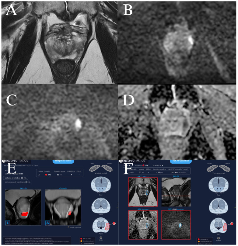 Figure 1
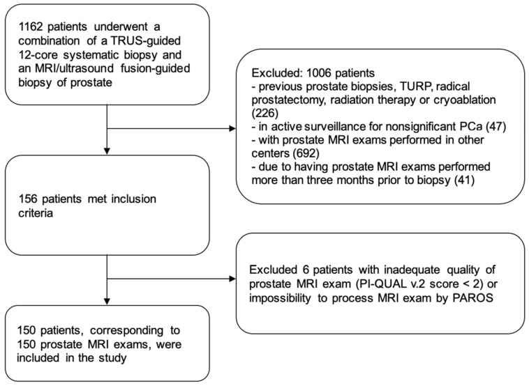 Figure 2
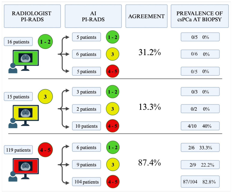 Figure 3
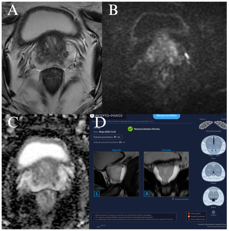 Figure 4
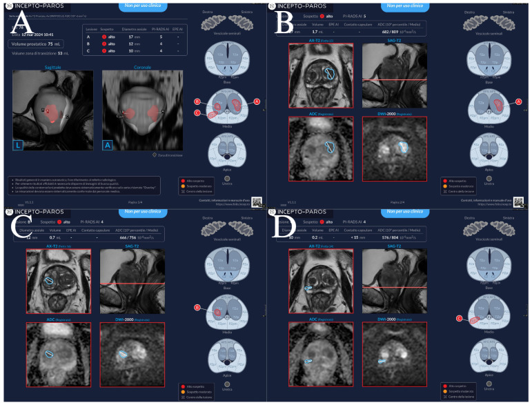 Figure 5
 Figure 6
Peer Reviews
No public reviews on file for this paper yet. If you reviewed it on a platform where reviews are public (OpenReview, ICLR, NeurIPS, ICML), you can paste yours below so the community can read it here.
Videos
No videos yet. Explain this paper in a talk, walkthrough, or lecture? Add one.
Taxonomy
TopicsProstate Cancer Diagnosis and Treatment · Artificial Intelligence in Healthcare and Education · Radiomics and Machine Learning in Medical Imaging