Necrotizing (Abscessing) Lymphadenopathy and the Diagnostic Value of Contrast-Enhanced Ultrasound (CEUS): A Review with Clinical Vignettes
Christian Görg, Yi Dong, Görg Friedemann, Christian Jenssen, Michael Kallenbach, Kathleen Möller, Findeisen Hajo, Nitin Chaubal, Christoph Frank Dietrich

TL;DR
This review discusses how contrast-enhanced ultrasound can help diagnose and manage lymph node abscesses by improving the accuracy of identifying necrotic or abscessed tissue.
Contribution
The paper highlights the novel diagnostic potential of contrast-enhanced ultrasound in characterizing necrotizing lymphadenopathy compared to conventional methods.
Findings
Contrast-enhanced ultrasound (CEUS) provides real-time visualization of nodal microvascular perfusion, aiding in differentiation between viable and necrotic tissue.
CEUS patterns correlate with underlying pathology, such as central non-enhancement in abscesses and irregular avascular cores in tuberculosis.
CEUS supports biopsy targeting and drainage procedures, offering advantages over conventional sonography in diagnosing necrotizing lymphadenopathy.
Abstract
Necrotizing (abscessing) lymphadenopathy is a clinically relevant condition with a broad differential diagnosis, including acute bacterial infections, mycobacterial disease, zoonoses, fungal and parasitic infections, autoimmune disorders, and malignancies with central necrosis. Early and reliable differentiation between these causes is important to avoid misdiagnosis and to guide appropriate therapy. This review summarizes the pathophysiological mechanisms, typical imaging features, and diagnostic value of contrast-enhanced ultrasound (CEUS) in necrotizing lymphadenopathy. Representative clinical vignettes illustrate the disease spectrum and correlate CEUS patterns with underlying pathology. The literature review was narrative and based on targeted searches of PubMed/MEDLINE and Google Scholar focusing on CEUS in necrotizing lymphadenopathy. A brief literature overview highlights…
Genes, proteins, chemicals, diseases, species, mutations and cell lines named across the full text — each resolved to its canonical identifier and authoritative record.
Click any figure to enlarge with its caption.
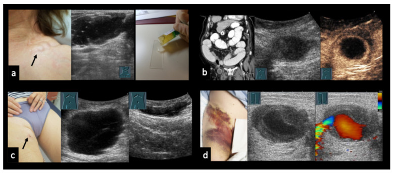 Figure 1
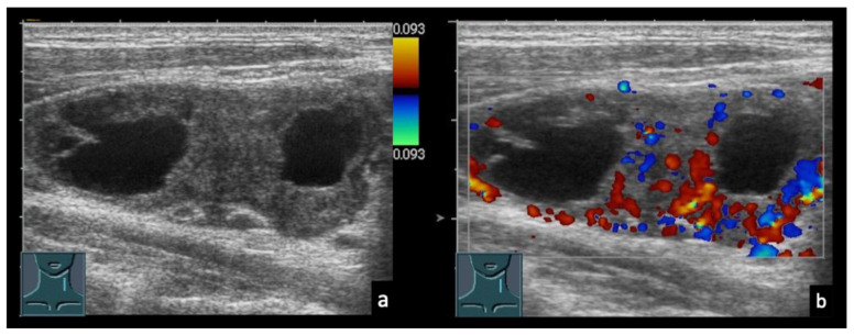 Figure 2
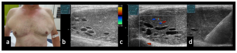 Figure 3
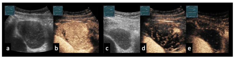 Figure 4
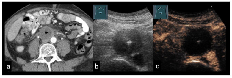 Figure 5
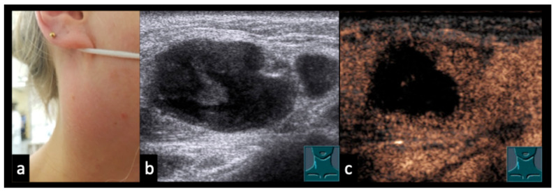 Figure 6
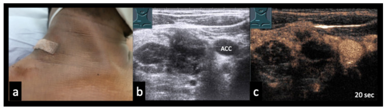 Figure 7
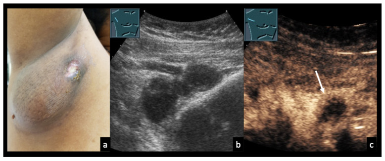 Figure 8
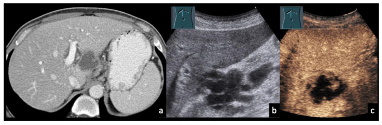 Figure 9
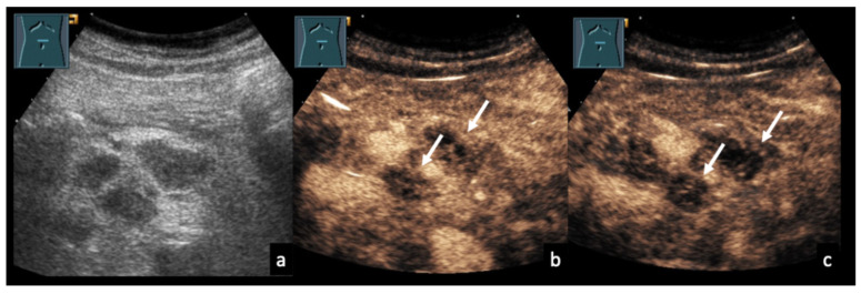 Figure 10
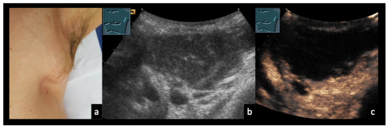 Figure 11
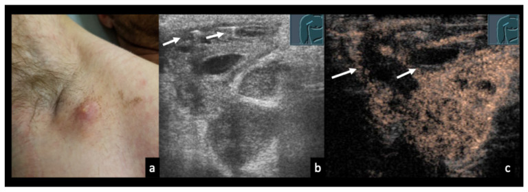 Figure 12
 Figure 13
 Figure 14
 Figure 15
 Figure 16
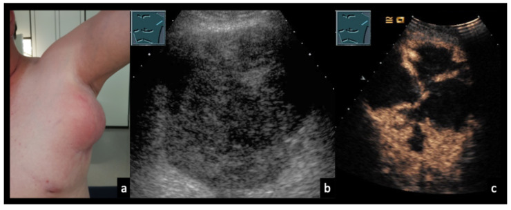 Figure 17
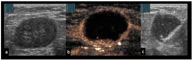 Figure 18
Peer Reviews
No public reviews on file for this paper yet. If you reviewed it on a platform where reviews are public (OpenReview, ICLR, NeurIPS, ICML), you can paste yours below so the community can read it here.
Videos
No videos yet. Explain this paper in a talk, walkthrough, or lecture? Add one.
Taxonomy
TopicsLymphadenopathy Diagnosis and Analysis · Lymphatic Disorders and Treatments · Salivary Gland Tumors Diagnosis and Treatment