E-Cadherin Is an Accurate Target for Fluorescence-Guided Imaging of Lymph Nodes
Kelly A. McGovern, Katherine O. Welch, Jake Mlakar, Ryan Krouse, Michael Brown, Lydia Chen, Kevin Guo, Jeffrey Huang, Edward J. Delikatny, Viktor Gruev, Paul Zhang, Sunil Singhal

TL;DR
This study identifies E-cadherin as a promising biomarker for fluorescence-guided imaging to distinguish cancerous from non-cancerous lymph nodes in lung cancer patients.
Contribution
The study introduces E-cadherin as a novel and accurate imaging target for identifying metastatic lymph nodes in lung cancer.
Findings
E-cadherin fluorescence was significantly higher in metastatic lymph nodes compared to non-metastatic ones.
High fluorescence was observed in both hilar and mediastinal lymph nodes across all primary tumor histologies.
The anti-E-cadherin monoclonal antibody showed strong potential for targeted imaging in surgical settings.
Abstract
Lymph node (LN) dissection is a necessary part of every oncologic surgery in order to provide important information for staging, predicting prognosis and improving survival. To do this, surgical oncologists strive to localize and dissect every pathologically positive LN while avoiding the increased morbidity of removing true negative LNs. The goal is to develop an imaging method to distinguish positive and negative LNs, but a specific biomarker is missing. Thus, our aim is to identify a reliable imaging marker for identifying LNs with lung cancer cells. After screening many epithelial markers, we identified E-cadherin, a membrane protein normally expressed in epithelial cells, including in the lung. To follow up on our potential target, we performed immunofluorescence staining on 48 human LNs with a conjugated anti-E-cadherin monoclonal antibody. Fluorescence was significantly higher in…
Genes, proteins, chemicals, diseases, species, mutations and cell lines named across the full text — each resolved to its canonical identifier and authoritative record.
Click any figure to enlarge with its caption.
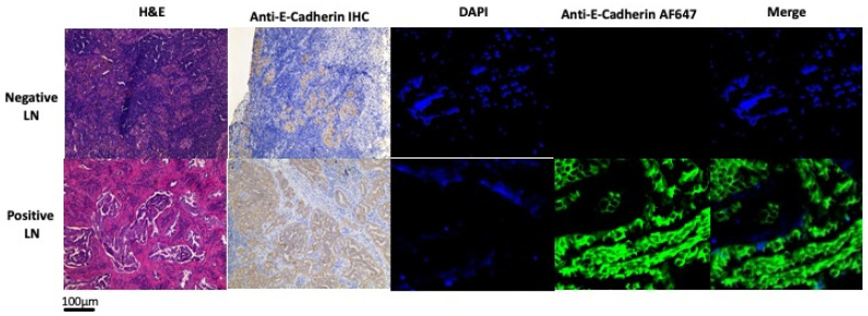 Figure 1
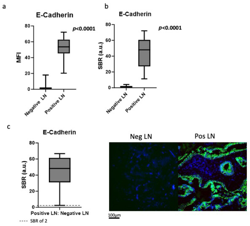 Figure 2
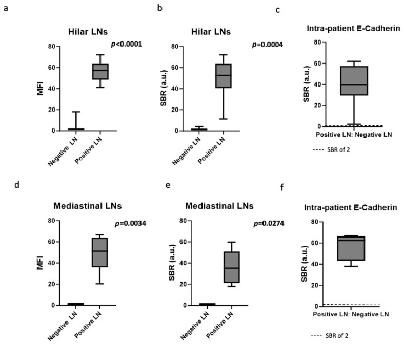 Figure 3
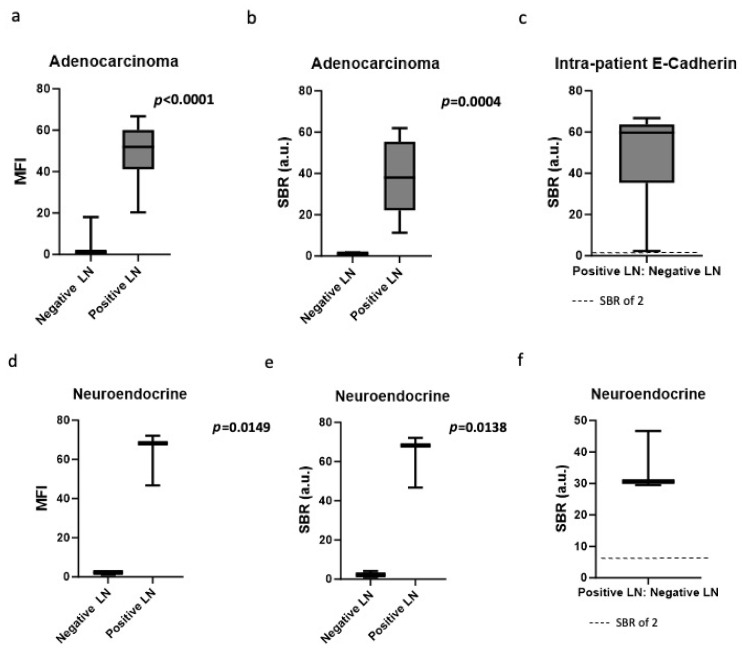 Figure 4
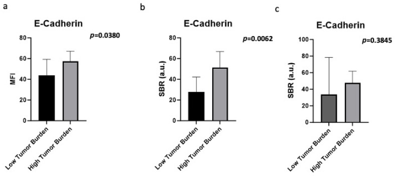 Figure 5
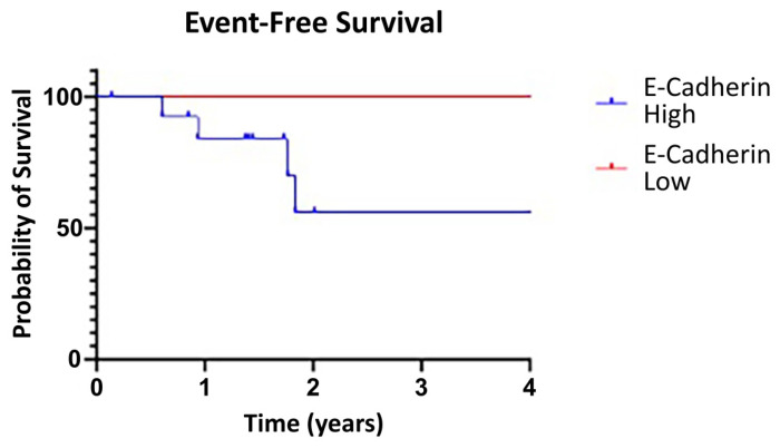 Figure 6
Peer Reviews
No public reviews on file for this paper yet. If you reviewed it on a platform where reviews are public (OpenReview, ICLR, NeurIPS, ICML), you can paste yours below so the community can read it here.
Videos
No videos yet. Explain this paper in a talk, walkthrough, or lecture? Add one.
Taxonomy
TopicsWnt/β-catenin signaling in development and cancer · Lymphatic System and Diseases · Esophageal Cancer Research and Treatment