Quantitative Autofluorescence Imaging of Oral Mucosa and Lesions: A Proof-of-Concept Study
Keerthi Gurushanth, Sumsum P. Sunny, Shubha Gurudath, Harshita Thakur, Kripa Adlene Edith, Keerthi Krishnakumar, Shikha Jha, Pavitra Chandrashekhar, Satyajit Topajiche, Lynette Linzbuoy, Sanjana Patrick, Ramyashree Rao, Simranjeet Kaur, Umeshgouda Patil, Ananya Nagaraj

TL;DR
This study shows that autofluorescence imaging can help distinguish between healthy and diseased oral tissues by measuring fluorescence intensity differences.
Contribution
The study introduces a smartphone-based method for quantitative autofluorescence imaging of oral mucosa and lesions as a potential diagnostic tool.
Findings
AFI intensity varied significantly across different oral subsites, with the lateral tongue showing the lowest and the ventral tongue the highest.
AFI intensity decreased with increasing disease severity, showing lower values in oral cancer compared to benign lesions.
Benign lesions showed intermediate AFI intensity, supporting AFI's potential to differentiate between lesion types.
Abstract
Background/Objectives: This study aimed to quantitatively assess site-specific mean autofluorescence intensity across normal oral mucosal subsites and to evaluate the effectiveness of Autofluorescence Imaging (AFI) as an adjunct tool for distinguishing benign lesions, OPMDs, and oral cancers by comparing lesion intensity with anatomically matched healthy subsites. Methods: This observational study employed dual-mode imaging, comprising paired White Light Imaging (WLI) and AFI, captured from different oral cavity subsites using a smartphone-based point-of-care device. The Region of Interest (ROI) was annotated on WLI and automatically mapped to the corresponding AFI for both normal mucosa and lesions. WLI and AFI images were separated into their constituent red, green, and blue (RGB) channels, and AFI intensity was quantified via ImageJ. Results: A total of 1380 dual-mode images were…
Genes, proteins, chemicals, diseases, species, mutations and cell lines named across the full text — each resolved to its canonical identifier and authoritative record.
Click any figure to enlarge with its caption.
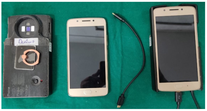 Figure 1
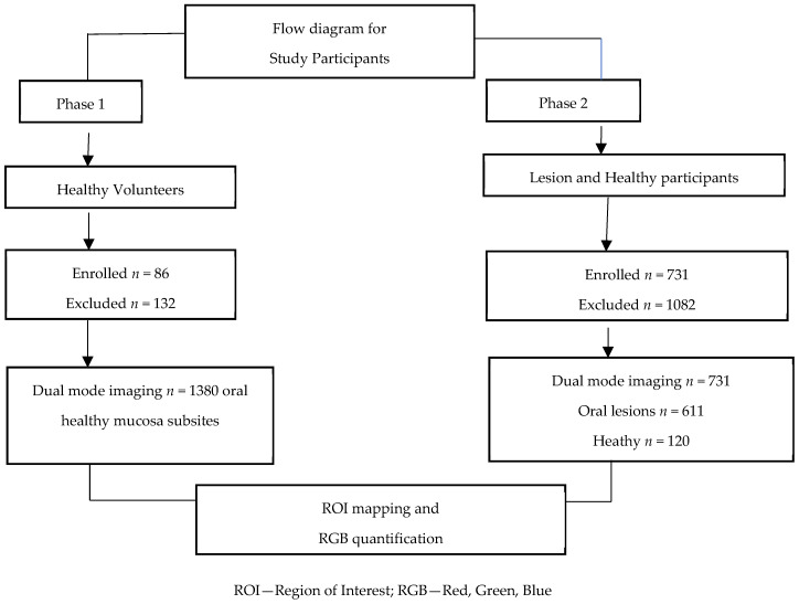 Figure 2
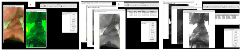 Figure 3
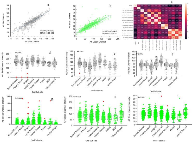 Figure 4
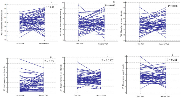 Figure 5
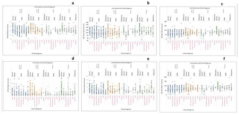 Figure 6
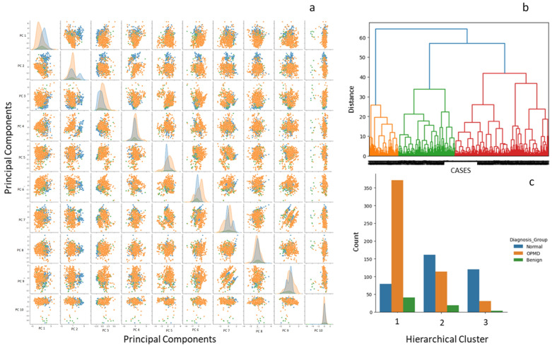 Figure 7
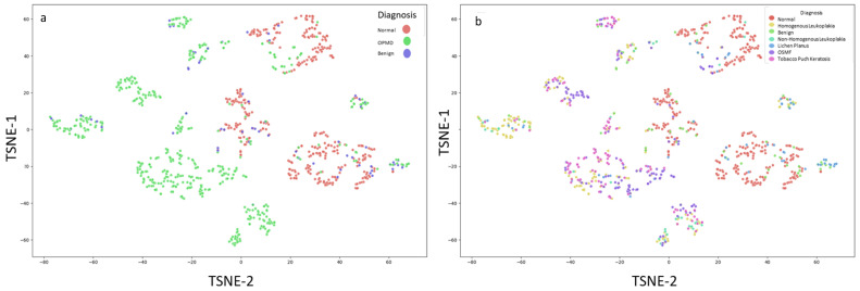 Figure 8
Peer Reviews
No public reviews on file for this paper yet. If you reviewed it on a platform where reviews are public (OpenReview, ICLR, NeurIPS, ICML), you can paste yours below so the community can read it here.
Videos
No videos yet. Explain this paper in a talk, walkthrough, or lecture? Add one.
Taxonomy
TopicsOral Health Pathology and Treatment · Photodynamic Therapy Research Studies · Head and Neck Cancer Studies