The Possible Relationship Between Sigmoid Dehiscence, Degree of Mastoid Pneumatization, and Sigmoid Sinus Position in Patients with Pulsatile Tinnitus
Burak Bilecenoğlu, Tuğçe Akın, Berin Tuğtağ Demir, Ömer Korkmazyürek, Ali Köksal, Kaan Orhan

TL;DR
This study explores how anatomical features of the sigmoid sinus and mastoid pneumatization relate to pulsatile tinnitus, finding that dehiscence and positioning are more common in affected patients.
Contribution
The study identifies specific anatomical correlations with pulsatile tinnitus using CBCT scans and introduces potential diagnostic and surgical implications.
Findings
Sigmoid sinus dehiscence was significantly more common in patients with pulsatile tinnitus compared to controls.
The sigmoid sinus was closer to key anatomical landmarks in patients with pulsatile tinnitus.
Quantitative CBCT measurements could aid in diagnosing and planning treatment for pulsatile tinnitus.
Abstract
Objective: This study aimed to determine the relationship between sigmoid sinus dehiscence (SSD), sigmoid sinus topography, mastoid pneumatization, and adjacent temporal bone structures in patients with pulsatile tinnitus (PT). Methods: A retrospective analysis was performed on 344 temporal bone cone-beam computed tomography (CBCT) scans (172 PT patients and 172 age- and sex-matched controls). The degree of mastoid pneumatization, presence and size of SSD, sinus topography, and distances between the sigmoid sinus and key landmarks—the lateral semicircular canal (LSCC), jugular bulb (HJB), and external auditory canal (EAC)—were measured. Quantitative and qualitative characteristics were compared between groups, and independent predictors of PT were identified using multivariate logistic regression. Results: Compared to controls, SSD was substantially more common in the PT group (115/172…
Genes, proteins, chemicals, diseases, species, mutations and cell lines named across the full text — each resolved to its canonical identifier and authoritative record.
Click any figure to enlarge with its caption.
 Figure 1
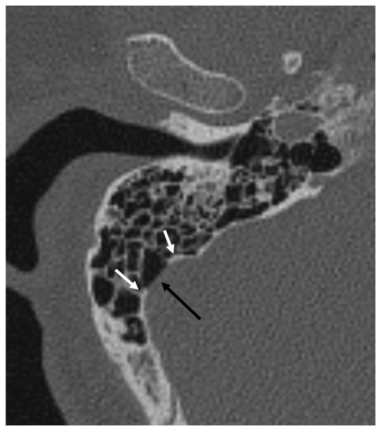 Figure 2
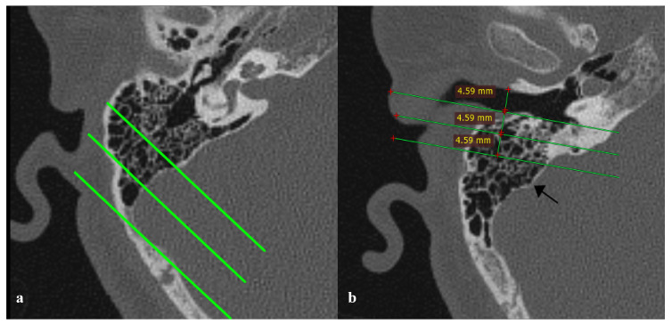 Figure 3
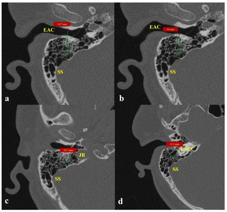 Figure 4
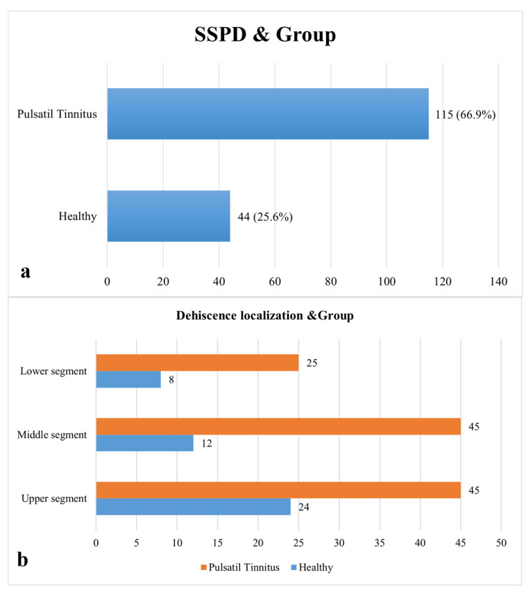 Figure 5
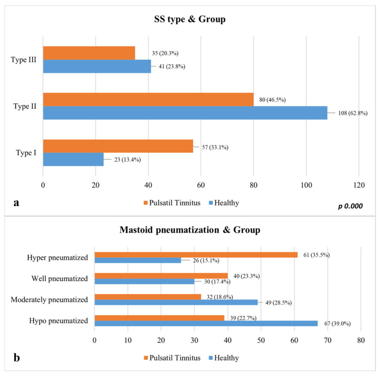 Figure 6
Peer Reviews
No public reviews on file for this paper yet. If you reviewed it on a platform where reviews are public (OpenReview, ICLR, NeurIPS, ICML), you can paste yours below so the community can read it here.
Videos
No videos yet. Explain this paper in a talk, walkthrough, or lecture? Add one.
Taxonomy
TopicsEar Surgery and Otitis Media · Sinusitis and nasal conditions · Cerebral Venous Sinus Thrombosis