Local Versus Global Binarization Techniques After Frangi Filtering for Optical Coherence Tomography Angiography Based Retinal Vessel Density Assessment in Diabetic Retinopathy
Andrada-Elena Mirescu, Ioana Teodora Tofolean, Sanda Jurja, Florian Balta, Alina Popa-Cherecheanu, Ruxandra Angela Pirvulescu, Gerhard Garhofer, George Balta, Irina-Elena Cristescu, Dan George Deleanu

TL;DR
This study compares local and global binarization methods for analyzing retinal vessel density in diabetic retinopathy using OCTA, finding that local methods perform better.
Contribution
The study introduces a comparison of local and global binarization techniques after Frangi filtering for OCTA-based vessel density assessment in diabetic retinopathy.
Findings
Local binarization methods showed significant differences in vessel density between PDR and control/no DR groups.
Global Otsu thresholding was the only global method to detect significant differences between PDR and control.
Nasal and inferior quadrants showed the most robust differences in vessel density.
Abstract
Background/Objectives: Optical coherence tomography angiography (OCTA) enables noninvasive quantitative assessment of the retinal microvasculature and is widely used in diabetic retinopathy (DR). However, OCTA-derived metrics are highly dependent on post-processing techniques, particularly vessel binarization. This study aimed to compare local and global binarization methods applied after Frangi filtering for vessel enhancement in parafoveal vessel density analysis. Methods: This cross-sectional study included 69 participants: 17 healthy controls and 52 diabetic patients, classified as the following: no DR (n = 14), non-proliferative DR (NPDR, n = 18), or proliferative DR (PDR, n = 20). All subjects underwent comprehensive ophthalmological examination and OCTA imaging of the superficial capillary plexus using a Topcon OCTA system. Images were processed using a custom MATLAB protocol.…
Genes, proteins, chemicals, diseases, species, mutations and cell lines named across the full text — each resolved to its canonical identifier and authoritative record.
Click any figure to enlarge with its caption.
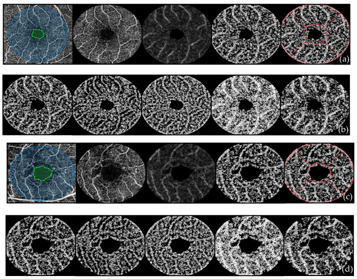 Figure 1
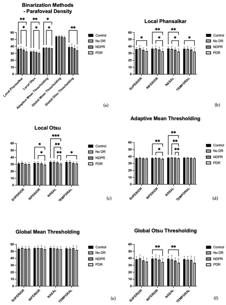 Figure 2
Peer Reviews
No public reviews on file for this paper yet. If you reviewed it on a platform where reviews are public (OpenReview, ICLR, NeurIPS, ICML), you can paste yours below so the community can read it here.
Videos
No videos yet. Explain this paper in a talk, walkthrough, or lecture? Add one.
Taxonomy
TopicsRetinal Imaging and Analysis · Retinal Diseases and Treatments · Retinal and Optic Conditions