Optical Coherence Tomography (OCT) Evaluation of Thermal Tissue Alterations After Diode Laser Excision of Oral Leukoplakia (OL)
Alessio Gambino, Alessandro Magliano, Giorgia El Haddad, Marta Bezzi, Adriana Cafaro, Dora Karimi, Roberto Broccoletti, Paolo Giacomo Arduino

TL;DR
This study shows that optical coherence tomography (OCT) can effectively assess laser-induced tissue changes during oral leukoplakia excision, helping ensure accurate diagnosis and margin evaluation.
Contribution
The study introduces OCT as a reliable, non-invasive method for real-time evaluation of thermal effects during diode laser excision of oral leukoplakia.
Findings
OCT provided high-resolution visualization of tissue microarchitecture and detected laser-induced thermal effects at surgical margins.
OCT measurements showed strong agreement with histological findings, with no significant differences between OCT and histology assessments.
Laser-induced thermal effects did not impair histopathological diagnosis in any specimen.
Abstract
Objectives: Oral leukoplakia (OL) is the most prevalent oral potentially malignant disorder and requires accurate diagnosis, safe excision, and reliable margin evaluation to minimize recurrence and malignant transformation. Diode laser excision is increasingly adopted due to its precision and favorable clinical outcomes; however, laser-induced thermal effects at surgical margins raise concerns regarding tissue integrity and histopathological reliability. This study aimed to evaluate optical coherence tomography (OCT) as a real-time, high-resolution, non-invasive imaging modality for assessing peri-incisional thermal effects during diode laser excision of non-dysplastic OL. The primary objective was to validate OCT for ultrastructural and morphometric tissue analysis while ensuring preservation of diagnostic readability. Methods: A single-center observational case series was conducted at…
Genes, proteins, chemicals, diseases, species, mutations and cell lines named across the full text — each resolved to its canonical identifier and authoritative record.
Click any figure to enlarge with its caption.
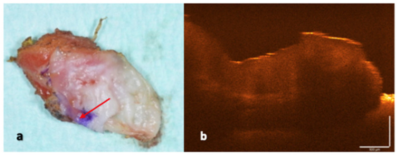 Figure 1
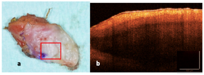 Figure 2
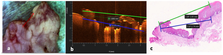 Figure 3
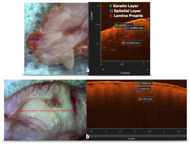 Figure 4
Peer Reviews
No public reviews on file for this paper yet. If you reviewed it on a platform where reviews are public (OpenReview, ICLR, NeurIPS, ICML), you can paste yours below so the community can read it here.
Videos
No videos yet. Explain this paper in a talk, walkthrough, or lecture? Add one.
Taxonomy
TopicsOptical Coherence Tomography Applications · Oral Health Pathology and Treatment · Laser Applications in Dentistry and Medicine