U-Net Optimization for Hyperreflective Foci Segmentation in Retinal OCT
Pavithra Kodiyalbail Chakrapani, Preetham Kumar, Sulatha Venkataraya Bhandary, Geetha Maiya, Shailaja Shenoy, Steven Fernandes, Prakhar Choudhary

TL;DR
This paper explores the best U-Net configurations for segmenting hyperreflective foci in retinal OCT images, finding that standard U-Net with specific preprocessing improves detection accuracy.
Contribution
The study identifies optimal U-Net settings and preprocessing for hyperreflective foci segmentation in retinal OCT.
Findings
Standard U-Net with CLAHE and focal Tversky loss achieved a dice score of 0.5207 and improved recall by 19.4%.
Attention U-Net with preprocessing showed satisfactory performance but lower metrics compared to the standard U-Net.
The best configuration reduced false negatives by 23%, indicating higher sensitivity for HRF detection.
Abstract
Background/Objectives: Hyperreflective foci (HRF) are supportive optical coherence tomography (OCT) imaging biomarkers that have been examined for their association with disease progression and severity in various retinal disorders. The accurate identification and segmentation of these tiny structures of lipid extravasation remain complicated because of their small size, class imbalance, similarity in the reflectivity patterns with the surrounding structures and imaging artifacts. While U-Net-based models have promised exceptional results for medical image segmentation, optimal architectural settings and suitable preprocessing methods for HRF detection remain unclear. Methods: This research assessed optimal settings for U-Net-based models for HRF segmentation by evaluating standard U-Net and attention U-Net under different preprocessing regimes. Attention U-Net employed Z-score…
Genes, proteins, chemicals, diseases, species, mutations and cell lines named across the full text — each resolved to its canonical identifier and authoritative record.
Click any figure to enlarge with its caption.
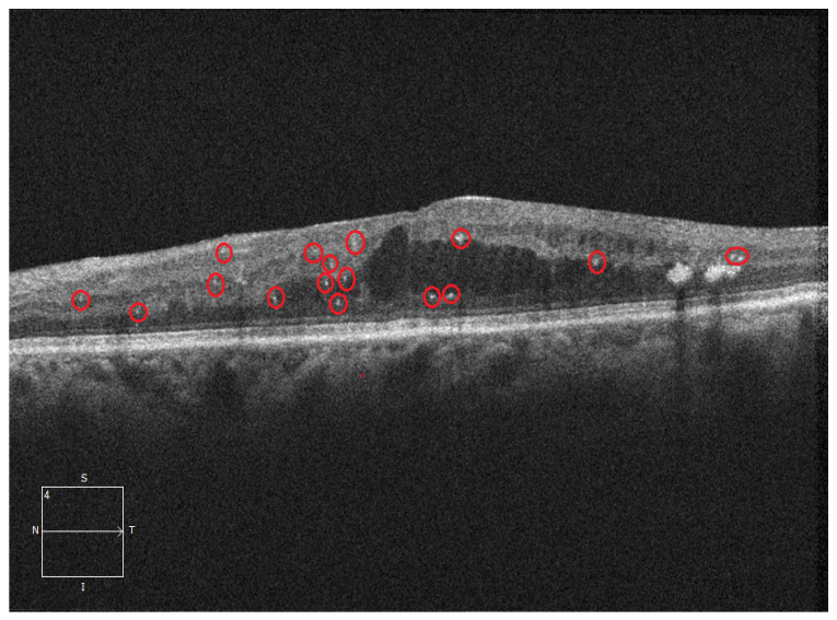 Figure 1
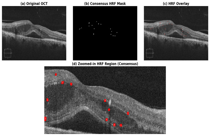 Figure 2
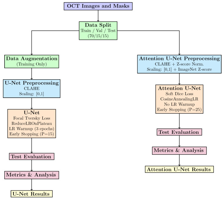 Figure 3
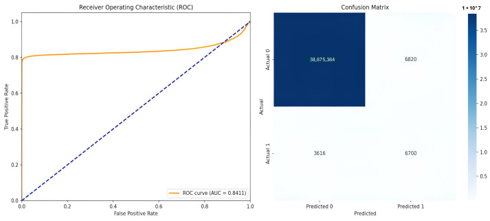 Figure 4
 Figure 5
 Figure 6
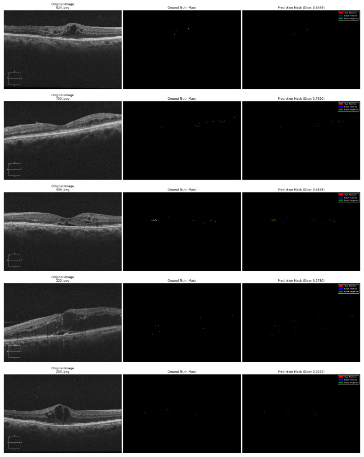 Figure 7
Peer Reviews
No public reviews on file for this paper yet. If you reviewed it on a platform where reviews are public (OpenReview, ICLR, NeurIPS, ICML), you can paste yours below so the community can read it here.
Videos
No videos yet. Explain this paper in a talk, walkthrough, or lecture? Add one.
Taxonomy
TopicsRetinal Imaging and Analysis · Optical Coherence Tomography Applications · Retinal Diseases and Treatments