Comprehensive Characterization of Lantana camara Essential Oil from Angola: GC-MS Profiling, Antioxidant Capacity, and Drug-likeness Prediction
Nswadi Kinkela, Abdy Morales, Hugo A. Sánchez-Martínez, Maricselis Díaz, Nsevolo Samba, Monizi Mawunu, Juan A. Morán-Pinzón, Lúcia Silva, Jesus M. Rodilla, Estela Guerrero De León

TL;DR
This study analyzed the essential oil from Lantana camara in Angola, finding it has strong antioxidant and anti-inflammatory properties, but also notable cytotoxic effects.
Contribution
The study provides a comprehensive chemical and bioactivity profile of Lantana camara essential oil from Angola, including in silico drug-likeness predictions.
Findings
The essential oil contains 96 volatile compounds, dominated by sesquiterpenes and monoterpenes like β-caryophyllene and sabinene.
The oil showed strong antioxidant activity in DPPH and lipid peroxidation assays but minimal effect on superoxide anion radicals.
In silico ADMET predictions suggest drug-like properties for major compounds, but the oil exhibited moderate to high cytotoxicity in some assays.
Abstract
Lantana camara L. (Verbenaceae) is a medicinal plant widely used in traditional medicine in Angola, especially for its anti-inflammatory effects. This study evaluated the chemical composition of L. camara essential oil from leaves (Lc-EO) collected in Uíge Province, Angola. GC–MS analysis enabled the identification of 96 volatile compounds, with sesquiterpenes and monoterpenes as the predominant constituents. Among them, β-caryophyllene (14.49%), sabinene (9.13%), bicyclogermacrene (8.18%), α-humulene (5.66%), nerolidol (5.29%), and 1,8-cineole (5.14%) were identified as major components. The antioxidant activity of Lc-EO was assessed using DPPH, ABTS, and superoxide anion (O2•−) assays. Lc-EO showed strong activity in the DPPH assay (IC50 = 0.72 µg/mL), moderate activity in the ABTS assay (IC50 = 87.5 µg/mL), but minimal effect on O2•− radicals (IC50 = 1491 µg/mL). It also…
Genes, proteins, chemicals, diseases, species, mutations and cell lines named across the full text — each resolved to its canonical identifier and authoritative record.
Click any figure to enlarge with its caption.
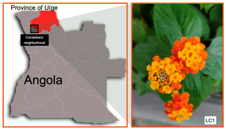 Figure 1
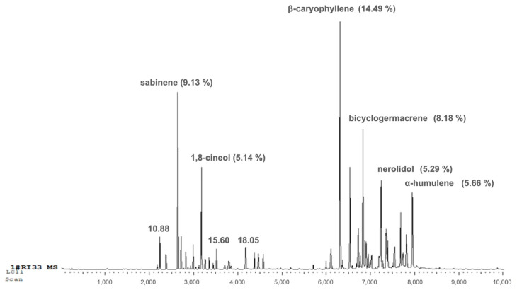 Figure 2
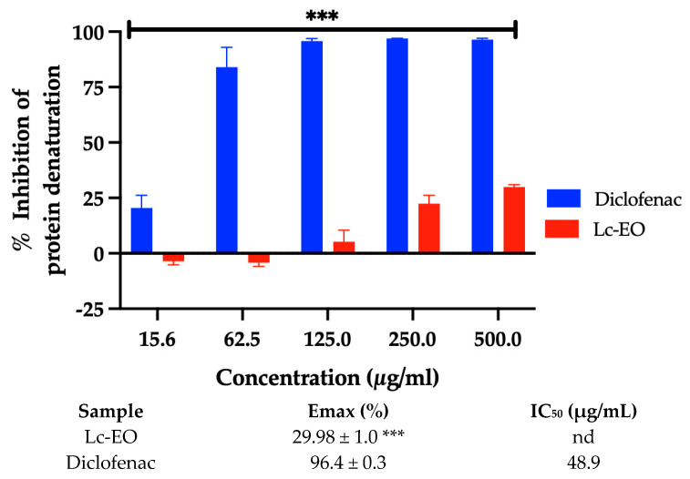 Figure 3
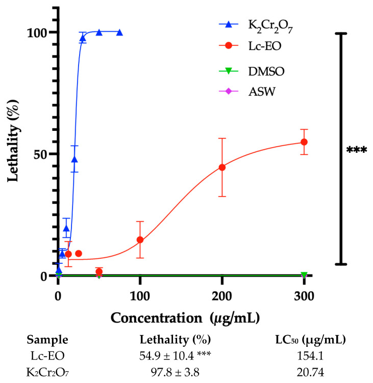 Figure 4
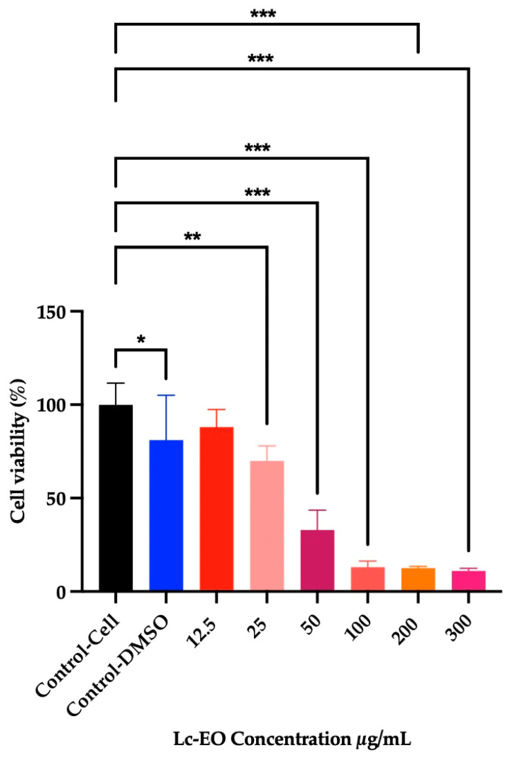 Figure 5
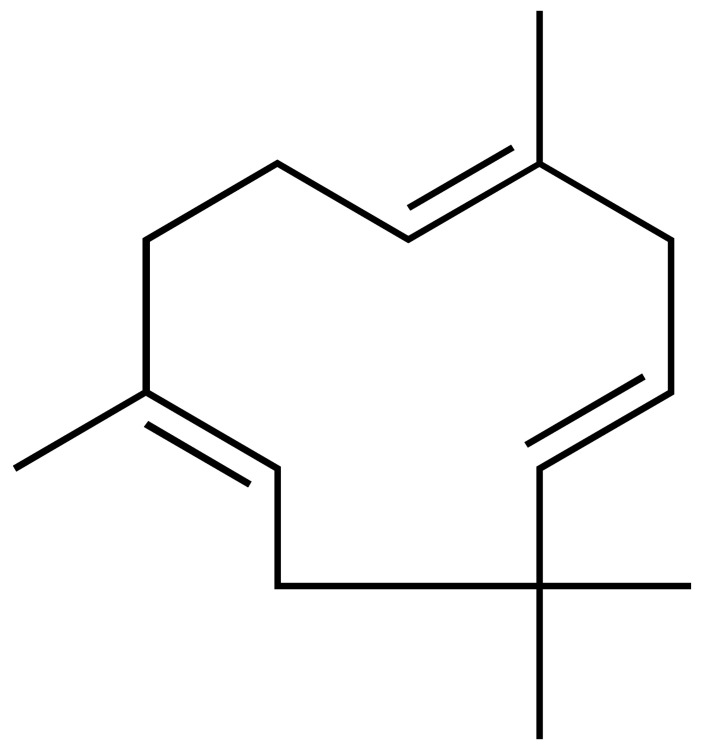 Figure 6
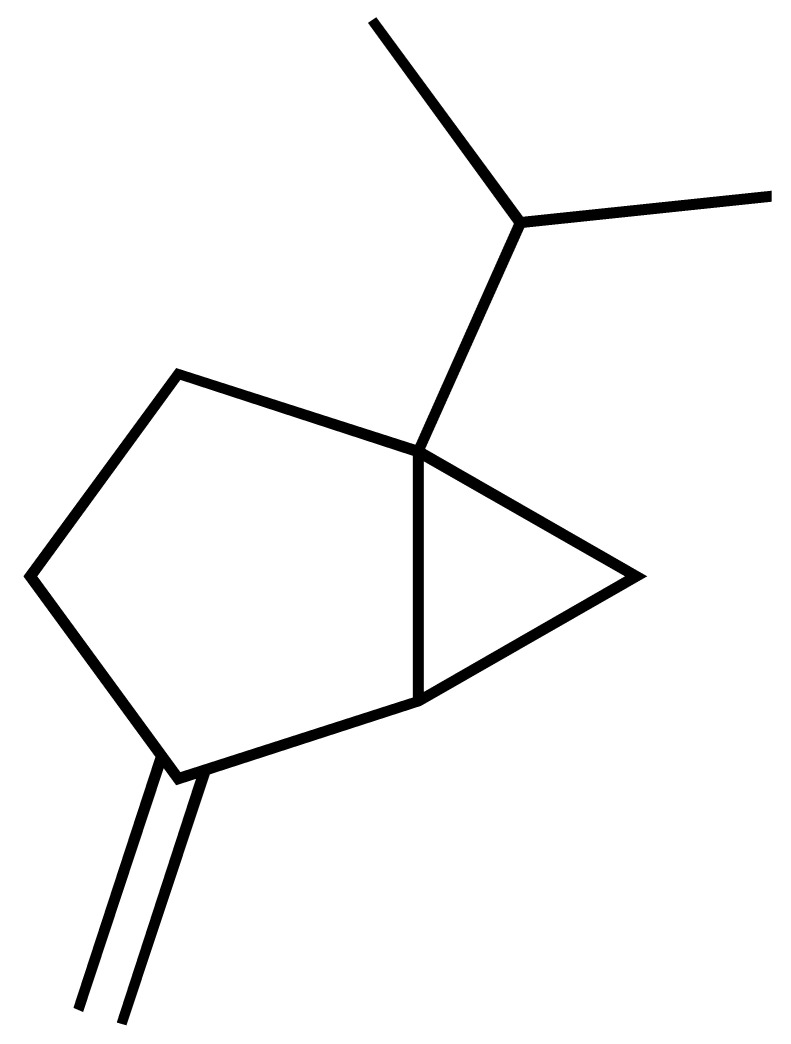 Figure 7
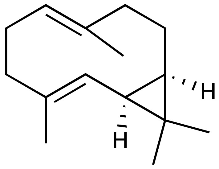 Figure 8
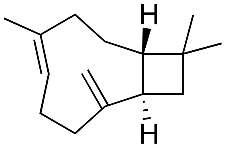 Figure 9
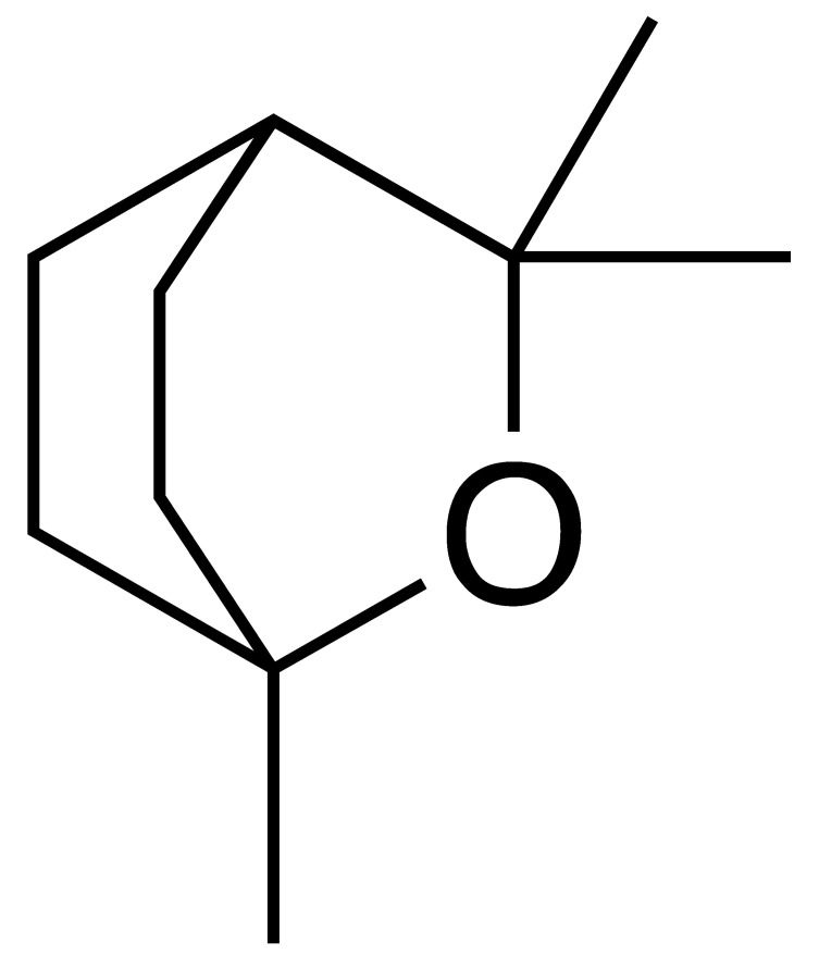 Figure 10
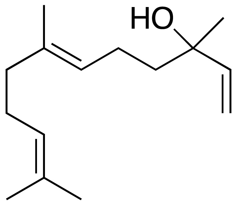 Figure 11
- —Secretaría Nacional de Ciencia Tecnología e Innovación
- —Research Unit of Fiber Materials and Environmental Technologies (FibEnTech-UBI)
- —Fundação para a Ciência e a Tecnologia
Peer Reviews
No public reviews on file for this paper yet. If you reviewed it on a platform where reviews are public (OpenReview, ICLR, NeurIPS, ICML), you can paste yours below so the community can read it here.
Videos
No videos yet. Explain this paper in a talk, walkthrough, or lecture? Add one.
Taxonomy
TopicsEssential Oils and Antimicrobial Activity · Phytochemistry and Biological Activities · Ethnobotanical and Medicinal Plants Studies
1. Introduction
L. camara L. is an invasive plant species widely distributed across multiple regions worldwide [1]. In Angola, it is known by vernacular names such as “Cambumbulu,” “Cambumbe,” and “Flor-de-cerca,” particularly in areas where Kikongo, Kimbundu, and Umbundu are spoken. Although often regarded as a weed, the species is traditionally used in folk medicine primarily for the management of inflammatory conditions and topical skin applications [2,3], uses that are consistent with the biological activities investigated in the present study. In addition to its medicinal uses, L. camara has attracted scientific interest for its pesticidal, antimicrobial, and larvicidal activities, which have been validated in several studies [4,5,6].
Phytochemical analyses of L. camara essential oil, especially from leaves, have consistently revealed a predominance of mono- and sesquiterpenes, including β-caryophyllene, α-humulene, bicyclogermacrene, and related constituents [7,8,9]. These terpenoid compounds have been associated with antioxidant and anti-inflammatory properties [10,11,12,13], while their lipophilic nature may also contribute to cytotoxic or irritant effects, underscoring the importance of concurrent safety evaluation. However, previous studies have shown that different varieties of L. camara, influenced by geographical origin, plant parts, and environmental conditions, may contain varying types and concentrations of lantadenes and other phytochemicals, resulting in distinct bioactive profiles [14,15,16].
Despite the widespread traditional use of L. camara, information regarding the chemical variability, antioxidant activity, and safety profile of its essential oil from Angola remains limited. We hypothesized that essential oil obtained from leaves collected in Uíge Province exhibits chemical compositions associated with measurable antioxidant activity and model-dependent cytotoxic effects. Therefore, this study aimed to (i) characterize the chemical composition of leaf essential oil, (ii) evaluate its antioxidant and anti-inflammatory activities, and (iii) assess preliminary safety through in silico ADMET predictions and in vitro cytotoxicity assays. This approach allows a balanced evaluation of both bioactivity and safety within the analytical scope of the study.
2. Materials and Methods
2.1. Location and Origin of Samples
Leaves of L. camara L. were collected in November 2022 from three individual plants located in the Condobenz neighborhood, Uíge Province, northern Angola (7°35′59″ S, 15°00′13″ E) (Figure 1). After collection, the leaves were carefully homogenized to form a composite sample representative of the local population. Taxonomic identification was performed by Professor Mawunu Monizi, Department of Agronomy, Kimpavita University (Uíge, Angola).
2.2. Preparation of Essential Oil
Fresh L. camara leaves were air-dried in a shaded environment protected from direct sunlight for 5 days to preserve the integrity of the active compounds and the natural coloration. After drying, the material was stored in plastic bags until distillation.
The essential oil was obtained by hydrodistillation using a Clevenger-type apparatus, with a solid-to-liquid ratio of 1:8 (g/mL). Distillation was carried out in triplicate, each run lasting three hours. At the end of each distillation, the essential oil was carefully separated from the aqueous phase, dried over anhydrous sodium sulfate, and filtered to remove residual moisture. Then the essential oil from L. camara leaves (Lc-EO) was stored in amber glass vials with airtight seals and kept under refrigeration (4 °C) until analysis.
Since the three distillations were performed using the same plant material and yielded similar results, only one representative oil sample was used for subsequent chemical and biological analyses. The yield was calculated based on the dry weight of the plant material and the volume of oil obtained.
2.3. Gas Chromatography-Mass Spectrometry Analysis
The chemical composition of Lc-EO was analyzed using Gas Chromatography coupled to Mass Spectrometry (GC-MS), a technique that enables efficient separation and precise identification of volatile and semi-volatile compounds.
GC-MS analyses were performed using an Agilent 7890A gas chromatograph equipped with a DB-5 capillary column (30 m × 0.25 mm, film thickness: 0.25 µm, J&W Scientific, Folsom, CA, USA) and coupled to an Agilent 5975C Inert XL MSD mass spectrometer with a triple-axis quadrupole detector. A 1 µL aliquot of each essential oil sample was injected in splitless mode at an injector temperature of 250 °C. The oven temperature program started at 50 °C (held for 2 min), followed by a temperature ramp of 3 °C/min to 190 °C and 10 °C/min to 300 °C, for each sample, to ensure optimal separation of the components. Helium was used as the carrier gas at a constant flow rate of 1 mL/min.
Mass spectra were acquired using electron impact (EI) ionization at 70 eV, with the ion source temperature set at 230 °C. Data acquisition and processing were carried out using ChemStation software C.01.10 (Agilent Technologies). The identification of the compounds was performed based on the Kováts Index (KI), which considers the chromatographic behavior of the substances. This index was used as a tool to characterize and differentiate the compounds present in essential oils, ensuring accurate identification. Compound identification was performed by comparing the obtained mass spectra with reference spectra from the NIST Mass Spectral Library (NIST/EPA/NIH).
2.4. In Silico Prediction of ADME and Toxicity Properties
The absorption, distribution, metabolism, and excretion (ADME) properties of the identified compounds were evaluated using the OSIRIS Property Explorer (https://www.organic-chemistry.org/prog/) (accessed on 12 May 2025) and the SwissADME platform (http://www.swissadme.ch) (accessed on 2 May 2025) [17]. OSIRIS was employed to predict key physicochemical and drug-likeness parameters, including calculated lipophilicity (cLogP), aqueous solubility (cLogS), molecular weight (MW), topological polar surface area (TPSA), drug-likeness, and drug score. Gastrointestinal absorption (GIA) and blood–brain barrier (BBB) permeability were predicted using the BOILED-Egg model available on the SwissADME platform [18].
Assessment of oral bioavailability was conducted according to Lipinski’s Rule of Five (Ro5) [19], which considers a compound drug-like if it fulfills the following criteria: molecular weight < 500 Da, logP ≤ 5, hydrogen-bond donors (HBD) ≤ 5, hydrogen-bond acceptors (HBA) ≤ 10, and no more than one violation of these rules.
Toxicity risk prediction was performed using two independent web-based platforms: PASS (http://www.way2drug.com/passonline) (accessed on 19 May 2025) and ADMETlab 3.0 (https://admetlab3.scbdd.com) (accessed on 13 May 2025) [20]. A compound was considered to have a potential toxicological risk if the predicted outcome was classified as “active” or if the model’s confidence score was ≥0.7 (ADMETlab 3.0). Additional predictive platforms were employed for comparative purposes, and their extended outputs are provided in the Supplementary Materials.
2.5. Evaluation of Antioxidant Activity of Lc-EO
The Lc-EO was dissolved in DMSO and vortex-mixed at 2000 rpm for 2 min to ensure homogeneous dispersion prior to the in vitro assays.
2.5.1. DPPH Radical Scavenging Assay
The percentage inhibition of the DPPH (2,2-diphenyl-1-picrylhydrazyl), ABTS (2,2′-azino-bis(3-ethylbenzothiazoline-6-sulfonic acid)) radical was determined following the methodology proposed by Jianu et al. [21]. In each well of a 96-well microplate, 100 µL of DPPH solution and 100 µL of Lc-EO, previously dissolved in DMSO, were added at concentrations ranging from 15.6 to 500 µg/mL. Quercetin was used as the positive control. A negative control consisting of DMSO at concentrations equivalent to those used in the test samples was included and showed no antioxidant activity under the experimental conditions. The microplate was incubated in the dark at room temperature for 30 min, and the absorbance was measured at 492 nm using an EPOCH™).
All assays were performed in triplicate. The DPPH radical scavenging activity was expressed as the percentage of inhibition, calculated using Formula (1).
where A_control_ is the absorbance of the reaction media without the test sample, and A_test_ is the absorbance in the presence of the essential oil or Quercetin.
2.5.2. ABTS Radical Cation Scavenging Assay
The free radical scavenging capacity of Lc-EO was evaluated as described by Valarezo et al. [22]. Two stock solutions were prepared: ABTS (2,2′-Azino-bis(3-ethylbenzthiazoline-6-sulfonic Acid)) (7.4 µM) and potassium persulfate (2.6 µM). Equal volumes of both solutions were mixed under constant stirring and incubated in the dark for 12 h to generate the ABTS working solution. Subsequently, 333 µL of the resulting solution was diluted with 20 mL of methanol to adjust the absorbance to 1.1 ± 0.02 at 734 nm, as measured using a UV–Vis spectrophotometer (EPOCH™ microplate reader, model M491, BioTek Instruments, Inc., Winooski, VT, USA).
For the assay, 10 µL of Lc-EO was mixed with 190 µL of the ABTS working solution in a 96-well plate and tested at various concentrations (15.6–500 µg/mL). The mixture was incubated at room temperature for 2 h, after which the absorbance was recorded at 734 nm. Quercetin was used as the positive control, and DMSO at a maximum concentration of 5% served as the negative control. The ABTS radical scavenging activity (%) was calculated using Formula (1).
2.5.3. NBT Superoxide Radical Scavenging Assay
The superoxide anion radical (O_2_^•−^) scavenging activity was evaluated using a non-enzymatic system, following the method described by Saha et al. [23]. The assay was conducted in 96-well microplates. A volume of 50 μL of the Lc-EO, prepared to assay various concentrations (15.6 to 500 µg/mL), was mixed with 50 µL of each of the following reagents: phenazine methosulfate (PMS, 120 µM), nicotinamide adenine dinucleotide (NADH, 936 µM), and nitroblue tetrazolium (NBT, 300 µM). The reaction mixtures were incubated at 25 °C for 5 min, after which the absorbance was measured at 560 nm using a microplate reader as previously described.
All experiments were performed in triplicate for each concentration tested. The percentage of superoxide scavenging activity was calculated using the same formula applied for the DPPH radical inhibition assay.
2.5.4. Lipid Peroxidation Inhibition Assay
The effect of Lc-EO (15.6 to 500 µg/mL) on egg yolk lipid peroxidation was assessed using the method described by Ruberto et al. [24], which quantifies malondialdehyde (MDA), a marker of fatty acid peroxidation. This assay was selected as a preliminary screening model for lipid peroxidation inhibition, acknowledging its simplified nature and limited extrapolation to in vivo systems. Briefly, 100 μL of egg yolk homogenate (1:25 v/v in phosphate-buffered saline, PBS, pH 7.4) was mixed with 10 µL of the essential oil, 50 μL of FeSO_4_ (25 mmol/L), and 300 µL of PBS. The mixture was incubated at 37 °C for 15 min, after which 50 µL of 15% (w/v) trichloroacetic acid (TCA) was added to stop the reaction. The samples were centrifuged at 3500 rpm for 15 min, and the resulting supernatants were collected. The absorbance of the sample (15.6 to 500 µg/mL) was measured at 532 nm to quantify the MDA levels.
The percentage inhibition of lipid peroxidation was calculated using Formula (2).
where A_control_ is the absorbance of an egg yolk emulsion in a blank buffer without the test sample, and A_test_ is the absorbance of the egg yolk emulsion containing either the Lc-EO or the standard substance (quercetin).
2.6. Anti-Inflammatory Activity by Inhibition of Protein Denaturation
The in vitro anti-inflammatory activity of Lc-EO was evaluated using a method adapted from Gîlcescu Florescu et al. [25], optimized for use in 96-well microplates. In this assay, 5 µL of Lc-EO (at concentrations ranging from 15.6 to 500 µg/mL) was mixed with 245 µL of bovine serum albumin (0.4% BSA) prepared in phosphate-buffered saline (PBS, pH 6.4). The mixtures were incubated at 37 °C for 10 min, followed by heating at 70 °C for 5 min to induce protein denaturation. After cooling, the absorbance was measured at 660 nm using a microplate reader as previously described. Diclofenac was used as a positive control. A negative control was prepared under identical conditions, replacing Lc-EO with a solution of DMSO in proportions equivalent to those used in the test samples.
The percentage inhibition of albumin denaturation, which reflects the anti-inflammatory potential of the Lc-EO, was calculated using Formula (3).
where A_control_ is the absorbance of the negative control, and A_test_ is the absorbance of the sample containing either Lc-EO or diclofenac.
2.7. Toxicity Test of Lc-EO
2.7.1. Toxicity Test with Artemia salina
The toxicity of Lc-EO was evaluated using the Artemia salina (EG Artemia, SEP-Art^®^, Great Salt Lake, UT, USA) bioassay, adapted for 96-well microplates as described by Mesquita et al. [26]. In each well, 100 µL of seawater containing 10 to 15 A. salina larvae was combined with 98 µL of artificial seawater (ASW) Instant Ocean (United Pet Group, Blacksburg, VA, USA) (prepared by dissolving 35 g/L in distilled water) and 2 µL of Lc-EO or DMSO (control), to achieve final concentrations ranging from 15.5 to 300 µg/mL. After 24 h of exposure, toxicity was assessed by determining the percentage of dead larvae in each well.
The median lethal dose (LD_50_) was defined as the concentration required to cause 50% mortality of Artemia salina nauplii. Based on established criteria, samples with LC_50_ values greater than 1000 µg/mL were considered non-toxic, whereas those with LD_50_ values between 500 and 1000 µg/mL were classified as weakly toxic, values between 100 and 500 µg/mL as moderately toxic, and values below 100 µg/mL as strongly toxic, according to Meyer et al. (1982) and Nguta et al. (2012) [27,28].
2.7.2. MTT-Based Cytotoxicity Screening in RAW 264.7 Macrophages
RAW 264.7 murine macrophages were obtained from the National Centre for Cell Science (NCCS, Pune, India) and cultured in high-glucose DMEM as described by Taciak et al. [29]. Cytotoxicity was evaluated using the MTT (3-[4,5-dimethylthiazol-2-yl]-2,5 diphenyl tetrazolium bromide) assay, which quantifies mitochondrial dehydrogenase activity via the reduction of MTT to formazan. Cells were seeded in 96-well plates (1.2 × 10^5^ cells/mL) and incubated for 24 h to allow adherence and proliferation, following protocols by Selvaraj et al. [30] and Marques et al. [31]. Subsequently, cells were treated with Lc-EO (12.5–300 µg/mL in DMSO) for 24 h at 37 °C and 5% CO_2_. In all experiments, the final DMSO concentration was kept below 0.5%, and DMSO at equivalent proportions was used as the negative control. After treatment, MTT solution (0.5 mg/mL) was added and incubated for 12 h. Formazan crystals were solubilized in acidified isopropanol, and absorbance was measured at 570 nm using a microplate reader (EPOCH, BioTek Instruments, Inc., USA). Cytotoxicity was expressed relative to untreated controls.
2.8. In Silico Prediction of Biological Activity
To predict the potential pharmacological effects and molecular targets associated with the compounds identified in the Lc-EO, the PASS (Prediction of Activity Spectra for Substances) online platform was employed (http://www.way2drug.com/passonline) (accessed on 19 May 2025) [32].
For each compound, predicted activities are expressed as probability values: Pa (probability to be active) and Pi (probability to be inactive). In this study, only those predicted activities with Pa ≥ 0.7 were considered significant and retained for further analysis, as this threshold indicates a high likelihood of biological relevance.
2.9. Statistical Analysis
Data are expressed as mean ± standard deviation (SD). Statistical analyses were performed using GraphPad Prism software (version 10.0; GraphPad Software Inc., San Diego, CA, USA). Differences among groups were assessed by one-way analysis of variance (ANOVA), followed by Tukey’s multiple comparisons test. Dose-response curves were fitted by nonlinear regression using a four-parameter logistic model with variable slope. In the anti-inflammatory activity assay, IC_50_ values were estimated relative to the fitted Emax due to the limited maximal efficacy of Lc-EO (~72%), rather than by direct interpolation. Statistical significance was denoted as * p < 0.05, ** p < 0.01, and *** p < 0.001.
3. Results
3.1. Yields and Phytochemical Characterization of Essential Oil
The essential oil yields from leaf samples of L. camara were comparable, exhibiting minor variations in volume and yield (Table 1).
3.2. Phytochemical Characterization of Essential Oil
GC–MS analysis allowed the identification of 96 compounds in the Lc-EO, excluding non-identified peaks. The composition was dominated by sesquiterpenes (51.5%), followed by monoterpenes (27.3%), with minor proportions of aromatic compounds and other trace constituents.
The chromatogram (Figure 2) showed well-defined and high-intensity peaks corresponding to the major constituents. Among them, β-caryophyllene (14.49%), sabinene (9.13%), bicyclogermacrene (8.18%), α-humulene (5.66%), nerolidol (5.29%), and 1,8-cineole (5.14%) were the most abundant compounds, together accounting for 47.89% of the total oil composition.
In addition to the major constituents, other characteristic components were identified in the Lc-EO sample, including α-pinene, β-pinene, δ-3-carene, 2-bornanone, 1-octen-3-ol, and Davana ether 1. Table 2 presents the complete list of volatile constituents identified in the analyzed essential oil.
3.3. Prediction of ADME and Toxicity Properties
Computational ADMET analysis indicated that major compounds possess drug-like properties, with only one violation of Lipinski’s Rule of Five (Table 3). The compounds displayed a range of lipophilicity (cLogP 2.11–6.24), with α-humulene being the most lipophilic (6.24) and 1,8-cineole the least (2.11), while all showed low water solubility (cLogS −2.48 to −3.66). 1,8-cineole (eucalyptol) and nerolidol demonstrated high gastrointestinal absorption (GIA), and three compounds (sabinene, 1,8-cineole, and nerolidol) were predicted to cross blood–brain barrier (BBB). All compounds were predicted not to be substrates of P-glycoprotein (P-gp), suggesting a lower likelihood of active efflux, which may enhance their intracellular bioavailability.
On the other hand, the major compounds of Lc-EO showed no predicted structural alerts for pan-assay interference (PAINS) and yielded synthetic accessibility scores ranging from 2.87 to 4.51. However, sabinene exhibited the highest predicted drug score (0.45).
Toxicological prediction identified six compounds with potential irritant effects, with skin sensitization, carcinogenicity, respiratory effect, and hepatotoxicity being the most frequently predicted toxicological endpoints.
Despite the utility of tools for ADMET profiling, these results must be interpreted as theoretical predictions requiring experimental validation. The predictive values are inherently constrained by the underlying algorithms and the varying quality of training data, which may suffer from class imbalance or subjective annotations.
3.4. Antioxidant Activity of Lc-EO
The antioxidant capacity of Lc-EO was evaluated using DPPH, ABTS, O_2_^•−^, and lipid peroxidation inhibition assays. The results are expressed as half-maximal inhibitory concentrations (IC_50_, µg/mL) and compared with the reference antioxidant quercetin (Table 4).
The Lc-EO exhibited a strong radical-scavenging effect against DPPH with an IC_50_ value of 0.72 ± 0.2 µg/mL, markedly lower than that of quercetin (17.32 ± 3.2 µg/mL). This finding highlights a high efficiency of the oil in neutralizing DPPH radicals. Similarly, in the ABTS assay, the essential oil showed an IC_50_ of 87.5 ± 4.6 µg/mL, which, although higher than that of quercetin (15.8 µg/mL), still demonstrates relevant antioxidant potential.
In contrast, Lc-EO displayed limited scavenging activity against O_2_^•−^, with a high IC_50_ of 1491 µg/mL, indicating weak activity compared to quercetin (13.92 ± 9.1 µg/mL).
The lipid peroxidation assay further corroborated the antioxidant potential of Lc-EO, in agreement with the results obtained in the DPPH and ABTS radical scavenging assays. The Lc-EO sample achieved a maximum inhibition of 72.2 ± 1.4%, which, although lower than that observed for the reference compound quercetin (92.4 ± 0.1%), confirms its relevant antioxidant activity.
3.5. Anti-Inflammatory Activity of L. camara Essential Oil
The anti-inflammatory activity of Lc-EO was evaluated based on its ability to inhibit protein denaturation (in vitro model). As shown in Figure 3, Lc-EO exhibited a moderate inhibitory effect compared to the reference drug diclofenac sodium (p < 0.05). While diclofenac achieved nearly complete inhibition (~100%) of protein denaturation, Lc-EO showed a dose-dependent effect, reaching a maximum inhibition of approximately 28% at the highest tested concentration. The IC_50_ value for Lc-EO could not be determined, as the maximum inhibition did not reach 50%, preventing reliable curve fitting. This was reported as “not determined” (nd), indicating limited anti-inflammatory activity within the tested concentration range.
3.6. Toxicity Assays
3.6.1. Preliminary Toxicity Assessment Using the Artemia Salina
No mortality was observed in the control group, indicating that 1% DMSO does not significantly affect nauplius viability (Figure 4). Lc-EO exhibited a concentration-dependent mortality, ranging from 10% to 60% across the tested concentrations. The Lc-EO induced a maximum mortality rate of 54.9 ± 10.4%, whereas the positive control (potassium dichromate) caused 97.8 ± 3.8% mortality. The calculated LC_50_ values were 154.1 µg/mL for the essential oil and 20.74 µg/mL for the standard.
3.6.2. Cytotoxicity Screening in RAW 264.7
The relative metabolic activity of RAW 264.7 macrophages decreased in a concentration-dependent manner following exposure to increasing concentrations of Lc-EO at 12.5, 25, 50, 100, 200, and 300 µg/mL (Figure 5). After 24 h of treatment, the IC_50_ value was determined to be 31.58 µg/mL, indicating a significant cytotoxic effect of Lc-EO. At the highest concentration tested (100 to 300 µg/mL), cell viability was reduced by up to 88% compared to untreated control cells.
3.7. Prediction of Biological Activity
Using the PASS online tool, we screened the six primary Lc-EO compounds for potential biological activities. Table 5 details the predicted activities for each compound. Our data indicate some interesting pharmacological activities, considering probably activity ≥ 0.7 as highly probable for experimental validation. The most highlighted evidence is targeted for anti-cancer, anti-inflammatory, and dermatologic applications. Some multiple-effect compounds, such as sabinene and β-caryophyllene, show broad activity (bone, skin, and cancer), and 1,8-cineole and nerolidol may benefit the liver, mental health, and some metabolic disorders.
4. Discussion
This study presents a comprehensive phytochemical, pharmacological, and toxicological evaluation of essential oil obtained from L. camara leaves collected in Uíge, Angola. The objective of determining the chemical composition of L. camara essential oil from Uíge Province, Angola, was motivated by previous reports indicating that chemical composition and relative abundance can vary according to geographical origin, climatic conditions, and soil characteristics [35]. Thus, the data from our study, consistent with previous reports, identify L. camara as a significant source of monoterpenes and sesquiterpenes, with β-caryophyllene, sabinene, and α-humulene as the main and most abundant compounds [5,6,36].
Several studies have shown that the chemical composition of L. camara exhibits high geographic variability, giving rise to different chemotypes. Satyal et al. (2016) [35] reported marked differences among samples from Yemen, Cuba, and Nepal, with significant changes in the proportions of monoterpenes and sesquiterpenes. Similarly, research conducted in Brazil identified distinct chemical profiles among populations of the species, highlighting relevant variations in the predominance of γ-curcumenes, zingiberene, and germacrenes [7,12]. In contrast, the essential oil from Uíge presents a sesquiterpene-rich chemotype characterized by high concentrations of β-caryophyllene and bicyclogermacrene, in addition to sabinene levels higher than those generally reported in the literature. This differentiation confirms the strong influence of environmental, edaphoclimatic, and genetic factors on the species’ ability to alter its chemical composition according to the surrounding conditions [6,26], indicating that the Uíge chemotype constitutes a unique phytochemical signature and reinforcing the importance of characterizing L. camara populations from different geographic regions.
The PASS online platform predicted various pharmacological activities for the major compounds identified in the Lc-EO sample. The most prominent predicted effects included antineoplastic, anti-inflammatory, and dermatological applications. Additional potential benefits included lipid metabolism regulation, antiulcer activity, and hepatoprotective effects. These computational predictions are consistent with findings from biological studies. For instance, anti-inflammatory activity has been previously described for α-humulene [10,37], sabinene [38], β-caryophyllene [39], 1,8-cineole [40], and nerolidol [41]. Nevertheless, these predicted activities should be regarded as exploratory indicators of potential biological relevance rather than confirmed therapeutic effects.
These compounds have also been reported to exhibit antioxidant properties [42,43,44], which are relevant to their proposed antitumor and anti-inflammatory activities. The findings regarding the individual pharmacological potential of L. camara constituents are further supported by this study, in which the Lc-EO demonstrated significant antioxidant activity. This activity was demonstrated by its strong capacity to scavenge the DPPH radical. Comparable results have been reported by other authors, who confirmed the antioxidant potential of L. camara leaf essential oil against DPPH [45,46,47]. The antioxidant capacity exhibited by Lc-EO is likely attributable to its principal constituents, and a similar synergistic antiradical effect has been documented in essential oils from other geographical origins [6,36]. Furthermore, individual contributions from major compounds such as β-caryophyllene are well-supported in the literature, as this sesquiterpene has demonstrated the ability to inhibit lipid peroxidation in both in vitro and in vivo models [48,49,50].
Regarding experimental anti-inflammatory activity, the essential oil exhibited a moderate effect, inhibiting 28% of protein denaturation, which was lower than the inhibition observed for diclofenac (Emax ≈ 100%). The protein denaturation assay has been previously employed by Khairan et al. (2024) to evaluate the anti-inflammatory potential of an ethanolic extract of L. camara, where the authors reported a concentration-dependent increase in inhibition of protein denaturation [2]. The anti-inflammatory properties described for the terpenoids present in L. camara are part of the findings that reinforce the results obtained in our study [51]. Collectively, these results demonstrate the potential anti-inflammatory effect of L. camara, whose magnitude appears to depend on the extraction method employed.
In terms of toxicity, previous studies have demonstrated that L. camara essential oil exhibits notable acaricidal activity, primarily attributed to major constituents such as γ-curcumene and nerolidol, both of which were also identified as abundant components in Lc-EO [52]. Beyond nerolidol, other predominant constituents, including bicyclogermacrene and α-humulene, have been reported to exert cytotoxic effects [43,53].
In our study, Lc-EO displayed moderate toxicity in the Artemia salina assay (IC_50_ = 154.1 μg/mL), according to the classification proposed by Rajabi et al. (2015) [54] and marked cytotoxicity in the MTT assay (IC_50_ = 31.58 μg/mL), suggesting a relationship with the antiparasitic action described for L. camara essential oil [6].
The pronounced cytotoxic response observed in the MTT assay, together with the presence of bioactive sesquiterpenes, provides strong support for the toxicological profile of Lc-EO and is consistent with the antitumoral and acaricidal activities previously reported for this species [11,52]. Thus, while the oil displays promising pharmacological potential, its therapeutic margin should be carefully evaluated. In this context, the detected cytotoxicity suggests that its practical applications may be limited, particularly for oral formulations, and reinforces the need for dose optimization and route-of-administration considerations.
Finally, it is important to emphasize that the use of L. camara by communities in Uíge is largely empirical and rooted in tradition, without formal scientific support. Although the plant is widely used for medicinal purposes, the findings of this study show that the local chemotype contains compounds with toxic potential, indicating that indiscriminate use may pose health risks. In this context, the data presented here provide an essential scientific basis to guide safer practices and support educational initiatives aimed at the population, contributing to a more informed and responsible use of the species.
5. Conclusions
This study highlights the importance of integrated phytochemical and biological characterization of L. camara in the context of its ethnobotanical use in Angola. The identification of bioactive terpenoid constituents, together with the observed antioxidant activity, moderate anti-inflammatory effects, and model-dependent cytotoxicity, provides partial pharmacological support for its traditional medicinal applications. Importantly, the detection of cytotoxic effects also emphasizes the need for careful safety assessment, dose consideration, and route-of-administration evaluation, as these factors may limit certain applications, particularly oral use. Collectively, these findings contribute mechanistic and experimental evidence that helps to contextualize the ethnomedicinal use of L. camara and supports a more rational, evidence-based, and informed utilization of this species.
The reference list from the paper itself. Each links out to its DOI / PubMed record.
- 1Bhagwat S.A. Breman E. Thekaekara T. Thornton T.F. Willis K.J. A Battle Lost? Report on Two Centuries of Invasion and Management of Lantana Camara L. in Australia, India and South Africa P Lo S ONE 20127 e 3240710.1371/JOURNAL.PONE.003240722403653 PMC 3293794 · doi ↗ · pubmed ↗
- 2Khairan K. Maulydia N.B. Faddillah V. Tallei T.E. Fauzi F.M. Idroes R. Uncovering Anti-Inflammatory Potential of Lantana Camara Linn: Network Pharmacology and in Vitro Studies Narra J.20244 e 89410.52225/NARRA.V 4I 2.89439280287 PMC 11391997 · doi ↗ · pubmed ↗
- 3Seyoum A. Pålsson K. Kung’a S. Kabiru E.W. Lwande W. Killeen G.F. Hassanali A. Knols B.G.J. Traditional Use of Mosquito-Repellent Plants in Western Kenya and Their Evaluation in Semi-Field Experimental Huts against Anopheles Gambiae: Ethnobotanical Studies and Application by Thermal Expulsion and Direct Burning Trans. R. Soc. Trop. Med. Hyg.20029622523110.1016/S 0035-9203(02)90084-212174767 · doi ↗ · pubmed ↗
- 4Braga F.G. Bouzada M.L.M. Fabri R.L. de O. Matos M. Moreira F.O. Scio E. Coimbra E.S. Antileishmanial and Antifungal Activity of Plants Used in Traditional Medicine in Brazil J. Ethnopharmacol.200711139640210.1016/j.jep.2006.12.00617234373 · doi ↗ · pubmed ↗
- 5Aisha K. Visakh N.U. Pathrose B. Mori N. Baeshen R.S. Shawer R. Extraction, Chemical Composition and Insecticidal Activities of Lantana Camara Linn. Leaf Essential Oils against Tribolium castaneum, Lasioderma serricorne and Callosobruchus chinensis Molecules 20242934410.3390/molecules 2902034438257257 PMC 10819012 · doi ↗ · pubmed ↗
- 6Barros L.M. Duarte A.E. Morais-Braga M.F.B. Waczuk E.P. Vega C. Leite N.F. De Menezes I.R.A. Coutinho H.D.M. Rocha J.B.T. Kamdem J.P. Chemical Characterization and Trypanocidal, Leishmanicidal and Cytotoxicity Potential of Lantana camara L. (Verbenaceae) Essential Oil Molecules 20162120910.3390/molecules 2102020926875978 PMC 6272997 · doi ↗ · pubmed ↗
- 7Kurade N.P. Jaitak V. Kaul V.K. Sharma O.P. Chemical Composition and Antibacterial Activity of Essential Oils of Lantana camara, Ageratum houstonianum and Eupatorium adenophorum Pharm. Biol.20104853954410.3109/1388020090319333620645797 · doi ↗ · pubmed ↗
- 8Passos J.L. Almeida Barbosa L.C. Demuner A.J. Alvarenga E.S. Da Silva C.M. Barreto R.W. Chemical Characterization of Volatile Compounds of Lantana camara L. and L. radula Sw. and Their Antifungal Activity Molecules 201217114471145510.3390/molecules 17101144723018920 PMC 6268859 · doi ↗ · pubmed ↗