Linking Clinical and Environmental Multidrug Resistance Plasmids Captured from the Tama River Flowing Through the Tokyo Megalopolis
Rin Yamazaki, Maho Tokuda, Singh Shweta, Koichiro Nakamichi, Ryota Moriuchi, Hideo Dohra, Hiroyuki Futamata, Kazuhide Kimbara, Masaki Shintani

TL;DR
This study shows that urban rivers like the Tama River in Tokyo can contain plasmids that spread antibiotic resistance between clinical and environmental settings.
Contribution
The study provides nucleotide-level evidence of clinically related and environmentally unique resistance plasmids in an urban river.
Findings
Eleven plasmids from the Tama River encode resistance to five antimicrobial classes and contain mobile genetic elements like integrons and ISCR1.
Some plasmids show structural similarity to clinical plasmids from distant regions, while others exhibit environmental characteristics and broad host-range transferability.
These plasmids significantly increase antimicrobial resistance in host bacteria, highlighting their functional impact.
Abstract
Background: Plasmid-mediated horizontal transfer of antimicrobial resistance genes (ARGs) is a major driver of resistance dissemination across clinical and environmental settings. Urban rivers flowing through densely populated megacities represent critical interfaces where human-associated and environmental microbiomes intersect; however, the genetic structures and functional characteristics of resistance plasmids circulating in such environments remain insufficiently resolved. Methods: In this study, we conducted detailed genomic and phenotypic analyses of 11 ARG-bearing plasmids previously captured from the Tama River, an urban river flowing through the Tokyo megalopolis. These plasmids belonged to IncN, IncU, IncQ2γ, IncC, and IncPγ groups. Whole-plasmid sequencing, comparative genomic analyses, conjugation assays, and antimicrobial susceptibility testing were employed to…
Genes, proteins, chemicals, diseases, species, mutations and cell lines named across the full text — each resolved to its canonical identifier and authoritative record.
Click any figure to enlarge with its caption.
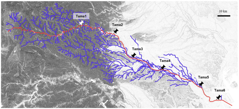 Figure 1
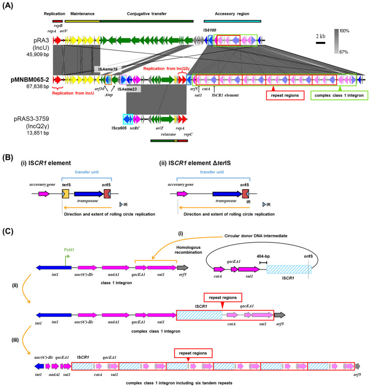 Figure 2
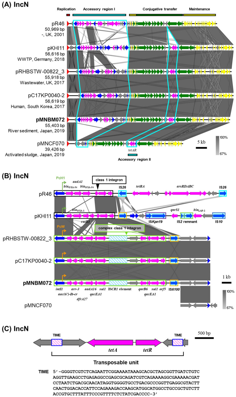 Figure 3
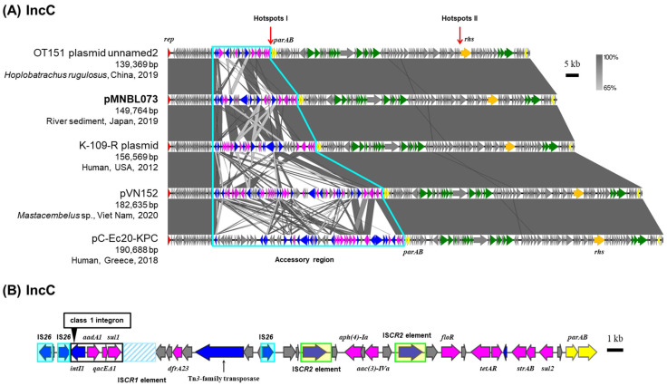 Figure 4
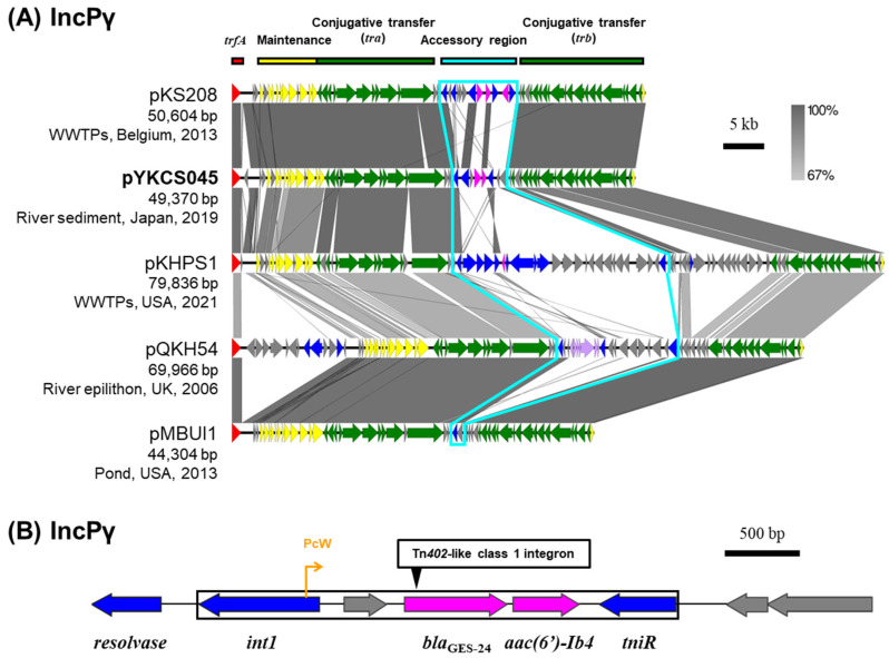 Figure 5
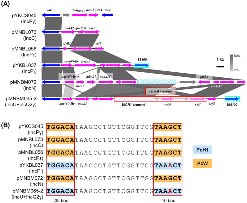 Figure 6
- —Tokyu Foundation
- —JSPS KAKENHI
- —Ministry of Education, Culture, Sports, Science, and Technology (MEXT), Japan
- —Japan Agency for Medical Research and Development (AMED)
- —Asahi Glass Foundation
- —Institute for Fermentation, Osaka
- —Toyota Physical and Chemical Research Institute
- —Research Institute of Green Science and Technology Fund for Research Project Support, Shizuoka University
- —Consortium for the Exploration of Microbial Functions of Ohsumi Frontier Science Foundation
Peer Reviews
No public reviews on file for this paper yet. If you reviewed it on a platform where reviews are public (OpenReview, ICLR, NeurIPS, ICML), you can paste yours below so the community can read it here.
Videos
No videos yet. Explain this paper in a talk, walkthrough, or lecture? Add one.
Taxonomy
TopicsPharmaceutical and Antibiotic Environmental Impacts · Antibiotic Resistance in Bacteria · Antimicrobial agents and applications
1. Introduction
The emergence and spread of antimicrobial-resistant bacteria poses a serious threat to global health. A particularly concerning mechanism behind this resistance is the spread of antimicrobial resistance genes (ARGs) via plasmids, one of the mobile genetic elements (MGEs), that can transfer between different bacterial species through conjugative transfer [1]. Plasmids carrying ARGs, known as resistance plasmids (R plasmids), are classified into incompatibility (Inc) groups based on the similarity of their replication systems. Many well-known R plasmids belong to groups such as IncC, IncF, IncN, IncL, among others, and IncC and IncN plasmids are capable of horizontal transfer across a wide range of bacterial hosts [2,3]. These plasmids play a central role in the dissemination of resistance traits not only in clinical settings but increasingly in the environment [1].
Recent studies have reported the presence of multidrug-resistant bacteria and ARGs in wastewater plants and urban rivers [4,5,6,7], as well as on agricultural products such as vegetables [8,9]. These findings suggest that ARG transfer is occurring in environments closely linked to daily human life. R plasmids originating from human clinical isolates have been well studied. However, such investigations in natural environments are still limited. Little is known about whether R plasmids are capable of propagating in environmental bacterial communities, or what kinds of bacteria are involved in such exchanges. Moreover, it is plausible that novel plasmids, distinct from those characterized in clinical strains, exist in natural environments and contribute to the emergence of resistance. It is therefore important to understand the linkage between R plasmids in clinical and natural environments.
The Tama River flows through the Tokyo megalopolis, one of the world’s most densely populated urban areas, where inputs from households, hospitals, industries, and wastewater treatment plants converge. This urban river therefore represents a critical human–environment interface in which clinical, anthropogenic, and environmental microbial communities can interact.
In our previous study, we collected 167 transconjugants from the Tama River using the exogenous plasmid capture method, revealing that diverse plasmids circulate in this urban river environment [10]. However, the genetic structures, MGEs, and antimicrobial resistance phenotypes of these plasmids remained largely unexplored. In the present study, we focused on 11 plasmids from this collection that were found to carry ARGs. These plasmids belonged to several incompatibility groups—IncN, IncU, IncQ2, IncC, and IncP—representing key vectors known to mediate multidrug resistance (MDR) in both clinical and environmental settings. We performed comprehensive genomic analyses of their backbone regions, accessory resistance regions, and associated MGEs, including integrons, insertion elements (ISs), transposons, and Tn3-derived inverted-repeat miniature elements (TIMEs) [11]. We additionally conducted conjugation assays and antimicrobial susceptibility testing to assess their transmissibility and resistance phenotypes. By comparing the plasmids with related sequences reported worldwide, we aimed to clarify the evolutionary connections between environmental and clinical plasmid pools and to better understand the role of urban rivers as reservoirs and dissemination points of MDR plasmids.
2. Results and Discussion
2.1. Various Plasmids Carrying ARGs Were Captured from the Tama River
Our previous exogenous plasmid captures using microbial communities collected from six sampling sites along the Tama River (Tama1 to Tama6, Figure 1) yielded 167 transconjugants through biparental matings and triparental matings, most of which were previously analyzed [10]. In principle, biparental matings allow the capture of plasmids that both confer the targeted resistance to the hosts and are self-transmissible between different cells. In contrast, triparental matings enable the acquisition of self-transmissible plasmids that do not carry the marker resistance genes but can mobilize non-self-transmissible plasmids as helper plasmids. Here, we specifically examine 11 plasmids carrying ARGs that were captured from midstream or downstream sites of the Tama River (Tama4, Tama5, and Tama6, Figure 1 and Table 1). There were seven IncP plasmids, including IncPβ, IncPγ, IncPε, IncPι and IncPκ subgroups, one IncN plasmid, and one IncC plasmid (Table 1). One plasmid, pMNBM065-2, was estimated to be classified both under the IncU and IncQ groups (a multi-replicon). In addition, a PromA plasmid (pMNBM065-1) was identified in the same host as pMNBM065-2; however, as it did not carry any ARGs, it was excluded from further analysis (Table 1). The 11 plasmids harbored resistance genes against aminoglycosides, β-lactams, tetracyclines, mercury, and other agents. Hosts carrying these ARG-bearing plasmids exhibited resistance to the tested antibiotics, including tetracycline (Tc), gentamicin (Gm), ampicillin (Ap), kanamycin (Km), chloramphenicol (Cm), streptomycin (Sm), and erythromycin (Em), as summarized in Table 2.
In the following sections, we present detailed nucleotide sequence-level analyses and comparative genomic analyses of each individual plasmid and the mobile genetic elements associated with ARGs on these plasmids.
2.2. Multi-Replicon Plasmid pMNBM065-2
Plasmid pMNBM065-2 was captured downstream (Tama5) along with pMNBM065-1 (PromAγ) through biparental matings (Table 1). pMNBM065-2 was shown to be a multi-replicon plasmid possessing genes encoding replication initiation protein (RIP) of both IncU and IncQ2γ (Figure 2A). A comparison of the nucleotide sequences of pMNBM065-2 with pRA3, an archetype plasmid of IncU group [12,13] and pRAS3-3759, a member of IncQ2γ plasmid [14,15,16], showed that pMNBM065-2 contained one region homologous to maintenance and accessory regions of pRA3 (IncU) and another region highly similar to almost the entire sequence of pRAS3-3759 (IncQ2γ) (Figure 2A). This plasmid additionally had six tandem 5134 bp repeat regions, containing resistance genes for sulfonamide (sul1) and chloramphenicol (catA) and a putative gene encoding a transposase of ISCR1, a member of the IS91 family (Table 1, Figure 2A).
ISCR1 elements encode a transposase that catalyzes transposition through a rolling-circle mechanism [17,18,19]. They possess two imperfect terminal 8 bp inverted repeats, terIS (the left end) and oriIS (the right end), each associated with characteristic dyad-symmetry sequences that serve as recognition sites for the transposase [20] (Figure 2B(i)). The oriIS region, located downstream of the transposase gene, is essential for initiating rolling-circle replication during transposition [18] (Figure 2B(i)). A notable feature of the ISCR1 element is that a single copy of the element is sufficient to mobilize adjacent DNA sequences when the ~100 bp oriIS region is present [18] (Figure 2B(ii)). This property enables one-ended transposition, in which transposition proceeds from the oriIS end without requiring a second IS end (terIS) [19] (Figure 2B(ii)).
Previous studies have shown that ISCR1 elements are almost exclusively associated with class 1 integrons [21]. A genetic model has been proposed in which the ISCR1 element is fused to the class 1 integron. The 3′-conserved segment (3′-CS) of the integron is formed by the fusion of the sul1 and qacEΔ1 genes [21]. In this structure, fusion of ISCR1 with the 3′-CS results in deletion of the terIS site, and the start codon of the ISCR1 ORF is located 404 bp downstream of the stop codon of the sul1 gene [20]. In addition, the gene orf5, which is typically found downstream of qacEΔ1-sul1, is absent [20]. The fusion of ISCR1 to the 3′-CS, together with the absence of terIS, allows the ISCR1 element to undergo rolling-circle replication encompassing all or part of the class 1 integron. This process enables mobilization of the class 1 integron [20]. Mobilized class 1 integrons can subsequently acquire non-cassette resistance genes, and recombination with the 3′-CS of another class 1 integron leads to the formation of a complex class 1 integron [20] (see Figure 2C as follows).
In pMNBM065-2, we identified two sequence features relevant to ISCR1-mediated rearrangements. First, a highly conserved 22 bp sequence (5′-GTGGTTTATACTTCCTATACCC-3′) [21,22] was detected at the right end of the ISCR1 element, which is consistent with an oriIS-like site (Figure 2C). Second, the stop codon of sul1 and the start codon of the ISCR1 ORF were separated by 404 bp (Figure 2C), a spacing previously reported for ISCR1 elements fused to the 3′-CS of class 1 integrons. These observations support a plausible scenario in which ISCR1 element, together with catA and qacEΔ1-sul1, generated a circular donor DNA intermediate via one-ended transposition, followed by recombination into the 3′-CS of the class 1 integron on pMNBM065-2 (Figure 2C(i)). If this scenario occurred, the architecture shown in Figure 2C(i) would be expected, namely a complex class 1 integron containing aminoglycoside resistance genes [aac(6′)-IIc and aadA1] plus the ISCR1-associated region (qacEΔ1-sul1-ISCR1-catA) with partial copies of qacEΔ1 and sul1 (Figure 2C(ii)). Reiteration of this recombination process could account for the presence of six tandem repeats of the region containing the ISCR1 element, catA, and partial qacEΔ1-sul1 sequences (Figure 2C(iii)). However, subsequent sequencing after cultivation indicated that this region was detected in only one or two copies, suggesting this repeat region may be unstable or subject to rearrangement under the cultivation condition used.
For the minimum inhibitory concentration (MIC) analysis, the plasmid pMNBM065-2 containing two copies of the repeat region was used. The MIC of the parental E. coli MG1655RGFP, which carries a single chromosomal catA gene inserted with a GFP gene by a mini-transposon [23], against chloramphenicol (Cm), determined by broth microdilution, was 250 µg/mL, while that of E. coli MG1655RGFP harboring pMNBM065-1 and pMNBM065-2 was 2000 µg/mL, representing an eightfold increase (this assay was conducted using two biological replicates with two technical replicates each). The pMNBM065-2 also carried tetracycline (tetRC) and conferred resistance to its host (Table 2). It should be noted that no known ARGs were detected on pMNBM065-1 (Table 1).
2.3. Mobilization of pMNBM065-2
Plasmid pMNBM065-2 possessed partial gene sets for conjugative transfer found in IncU plasmids, namely orf30-top genes [13], but the top gene was disrupted by the insertion of ISAeme19, and additional putative relaxosome-related genes (MOB_P_ family gene) and oriT region (137 bp) found in IncQ2γ plasmids (Figure 2A). The co-resident plasmid pMNBM065-1 (PromAγ) is self-transmissible, which has MOB_P_ and MPF_T_ family genes. To assess whether the pMNBM065-2 could be mobilized by pMNBM065-1, filter mating assays with E. coli JM109(pUC19) were conducted. As a result, both plasmids were transferred to the recipient with a transfer frequency of 1.7 × 10^–2^ per donor (two independent experiments). Although IncU and IncQ2γ plasmids were mainly isolated from Enterobacteriaceae, the transfer range of the PromAγ plasmid is known to be broad [24]. Therefore, pMNBM065-2 is likely to transfer among different bacterial families beyond its native host range, thereby conferring antimicrobial resistance and enhancing the survival of recipient bacteria in the Tama River.
2.4. IncN Plasmid pMNBM072
Plasmid pMNBM072 was captured downstream (Tama5) through biparental matings and classified under the IncN plasmid group (Table 1). IncN plasmids were detected in clinical and environmental sources worldwide, and their nucleotide sequences were highly conserved with the IncN archetype plasmid pR46 [25] (Figure 3A). Two accessory regions were identified, each probably inserted at specific regions, between the replication and conjugative transfer region (accessory region I) or downstream of the relaxase gene (accessory region II) [6]. pMNBM072 possessed a complex class 1 integron with gene cassettes aac(6′)-Ib-cr, arr-3, dfrA27, aadA16, qacEΔ1, sul1, ISCR1 element, qnrB6, qacEΔ1 and sul1 in the accessory region I (Figure 3B). Among them, a single copy of qacEΔ1-sul1, ISCR1 element and qnrB6 were likely inserted through transposition and recombination mediated by the ISCR1 element fused to the 3′-CS of the class 1 integron. In addition, the IS6100 element was located within the same region and 25 bp inverted repeats (IRs) (5′-TGTCRTTTTCAGAAGACGRCTGCAC-3′) and 5 bp direct repeats (DRs) (5′-CTGTT-3′) were detected outside of the IS6100 element and the class 1 integron. Consistent with the presence of these resistance genes, pMNBM072 conferred resistance to kanamycin, streptomycin and tetracycline on its host (Table 2).
Moreover, pMNBM072 and the IncN plasmids isolated from the United Kingdom and South Korea were highly similar in terms of both their backbones and accessory genes (Figure 3A,B). Additionally, pMNBM072 carried resistance genes for tetracycline (tetAR) in the accessory region II and conferred resistance on its host (Table 2). A region containing tetAR was flanked by 244 bp Tn3-derived inverted-repeat miniature elements (244 bp TIMEs) and 5 bp DR (5′-AGCAA-3′), which is a short and non-autonomous transposable element characterized by terminal inverted repeats but lacking the capacity to encode a transposase (Figure 3C) [11,26]. This region is likely mobilized by transposases provided in trans, thereby facilitating the dissemination of resistance genes.
2.5. IncC Plasmid pMNBL073
Plasmid pMNBL073 was captured from midstream site (Tama4) through biparental matings and estimated to be classified under the IncC group based on PCR analyses (Table 1) [10]. Based on the full-length nucleotide sequences of RIP genes, pMNBL073 was a member of the IncC group. IncC plasmids have been isolated from a wide range of sources, including humans, animals, and natural environments. Their backbone sequences are highly similar (Figure 4A), and accessory regions are predominantly inserted at two specific regions’ ‘hotspots’ [27]. In pMNBL073 and its homologous plasmids, an accessory region was also identified upstream of parA gene, at one of the specific insertion regions (Figure 4A) [27]. A comparison of the nucleotide sequences of this accessory region showed that the antimicrobial resistance genes, transposons and IS present were diverse and complex (Figure 4A). The other ‘hotspot’ is upstream of the rhs gene. pMNBL073 carried genes conferring resistance to aminoglycosides [aadA2, aac(3)-IVa, aph(4)-Ia and strAB], sulfonamide (sul1 and sul2), trimethoprim (dfrA23), chloramphenicol (floR) and tetracycline (tetAR) (Figure 4B). Among these resistance genes, aadA2, qacEΔ1 and sul1 are included in a class 1 integron (Figure 4B). pMNBL073 conferred resistance to gentamicin, chloramphenicol, and tetracycline on its host (Table 2).
2.6. IncP Plasmids
Among the seven IncP plasmids captured from the Tama River, plasmids pYKCS045 (IncPγ), pMNBL056 (IncPε), and pYKBL037 (IncPι) carried class 1 integrons containing multiple ARGs (Table 1) [10]. pYKCS045 captured from the downstream site of the Tama River (Tama5) through triparental matings was predicted to be a IncPγ plasmid. Only four other IncPγ plasmids are registered in the plasmid database PLSDB [28,29,30], and they were isolated from natural environments such as rivers, wastewater treatment plants (WWTPs) and ponds [31,32]. A comparison of the nucleotide sequences of IncPγ plasmids showed that their core genes for replication, maintenance, and transfer were highly conserved (Figure 5A). The accessory regions including ARGs were inserted between tra gene sets and trb gene sets. Plasmid pYKCS045 carried the resistance genes for carbapenems (blaGES-24) and aminoglycosides [aac(6′)-Ib4] within a Tn402-like class 1 integron (Figure 5B) with 25 bp IRs (5′-TGTCRTTTTCAGAAGACGRYTGCAC-3′) and 5 bp DRs (5′-CCTAT-3′). By broth microdilution, the MIC of Metapseudomonas resinovorans CA10dm4RGFP harboring this plasmid against meropenem was 32 µg/mL.
2.7. Class 1 Integrons
Regardless of the Inc groups, class 1 integrons were found in six plasmids including pYKCS045 (IncPγ), pMNBL056 (IncPε), pYKBL037 (IncPι), pMNBM065-2 (IncU and IncQ2γ), pMNBM072 (IncN) and pMNBL073 (IncC). Plasmid pYKCS045 had Tn402-like class 1 integrons, which contained a part of transposase genes, tniR, blaGES-24 and aac(6′)-Ib4 (Figure 6A). Plasmids pMNBL056 and pYKBL037 had a class 1 integron 3′ conserved segment (3′-CS) with three well-conserved genes, qacEΔ1-sul1-orf5, while pMNBL073 contained a similar integron with aadA2 gene and 3′-CS, lacking an orf5 gene (Figure 6A) [33]. In addition, the integron of pMNBL056 contained the gene cassette dfrB1 encoding for trimethoprim resistance, whereas that of pYKBL037 contained aac(6′)-Ib and ere(A), encoding for erythromycin resistance (Figure 6A). pMNBM065-2 and pMNBM072 had a complex class 1 integron generated by an ISCR1 element. These class 1 integrons contained the gene cassettes aac(6′)-IIc and aadA1 in pMNBM065-2, and aac(6′)-Ib-cr, arr-3, dfrA27 and aadA16 in pMNBM072. In contrast, catA and qnrB6 were likely acquired through the recombination involving the ISCR1 element-class 1 integron 3′-CS fusion variants, respectively (Figure 6A). The four variants of the class 1 integron gene cassette promoter located in intI1 gene (Pc)—PcW, PcH1, PcH2 and PcS—are known to differ in transcriptional strengths: PcW (ancestral and the weakest form), PcS (the strongest form), PcH1 (stronger than PcW but weaker than PcH2) and PcH2 (between PcS and PcH1) [33,34,35]. Two kinds of promoters, PcW and PcH1, of the class 1 integrons were found in the captured plasmids (Figure 6B). The promoters in pMNBM065-2 and pYKBL037 exhibited higher predicted strength than those in pYKCS045, pMNBM072, pMNBL073 and pMNBL056 (Figure 6B).
Collectively, our findings highlight two major patterns among the captured plasmids. First, IncN and IncC plasmids exhibited high structural similarity, including conserved accessory resistance regions, to clinically derived plasmids reported from geographically distant regions, indicating that clinically associated plasmids are also present in the urban river environment. Second, plasmids such as the multi-replicon IncU+IncQ2γ plasmid and several IncP plasmids displayed accessory-region architectures characteristic of environmental plasmids, suggesting that urban rivers can also serve as sites where potentially novel MDR plasmids persist and diversify. Taken together, these results support the idea that urban rivers, such as the Tama River, can function as ecological hubs where clinically derived and environmentally adapted resistance plasmids coexist, interact, and potentially disseminate. However, the extent to which these interactions translate into plasmid dissemination, their abundance in situ, and their broader epidemiological significance cannot be directly inferred from the present study, as described below.
This study has several limitations that should be acknowledged. First, the plasmids analyzed here were obtained using an exogenous plasmid capture approach, which selectively enriches plasmids that are transferable under the applied experimental conditions. Consequently, this method does not allow quantitative estimation of the absolute abundance or copy numbers of specific plasmids or antimicrobial resistance genes in the original river sediment samples. Second, only a subset of plasmids identified in our previous study was subjected to detailed analysis, and the 11 ARG-bearing plasmids examined here may not fully represent the overall plasmid diversity present in the Tama River. In addition, antimicrobial susceptibility testing and conjugation assays were conducted using selected laboratory and environmental host strains, which may not reflect the full range of host–plasmid interactions occurring in situ. Finally, sampling was conducted at limited time points, and seasonal or temporal variations in microbial community composition and plasmid dynamics were not assessed. Future studies integrating culture-independent quantitative approaches, such as metagenomic analyses, together with broader temporal sampling will be essential to further elucidate the abundance, diversity, and dissemination dynamics of resistance plasmids in urban river environments.
3. Materials and Methods
3.1. Bacterial Strains, Plasmids, and Culture Conditions
Escherichia coli MG1655RGFP is a rifampicin-resistant derivative of E. coli MG1655, in which a mini-Tn5-Km-P_A1/O4/O3_-RBSII-gfpmut3* cassette was chromosomally inserted at position 578,584 nt (NC_000913), conferring kanamycin and chloramphenicol resistance [34]. E. coli DH5α was used as a standard laboratory strain for plasmid propagation and maintenance. Metapseudomonas resinovorans CA10dm4RGFP (former Pseudomonas resinovorans) is a rifampicin-resistant strain, in which a miniTn7(Gm)-P_A1/O4/O3_-gfp-a cassette was integrated into the chromosome immediately downstream of the glmS gene (position 6,265,580 nt, NC_021499), conferring gentamicin and chloramphenicol resistance [36]. E. coli and Metapseudomonas resinovorans strains were cultivated in Luria broth (LB) [37] at 30 °C or 37 °C with shaking at 180 rpm. R2A plates containing 1.5% agar were used for filter matings. Ampicillin (Ap, 50 μg/mL), kanamycin (Km, 30 μg/mL for plasmid capture and 50 μg/mL for the other experiments), gentamicin (Gm, 30 μg/mL), and rifampicin (Rif, 30 μg/mL for plasmid capture and 50 μg/mL for the others) were added to the medium. Cycloheximide (100 μg/mL) was added to prevent fungal growth. For plate cultures, LB was solidified using 1.5% agar (wt/vol). The broad-host-range plasmid pBBR1MCS-2 [38], carrying a kanamycin resistance marker and a mobilization (mob) region compatible with IncP, IncQ, and IncW plasmids, was used where appropriate.
3.2. Collection of Environmental Samples and Exogenous Plasmid Captures
River sediment samples were collected using sterile spatulas and transferred into sterile containers from six sites located in the upstream, midstream and downstream regions of the Tama River in Tokyo, Japan in 6 July 2019, 6 October 2019, and 15 February 2020 (Figure 1). The sampling sites were as follows: Tama1 (35.803872 N 139.194108 E), Tama2 (35.776822 N 139.287567 E), Tama3 (35.695053 N 139.363358 E), Tama4 (35.652894 N 139.504669 E), Tama5 (35.601667 N 139.624847 E), and Tama6 (35.544467 N 139.725906 E). Samples were transported to the laboratory within a few days at room temperature and subsequently used for exogenous plasmid capture experiments, as described previously [10]. In brief, exogenous plasmid capture was performed using GFP-tagged Escherichia coli MG1655RGFP or Metapseudomonas resinovorans CA10dm4RGFP as recipient strains. Environmental samples were mixed with recipient cells on membrane filters and incubated to allow conjugative transfer (filter mating). Biparental mating enabled direct capture of self-transmissible tetracycline-resistant plasmids from environmental bacteria, whereas triparental mating employed an intermediate donor strain to facilitate mobilization of non-self-transmissible plasmids by helper plasmids (pBBR1MCS-2). Transconjugants were selected on antibiotic-containing media and screened based on GFP fluorescence.
3.3. Antibiotic Resistance Testing
Antibiotic resistance testing for the host of plasmids pMNDW109 and pMNDX110 was performed. For this testing, ampicillin (Ap, 50 µg/mL), gentamicin (Gm, 30 µg/mL), and tetracycline (Tc, 12.5 µg/mL) were added to LB. Resistance testing for the other plasmids had already been performed in our previous report [10]. The antibiotics were chosen to correspond directly to the antimicrobial resistance genes identified on the analyzed plasmids, allowing targeted phenotypic assessment of plasmid-encoded resistance determinants. The qualitative resistance (R/S) was assessed based on growth or no growth in liquid LB medium supplemented with antibiotics.
For pYKCS045, the determination of minimum inhibitory concentrations (MICs) was additionally performed using the broth microdilution method with Mueller Hinton Broth, following the Clinical and Laboratory Standards Institute CLSI M07 protocol (three biological replicates) [39]. This assay was performed in two independent experiments, and the same MIC value was observed in all replicates.
3.4. Plasmid Sequencing and Annotation
The nucleotide sequences of pMNDW109 and pMNDX110 were determined using MiSeq platform (Illumina, San Diego, CA, USA). 151 bp paired-end libraries were prepared using the Nextera XT DNA Library Preparation Kit (Illumina). Assembly of the sequencing reads was performed using SPAdes v. 3.14.1 [40] with default parameters. In addition, PCR was performed to confirm the connection between sequence fragments. These plasmids were annotated using DFAST (https://dfast.ddbj.nig.ac.jp/, accessed on 29 August 2025) [41] and their accession numbers in DDBJ are LC895901.1 and LC895902.1 (listed in Table 1). Other plasmids were previously determined and annotated [10]; their accession numbers deposited in DDBJ are shown in Table 1. ARGs were detected using ResFinder database (https://bitbucket.org/genomicepidemiology/resfinder_db/src/master/, accessed on 9 August 2025). Plasmid maps were visualized using Easyfig v.2.2.5 [42]. The structures of plasmids were compared using Easyfig.
The reference list from the paper itself. Each links out to its DOI / PubMed record.
- 1Castañeda-Barba S. Top E.M. Stalder T. Plasmids, a Molecular Cornerstone of Antimicrobial Resistance in the One Health Era Nat. Rev. Microbiol.202422183210.1038/s 41579-023-00926-x 37430173 PMC 12440250 · doi ↗ · pubmed ↗
- 2Flach C.-F. Johnning A. Nilsson I. Smalla K. Kristiansson E. Larsson D.G.J. Isolation of Novel Inc A/C and Inc N Fluoroquinolone Resistance Plasmids from an Antibiotic-Polluted Lake J. Antimicrob. Chemother.2015702709271710.1093/jac/dkv 16726124213 · doi ↗ · pubmed ↗
- 3Yu Z. Wang Q. Pinilla-Redondo R. Madsen J.S. Clasen K.A.D. Ananbeh H. Olesen A.K. Gong Z. Yang N. Dechesne A. Horizontal Transmission of a Multidrug-Resistant Inc N Plasmid Isolated from Urban Wastewater Ecotoxicol. Environ. Saf.202427111597110.1016/j.ecoenv.2024.11597138237397 · doi ↗ · pubmed ↗
- 4Nguyen T.N. Kasuga I. Liu M. Katayama H. Occurrence of Antibiotic Resistance Genes as Emerging Contaminants in Watersheds of Tama River and Lake Kasumigaura in Japan IOP Conf. Ser. Earth Environ. Sci.201926601200310.1088/1755-1315/266/1/012003 · doi ↗
- 5Shintani M. Vestergaard G. MilakovićM. Kublik S. Smalla K. Schloter M. Udiković-KolićN. Integrons, Transposons and IS Elements Promote Diversification of Multidrug Resistance Plasmids and Adaptation of Their Hosts to Antibiotic Pollutants from Pharmaceutical Companies Environ. Microbiol.2023253035305110.1111/1462-2920.1648137655671 · doi ↗ · pubmed ↗
- 6Hauschild K. Suzuki M. Wolters B. Tokuda M. Yamazaki R. Masumoto M. Moriuchi R. Dohra H. Bunk B. Spröer C. The Transferable Resistome of Biosolids-Plasmid Sequencing Reveals Carriage of Clinically Relevant Antibiotic Resistance Genesm Bio 202516 e 020682510.1128/mbio.02068-2541104936 PMC 12607642 · doi ↗ · pubmed ↗
- 7Wolters B. Hauschild K. Blau K. Mulder I. Heyde B.J. Sørensen S.J. Siemens J. Jechalke S. Smalla K. Nesme J. Biosolids for Safe Land Application: Does Wastewater Treatment Plant Size Matters When Considering Antibiotics, Pollutants, Microbiome, Mobile Genetic Elements and Associated Resistance Genes?Environ. Microbiol.2022241573158910.1111/1462-2920.1593835192222 PMC 9306954 · doi ↗ · pubmed ↗
- 8Blau K. Bettermann A. Jechalke S. Fornefeld E. Vanrobaeys Y. Stalder T. Top E.M. Smalla K. The Transferable Resistome of Producem Bio 20189 e 01300-1810.1128/m Bio.01300-1830401772 PMC 6222124 · doi ↗ · pubmed ↗