A Retrospective Review of Wild and Zoo-Housed Platypus Medical Records (1991–2024)
Jessica Whinfield, Rebecca Vaughan-Higgins, Larry Vogelnest, Kristin Warren, Cheryl Sangster

TL;DR
This study reviews 34 years of platypus medical records to better understand their health and diseases, revealing new insights for conservation and zoo care.
Contribution
The study reports novel diseases in platypuses, including cancers and rat lungworm infection, using a multi-institutional retrospective review.
Findings
Juvenile platypuses in northern regions were more frequently presented to veterinarians, especially around weaning time.
Skin conditions were common in zoo-housed platypuses, suggesting a need for further research.
Novel diagnoses such as cancers and rat lungworm infection were identified in platypuses for the first time.
Abstract
Platypuses are unique and iconic Australian mammals. Because they are frequently nocturnal and spend significant time either in burrows or in water, it can be challenging to understand platypus health and diseases. We accessed and reviewed the veterinary records of 278 individual wild platypuses and 40 zoo-housed platypuses from 21 organisations and individuals, spanning 34 years. From these records, we looked at information including: age and sex; location and date of presentations; why the platypuses were presented to a veterinarian; whether they died or survived; and health and disease findings. Wild juvenile platypuses were more likely to be presented to veterinarians in the north of their range than in the south, with many juvenile presentations around the time of weaning. Skin conditions were common in zoo-housed platypuses. The records included diagnoses not previously reported…
Genes, proteins, chemicals, diseases, species, mutations and cell lines named across the full text — each resolved to its canonical identifier and authoritative record.
Click any figure to enlarge with its caption.
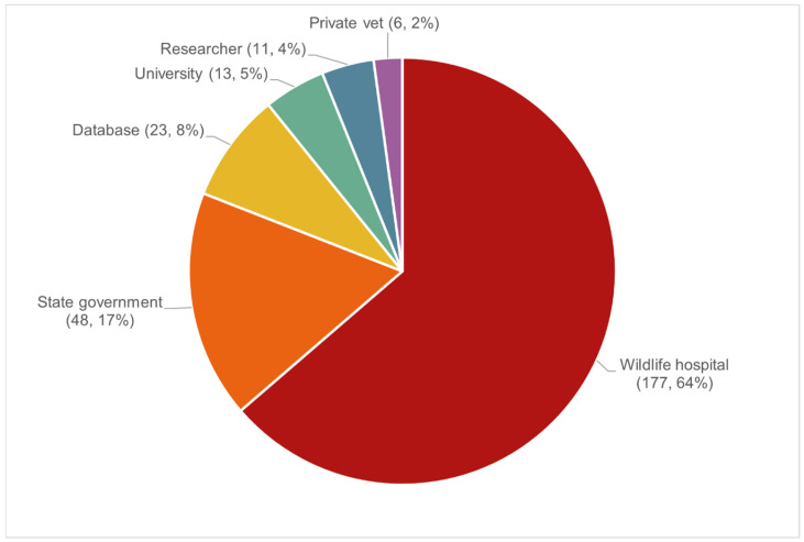 Figure 1
 Figure 2
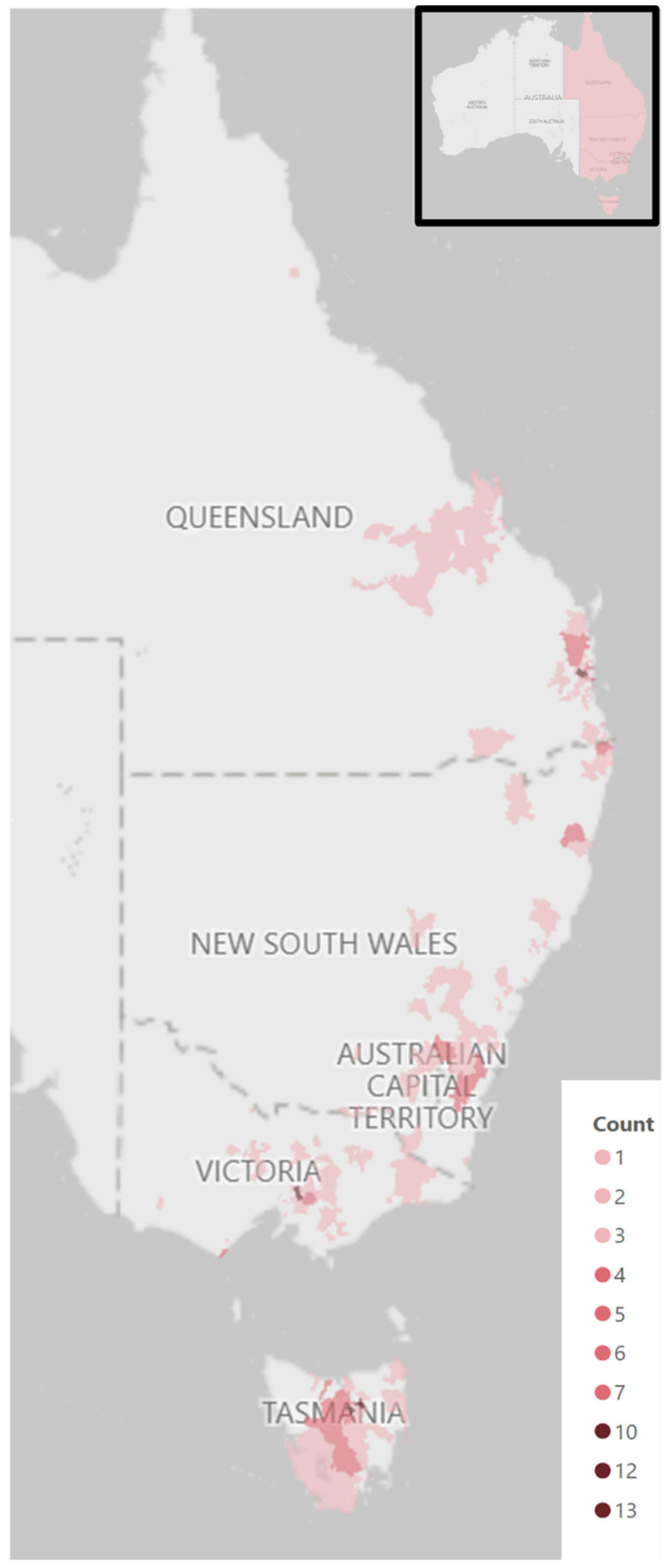 Figure 3
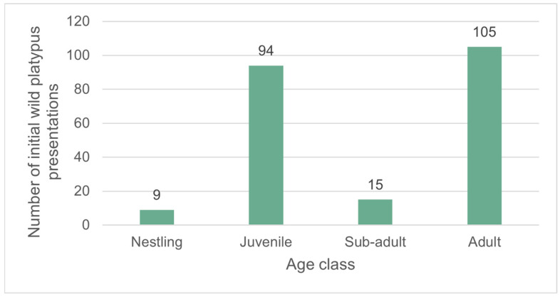 Figure 4
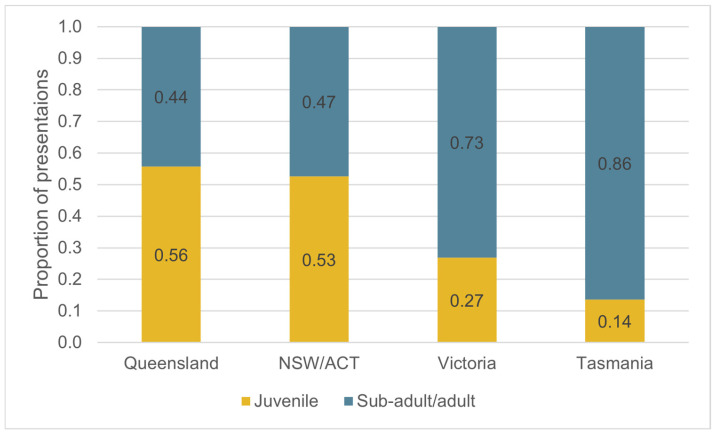 Figure 5
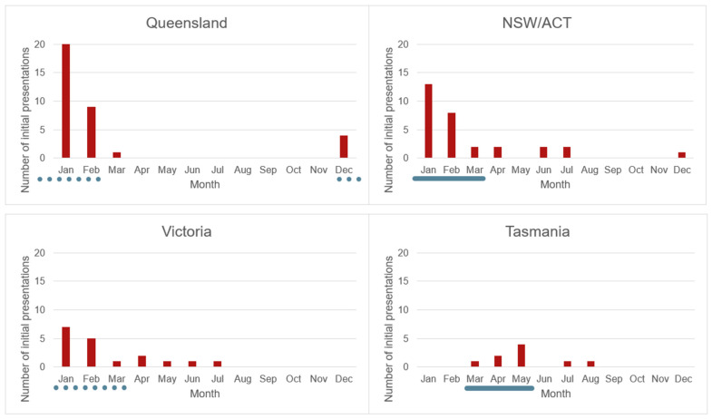 Figure 6
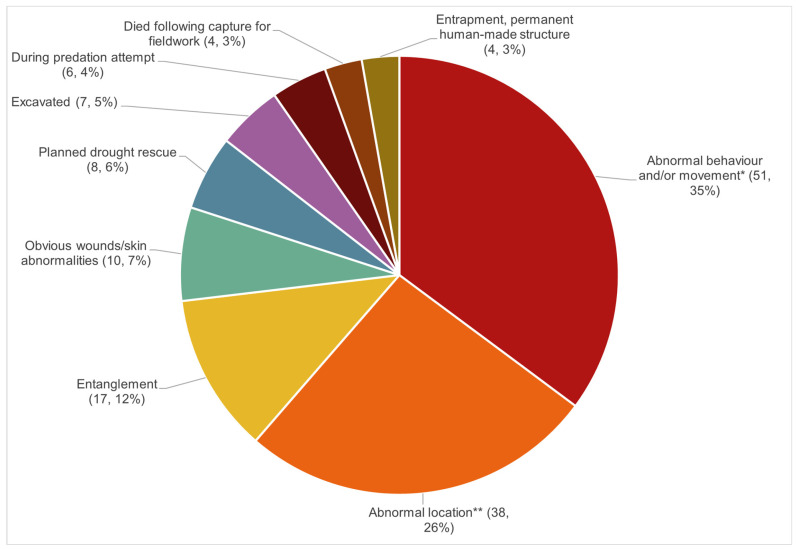 Figure 7
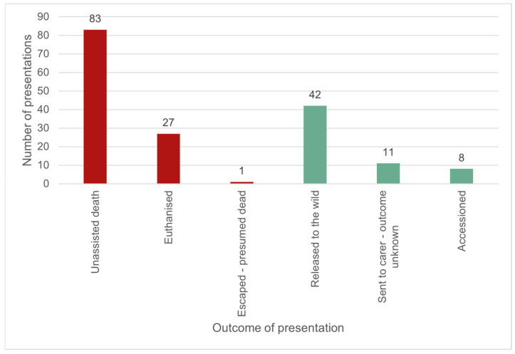 Figure 8
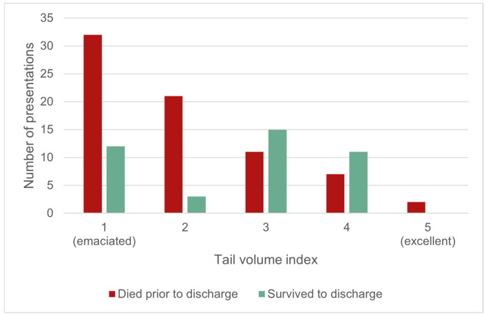 Figure 9
| History | Carcass Assessment (Continued) |
|---|---|
| Date found | Total spur length (mm) |
| Date submitted | Spur sheath length (mm) |
| Submitter details (name, mobile number, email, affiliation) | Tail volume index (1–5) (see |
| Location of carcass (address/GPS coordinates) | Bill width (mm) |
| Habitat type where found | Bill length (with and without bill shield) (mm) |
| Recent weather events | Total body length (mm) |
| Evidence of pollution at location found | Tail length (mm) |
|
| Tail width at midpoint (mm) |
| Microchip (present/absent) | Degree of decomposition (fresh; mild-moderate; advanced; mummified/skeletal) |
| Radiographs (subset of cases only) | Storage (fresh/chilled; frozen; other) |
| Weight | Gross necropsy |
| Sex | Histology (if carcass condition allowed) |
| Age | Additional testing (as indicated) |
| Variable | Values | Details of the Data Extraction Process |
|---|---|---|
| Local identification | The local identifier assigned by the submitter. | |
| Data submitter | Data submitter name (institution or individual). | |
| Data owner | The data owner name, where different to the data submitter. | |
| Submitter type | Database, private vet, researcher, state laboratory, university, wildlife hospital, zoo. | If a zoo facility had a free-ranging wildlife hospital, submitter type was classified as ‘zoo’ for zoo-housed platypus presentations and ‘wildlife hospital’ for wild platypus presentations. |
| Format | Digital—complete, digital—summary, paper. | ‘Digital—complete’: records were provided entirely in a digital format. |
| Ownership | Wild, zoo-housed. | ‘Wild’: free-living prior to presenting to veterinarians or pathologists, including those that were temporarily housed at zoos or wildlife sanctuaries whilst undergoing treatment or rehabilitation. |
| Date of presentation | Month, season, year. | Seasons: summer (December–February), autumn (March–May), winter (June–August), spring (September–November). |
| Location | Postcode, state/territory. | Postcode extrapolated from suburb. For zoo-housed platypuses, the address of the holding institution was considered the location. |
| Arrival status | Alive, found alive presented deceased, found dead. | |
| Sex | Male, female. | Sex determination in platypuses is possible on physical examination based on spur presence or absence, and morphology [ |
| Age class | Nestling, juvenile, subadult, adult ( | Age class was reclassified when either the age class was unspecified in the original record, but other supportive information was provided, or when there was a discrepancy between the descriptive information provided by the record’s author and the stated age class. Reclassification decisions were based on the following: Based on provided supporting information. For example, descriptions or images of the bill [ Individuals described as ‘orphaned’, ‘excavated from natal nest’, or similar, were classified as nestlings. Individuals described as ‘juvenile-subadult’ were classified as juveniles. Individuals were classified as juveniles if they weighed 35% less than the minimum recorded weight for an adult wild platypus of the relevant sex and geographic region *. This same criterion was also used to reclassify ‘sub-adult females’ (‘sub-adult’ is not recognised as an age class in female platypuses ( |
| Reason for presentation | Abnormal behaviour and/or movement (inc. lethargy, weakness, lameness, circling, inappetence etc.); abnormal location (inc. beaches, estuaries, roads and backyards—noting these might be close to expected platypus habitat, such as a creek); entanglement; obvious wounds/skin abnormalities; excavated; planned drought rescue; during predation attempt; entrapment in a permanent human-made structure; died following capture for fieldwork; found dead; weight loss (zoo-housed only); diarrhoea (zoo-housed only); egg—failure to hatch (zoo-housed only); fertility investigation (zoo-housed only); routine physical examination (zoo-housed only). | Values assigned based on the provided history. The presentation of ‘entrapment in a permanent, human-made structure’ was designated for platypuses found stuck in drains, water tanks, nest boxes, or similar. It did not include fishing gear, such as yabby traps. |
| Tail volume index (TVI) | 1 (emaciated)–5 (excellent) [ | Where a body condition scoring system other than TVI had been used, or the written description provided did not match the stated TVI, a TVI was assigned that matched [ |
| Outcome | Accessioned, unassisted death, found alive but died prior to presentation, euthanised, escaped—presumed dead, ongoing, procedure, released, resolved (zoo-housed only), sent to carer—lost to follow up. | The outcome was listed as ‘ongoing’ if the case was still in progress at the time the medical records were reviewed. |
| Diagnostics performed | Physical examination, haematology, biochemistry, imaging, urinalysis, faecal analysis, ante-mortem histology, post-mortem histology, microbiology, toxicology. | |
| Diagnostic confidence | High, low | Subjective assessment of the confidence in the reported diagnosis based on the information provided, such as whether diagnostic tests were performed and the reported results. |
| Morphological diagnosis, or clinical finding | Assigned by a specialist veterinary pathologist. | |
| Body system associated with morphological diagnosis/clinical finding | Generalised, integumentary, musculoskeletal, nervous, cardiovascular, respiratory, digestive, urinary, endocrine, sensory, reproductive, haemolymphatic. | Each morphological diagnosis/clinical finding was exclusively assigned to one system, however an individual could have multiple systems impacted simultaneously by the same aetiological process (e.g., a single fishing gear entanglement presentation could result in morphological diagnoses involving the generalised, respiratory and integumentary body systems). Characterisation was performed by a specialist veterinary pathologist. |
| Aetiological category of morphological diagnosis/clinical finding. (Subcategories in brackets). | Congenital, degenerative, environmental, iatrogenic, indeterminate, infectious (bacterial, fungal, metazoan parasite, protozoan parasite, viral, unidentified), inflammatory, metabolic, neoplastic, egg death (undiagnosed), nutritional, reproductive, toxic, trauma (entrapment in a permanent human-made structure, fishing gear—hook or line, fishing gear—trap or net, predation, rubbish (non-fishing), self or conspecific, vehicular), undetermined. | Categorisation was performed by a specialist veterinary pathologist, based on the categories suggested by [ |
- —WIRES Research Grants Program
- —Taronga Conservation Society Australia
- —Murdoch University
- —Australian Government Research Training Program (RTP)
- —WIRES Research Grants Program
Peer Reviews
No public reviews on file for this paper yet. If you reviewed it on a platform where reviews are public (OpenReview, ICLR, NeurIPS, ICML), you can paste yours below so the community can read it here.
Videos
No videos yet. Explain this paper in a talk, walkthrough, or lecture? Add one.
Taxonomy
TopicsEvolution and Paleontology Studies · Bat Biology and Ecology Studies · Ecology and biodiversity studies
1. Introduction
Platypuses (Ornithorhynchus anatinus) are a unique and iconic species, inhabiting freshwater ecosystems of eastern Australia, including the island state of Tasmania [1]. Platypuses are listed as near threatened by the IUCN, with a decreasing population trend [2]. This is reflective of their vulnerability to anthropogenic pressures, including changing land use (especially agriculturalisation and urbanisation), waterway regulation (including the construction of dams and weirs), entanglement and entrapment in rubbish and fishing gear, predation by introduced species (particularly dogs and foxes), and climate change-exacerbated natural disasters (such as drought, flooding, and fire) [1,2]. Platypuses are considered near extinct in South Australia, the western-most extent of the species’ distribution [1].
Platypuses are a cryptic species and inherently challenging to study, being semi-nocturnal, semi-aquatic, and semi-fossorial [1]. They are seasonal breeders, with the timing of reproduction earlier in the north of their range and later in the south. In New South Wales (NSW; approximately midway in their latitudinal distribution), courtship begins around August, followed by the female building the nest, and laying and incubating eggs. She then nurses the nestlings from hatching until their emergence from the nest at about four months of age, between late January and early March. In Tasmania (the most southerly extent of the platypuses’ range), emergence occurs two to three months later (late March to early May, i.e., autumn) [1,3,4]. At the time of writing, platypuses are held in zoos or sanctuaries at low numbers by nine institutions in Australia, and one in the United States of America [3]. To date, less than ten zoo-housed females have reproduced successfully.
Understanding platypus health and disease is important for: establishing a baseline of health risks; reducing risks associated with conservation actions such as translocations [5]; and improving welfare and outcomes for both rescued platypuses and those held in zoos or sanctuaries. However, understanding platypus health and disease is challenging due to difficulties in observing and sampling the species. This is further compounded by the relatively low numbers of individuals held in zoos or sanctuaries, and the significant limitations in extrapolating from similar species as a result of their unique biology; the closest living relative to platypuses are the echidnas (Family: Tachyglossidae), which are markedly different in their morphology, physiology, and ecology [6]. To date, peer-reviewed literature on platypus health and disease (excluding physiology studies) have included the following forms: case reports [7]; limited mortality case series (n = 20, [8]; n = 25, [9]); targeted surveillance for one or more pathogens or hazards in wild populations, coupled with health metrics [10,11,12,13]; and a limited number of retrospective reviews [14,15,16]. These studies have made significant advances in understanding platypus pathology; however, knowledge relating to platypus health remains poor when compared to other iconic Australian species, such as koalas [17], and rudimentary compared to domesticated species.
Retrospective medical record reviews are descriptive, observational studies that utilise data originally collected for purposes other than research, such as health care records [18,19]. The benefits of retrospective reviews include their low cost, lower resource requirements compared to other methodologies (for example, cohort studies), and the ability to study rare diseases or exposures, or uncommonly encountered species. This methodology is therefore popular in studies of human and animal health, including for both zoo-housed and free-ranging wildlife [18,19,20,21,22].
To date, there have been no systematic reviews of the medical records of zoo-housed platypuses, with the three previous retrospective reviews focusing on wild platypus morbidity and mortality records [14,15,16]. These three reviews have overlapping data sets and a limited geographic scope, focusing on Victoria either exclusively [14,15] or predominantly (78.0% of cases reviewed (305/391) in [16]). Only one of these reviews [14] included a veterinarian as an author, and none had a pathologist involved in authorship: the authors of [22] consider the involvement of veterinarians and pathologists essential for wildlife morbidity and mortality studies. [14] reviewed the veterinary medical records of 38 wild platypuses presenting to Healesville Sanctuary’s Australian Wildlife Health Centre (2000–2014). In comparison, the methodology used by [15,16] relied on “ad hoc” reporting of platypus mortalities from a range of sources (including members of the public, natural resource managers, biologists, and veterinarians), with or without supporting information (such as photos or the provision of carcasses). The latter study [16] included 23 mortality records from platypuses that presented to Healesville Sanctuary between 2005 and 2024, thus suggesting overlap with the records used in [14], although this study is not referenced. Despite the statement in [16] that they “develop(ed) a consolidated set of records to characterise the entire range of factors known to have contributed directly to platypus mortality in recent decades”, it is probable that there are additional morbidity and mortality causes beyond those reported in these three studies. Specifically, there is a risk of underestimating both the impact and diversity of disease on platypuses: between the three review studies performed to date, disease was only identified in three platypuses.
The aim of this project was to use the retrospective review methodology to conduct a distribution-wide study exploring morbidity and mortality trends for both zoo-housed and wild platypuses. This included trends relating to demography (age, sex), geographic location, season, reason for presentation, outcome of presentation, and clinical and pathological findings.
2. Materials and Methods
A retrospective medical record review was performed. The review had three main steps: record acquisition; record review and data extraction; and data analysis. The STROBE guidelines [23] are a checklist of recommendations designed to guide the reporting of observational research, and these were incorporated into the methodology where possible. The guidelines produced by [22] for performing retrospective morbidity and/or mortality studies in zoo-housed wildlife were also incorporated where possible.
2.1. Record Acquisition
The individuals and institutions considered most likely to hold platypus medical records were identified as those meeting one or more of the criteria described in Table 1 and were contacted by email. The platypus’s range states and territories were considered to be Queensland, New South Wales (NSW), the Australian Capital Territory (ACT), Victoria, Tasmania, and South Australia [1].
Organisations and individuals were initially contacted between April 2021 and January 2023. This extended period reflected additional points of contact being identified through both the record review process and through recommendations made by record holders. Individuals or institutions with relevant records who consented to participate shared records with the study’s first author in the format of electronic files or original paper documents. In some instances, travel to the institution by the first author was required to manually locate and copy paper records. Records were received for review between August 2021 and September 2024. This prolonged timeframe of record receipt was a product of multiple factors, including: the lag time between the request for records being made and the records being provided for review; individuals and institutions being contacted at different times; and some institutions continuing to submit additional records as cases presented, with these continuing to be included in the data set.
During the record acquisition period, a NSW-wide callout (through social media and by directly emailing key contacts) asked people to notify the first author of any dead wild platypuses found. Where possible, any platypuses that were found deceased were transported to Taronga Zoo, Sydney, where a gross necropsy and sample collection were performed by a wildlife veterinarian or veterinary pathologist. The information collected for each carcass is listed in Table 2, and these cases were included in the study’s data set. Sampling was performed under scientific licence (SL102578) issued by the Department of Planning, Industry and Environment, NSW Government. Ethics approval was not required for this study.
2.2. Record Review and Data Extraction
The record inclusion criteria were the medical records of all wild or zoo-housed platypuses in Australia. Medical records were defined as any clinical or pathology record produced by a clinical veterinarian or veterinary pathologist. Records were excluded based on the following criteria: healthy individuals captured during fieldwork or undergoing routine health examinations with no abnormalities detected; procedures performed on otherwise healthy individuals; autolysed carcasses or diagnostic testing (including blood tests or bacterial cultures) without accompanying information; anecdotal reports; records that were written without the direct involvement of a veterinarian or veterinary pathologist; or records where it was unclear if the platypus was wild or zoo-housed. Published case reports were only included when they accompanied the original medical record.
A standardised data extraction protocol and instrument were developed using Microsoft Excel (Microsoft 365 MSO, Redmond, WA, USA; Version 2301 Build 16.0.16026.20002). Record review and data extraction were performed by the first author. The study’s unit of observation was an individual platypus’s presentation to a clinician or pathologist (referred to as ‘presentation’ henceforth): each row in the data extraction instrument represented a single presentation by an individual platypus. For wild platypuses, presentations were considered separate if additional pathological processes developed whilst in care. For zoo-housed platypuses, presentations were considered separate if they met one or more of the following criteria: there were more than 12 months between the presentations; resolution of the problem was explicitly stated prior to its recurrence; two presentations occurred within 12 months but were considered separate aetiologies (e.g., sneezing noted for one week, and then an ulcer on the foot developed six months later). Where a single event resulted in multiple platypus presentations (e.g., multiple animals in a single abandoned trap), the individual platypuses involved were treated as a separate presentation. Each presentation was assigned a unique study identifier, composed of a unique whole number assigned to the individual platypus, and two decimal places reflecting the presentation number for that platypus.
The information extracted from records for each presentation is summarised in Table 3, including details of the data extraction procedures used for each variable (where required).
2.3. Data Analyses
Data were analysed in Microsoft Excel or SPSS Statistics (IBM, Armonk, NY, USA; Version 29.0.0.0 (241)). Most study variables were categorical, thus limiting statistical testing to non-parametric tests. Data analyses were primarily performed using descriptives, such as frequencies and chi-square analyses, with subsequent data visualisation. A p-value of <0.05 was considered statistically significant. Graphs and tables were produced in Microsoft Excel. Maps were created using Power BI (Microsoft Corporation, Redmond, WA, USA; Version 13.0.27228.39). Data were considered missing if information for a particular variable was not recorded on the medical record. Presentations with missing data were excluded from analyses where the missing data were relevant, unless otherwise stated (this resulted in a denominator that varied between analyses).
For wild platypuses, some analyses were performed only including the initial presentation, which is explicitly stated in the results section. Where specified, data from the ACT and NSW were combined, because the ACT is a small enclave within the much larger NSW. Where this occurred, the location was termed ‘NSW/ACT’. For all age-class analyses, the subadult age class (which is only recognised for male platypuses [4]; Table 4) was combined with the adult age class. This was done to improve consistency and comparability across sexes.
Where the objective of data analysis was to determine variables that could influence wild platypus presentations, only non-random reasons for presentation were included in analysis. The two presentation types that were considered random were ‘died following capture for fieldwork’ and ‘planned drought rescue’. It is specified in the results section when this occurred.
Only diagnoses with a ‘high’ diagnostic confidence (see Table 3 for explanation) were included in analyses of diagnoses. This study did not distinguish between incidental and clinically significant diagnoses due to challenges in interpreting clinical significance from retrospectively reviewed records. All diagnoses were reviewed by a Diplomate of the American College of Veterinary Pathologists (co-author CS).
3. Results
3.1. Medical Record Access
A total of 53 organisations or individuals were contacted regarding access to platypus medical records from wild or zoo-housed platypuses: Australia-wide = 1; NSW = 16; Queensland = 14; Tasmania = 9; South Australia = 6; Victoria = 6; ACT = 1. Of those contacted, 37.7% (20/53) had records that met the inclusion criteria and were shared in full, 47.2% (25/53) had no relevant records or provided records that did not meet the inclusion criteria. The remaining individuals or institutions (n = 8) either did not reply (n = 3), only partially shared records (n = 1), or indicated they had relevant records but did not share them (n = 4).
Records were provided for analyses in a complete digital format for 68.4% (262/383) of presentations. Almost one-fifth of presentations were provided in a paper format (18.0%, 69/383) across the entire study period (1991–2024), reducing only slightly over the most recent five-year period (2020–2024) covered by the study (13.2%; 12/90). Abbreviated digital summaries based on paper records that were no longer available were provided for 13.6% (52/383) of presentations.
3.2. Wild Platyuses
3.2.1. Records
Medical records were received from 15 institutions, representing 6 submitter types (Figure 1).
The records of 278 individual wild platypuses were reviewed, with a total of 293 presentations. Eight platypuses (2.9% (8/278) of individuals) presented more than once: six presented twice, one presented three times, and one presented eight times. Records spanned a 34-year period from 1991 to 2024. Figure 2 shows the number of wild platypus initial presentations by year.
3.2.2. Location
There were no records of wild platypus presentations in South Australia. Combining NSW/ACT, the numbers of initial wild platypus presentations in the states/territories with presentations were approximately equal, with a mean of 68 (Queensland = 63; NSW/ACT = 70; Victoria = 73; Tasmania = 66). There were nine initial presentations in the ACT. These statistics were repeated with the inclusion of only non-random reasons for presentation (i.e., drought rescue and fieldwork deaths were excluded, n = 12). With this data subset, the number of wild platypus initial presentations remained approximately equal, with an average of 65 cases per state/territory. The number of cases in the ACT decreased to two. Figure 3 shows the number of individual platypus initial presentations by postcode. The number of presentations by postcode ranged from 0 to 13.
3.2.3. Age
Figure 4 displays the total number of wild platypus initial presentations by the four recognised platypus age classes: nestling, juvenile, sub-adult, and adult.
The numbers of non-random initial presentations were compared between states/territories by age class. There was a significant difference in the proportion of juveniles and the sub-adult/adult age class presenting between the states/territories: χ^2^(3, 1; n = 251) = 33.8, p < 0.001 (Figure 5) (there were insufficient numbers of nestlings to include in the analysis). The northern states (Queensland and NSW) had a slight majority of juvenile presentations (Queensland: 55.7% juveniles, 34/61; NSW/ACT: 52.6% juveniles, 30/57). In contrast, the southern states (Victoria and Tasmania) had significantly fewer juvenile presentations than sub-adult/adult (Victoria: 26.9% juveniles; 18/67; Tasmania: 13.6% juveniles, 9/66).
3.2.4. Season
Including only non-random and initial reasons for presentation, the number of presentations varied by season. Summer was the most common season for wild platypuses to present (41.0% of presentations, 109/266) and winter the least common season (16.2% of presentations, 43/266). There was a significant relationship between season and the age class presenting (juvenile and sub-adult/adult age classes only; there were insufficient nestling presentations to include in the analysis): χ^2^(3, 1; n = 266) = 65.8, p < 0.001. The seasonality in presentations was driven by juvenile presentations, with 73.4% (69/94) of juvenile presentations occurring in summer. However, this varied between states (Figure 6), with no summer presentations of juveniles recorded in Tasmania: most juvenile Tasmanian presentations (7/9, 77.8%) occurred in autumn. The number of adult presentations was relatively consistent between seasons: summer 22.1% (36/163), autumn 27.0% (44/163), winter 20.9% (34/163), spring 30.1% (49/163).
3.2.5. Sex
Including only non-random initial reasons for presentation where the sex was known, male platypuses were more likely to present than females overall: males represented 60.2% of presentations (133/221). This difference was greater in juveniles (males: 64.8% of presentations (57/88)) than in sub-adults/adults (males: 57.1% (76/133) of sub-adults/adults), however this difference was not statistically significant (χ^2^(1,1; n = 221) = 1.3, p = 0.26).
There was a significant relationship between sex and the state/territory of presentation: χ^2^(3, 1; n = 219) = 8.4, p = 0.04). This was greatest in NSW/ACT, where males comprised 73.9% (34/46) of wild platypus presentations. Queensland was the only state/territory that did not have a male bias in presentations (48.1% males, 25/52). Sex could not be determined from the history in a significant number of cases: 16.6% (46/278).
3.2.6. Reason for Presentation
Platypuses were found deceased in 39.3% (94/239) of wild platypus initial presentations. Of those that were found alive, and where sufficient history was available to make a categorisation (n = 145), the reason for initial presentation is shown in Figure 7.
3.2.7. Diagnostics Performed
Of 293 wild platypus presentations, 72.7% (213/293) had a morphological diagnosis or clinical finding for which there was a high level of confidence. Table 5 presents the diagnostic tests that were performed for these high diagnostic confidence presentations. The same presentation could have multiple diagnostic tests performed.
3.2.8. Morphological Diagnoses/Clinical Findings
Table 6 displays the number of morphological diagnoses/clinical findings by body system and age class for the presentations with a high level of diagnostic confidence (n = 213). The five morphological diagnoses with the highest ratio of juvenile presentations to sub-adult/adult presentations were: anaemia (31.6%), heavy tick burden (subjective) (22.3%), enteritis (7.4%), splenic histiocytosis (7.4%), and emaciation (6.0%).
For the presentations with a high level of diagnostic confidence, diagnoses were also classified by aetiology: Table 7 displays the aetiological category and subcategory by age class and body system (n = 213). Collectively, the directly anthropogenic aetiologies (trauma from entrapment in a permanent human-made structure, trauma from fishing gear—hook or line, trauma from fishing gear—trap or net, trauma from rubbish (non-fishing), and trauma from vehicles) occurred in 44.1% all wild platypus presentations (94/213).
3.2.9. Outcome
The outcomes for the 172 wild platypuses (non-random reasons for presentation only) that were presented to veterinarians alive, and for which the outcome was known, are summarised in Figure 8. Mortality (due to euthanasia, unassisted death, or escaped—presumed dead) was the outcome for 64.5% (111/172) of cases. The remaining 35.5% (61/172) of cases survived to discharge (defined as either being released to the wild, discharged to a carer (outcome unknown), or being accessioned into permanent zoo housing).
There was a significant association between Tail Volume Index (TVI) and survival to discharge (χ^2^(4, 1; n = 114) = 18.6, p < 0.001; Figure 9). Animals that had an average to excellent TVI (TVI 3–5) had a survival rate of 56.5% (26/46); survival to discharge reduced to 22.1% (15/68) for animals with a poor or emaciated TVI (TVI 1–2). There was no statistically significant effect on survival to discharge between age class (nestling, juvenile, sub-adult/adult) (χ^2^(2, 1; n = 172) = 2.6, p = 0.28), or sex (χ^2^(1, 1; n = 150) = 0.2, p = 0.67).
3.3. Zoo-Housed Platypuses
The records of 40 individual zoo-housed platypuses were reviewed. Records spanned a 30-year period (1994–2023) and represented platypuses housed at nine institutions in four states (Queensland, NSW, Victoria, and Tasmania); there were no records that met the inclusion criteria received from South Australia or the ACT. There was a total of 90 presentations, with each platypus presenting between 1 and 7 times. The number of zoo-housed platypus presentations for a range of variables is displayed in Table 8.
The reasons zoo-housed platypuses presented are described in Table 9. Most zoo-housed platypuses (57.5%; 23/40) had at least one presentation for wounds or skin abnormalities.
Of the 90 presentations, 67.8% (61/90) had a diagnosis for which there was a high level of confidence. In Table 10, the diagnostic tests that were performed for these high diagnostic confidence cases are summarised.
Sixty-one presentations had morphological diagnoses/clinical findings with a high level of diagnostic confidence. These are displayed by body system in Table 11.
For the presentations with a high level of diagnostic confidence (n = 61), diagnoses were also classified by aetiology (Table 12).
Of the zoo-housed presentations for which an outcome could be determined (n = 88), resolution occurred in most cases (64.8%, 57/88); unassisted death occurred in 13.6% of cases (12/88); euthanasia occurred in 3.4% of cases (3/88); and 10.2% of cases were ongoing at the time the record was reviewed (9/88).
4. Discussion
This study revealed key trends in the reasons for the presentation of wild and zoo-housed platypuses to veterinarians and pathologists. For wild platypuses, amongst the most significant of these findings was a difference in the proportion of juvenile and sub-adult/adult presentations between states/territories, the timing of annual peaks in juvenile presentations, and the identification of previously unpublished causes of pathology in platypuses. For zoo-housed platypuses, the frequency with which presentations were attributable to wounds or skin abnormalities was a significant finding. These observations offer insights into wild platypus biology and highlight an important focus for zoo-housed platypus management, as described below.
An unexpected finding of the study was the significant difference in the proportion of wild juvenile and sub-adult/adult presentations between states (Figure 5). There was an apparent association between the proportion of juvenile presentations and latitude, with juveniles representing a minority of presentations in the southern states of Victoria and Tasmania, but a slight majority of all presentations in the northern states of Queensland and NSW. This is despite the juvenile age class likely only representing 6–16% of the wild population age structure: platypuses are classified as juveniles for ~8 months (from nest emergence—at ~4 months—to 12 months of age) [4], and the average wild platypus lifespan is 6–15 years [1]. Only in Tasmania was the ratio of juvenile to sub-adult/adult presentations (13.6%) proportional to the likely number of juveniles in the overall population.
The disproportionate number of juvenile presentations in the northern states can be interpreted alongside another of this study’s observations: that juvenile presentations were tightly clustered around the expected time of nest emergence in their respective states (Figure 6). Juvenile nest emergence occurs in late January to early March (summer to early autumn) on the mainland, and approximately two months later (late March to early May, i.e., autumn) in Tasmania [1,4]. Emergence from the nest corresponds with weaning, and the limited available evidence suggests weaning is abrupt in platypuses [32] and thus is likely to be a significant stressor for juveniles.
It is likely that recently weaned juvenile platypuses have less resilience than adults, and thus if they experience stressors beyond the baseline challenge of weaning they may rapidly fail to cope. One hypothesis for the latitudinal difference in the proportion of wild juvenile and sub-adult/adult presentations is that platypuses in the northern states are experiencing greater stressors during this vulnerable weaning period than those in the south. These stressors could include: reduced food availability (e.g., due to lower availability of suitable macroinvertebrate prey species); increased competition with other aquatic species (e.g., variations in fish numbers and/or species across the range); increased extreme weather events (such as flooding) that occur more frequently in summer in northern Australia; or other environmental stressors, such as high ambient temperatures. Juveniles may be physiologically less heat tolerant, or less able to employ behavioural thermoregulatory mechanisms. For example, they may be less capable than adults of finding or competing for suitable burrows [33,34], or competition with adults may force them to increase overland travel or to forage during warmer parts of the day. If increased juvenile presentations in the north is reflective of reduced juvenile survival in their northern range, it is possible this could impact population recruitment and thus have implications for platypus conservation.
The observation that the peak in juvenile presentations corresponds to the timing of nest emergence and weaning refutes a previously proposed hypothesis that juvenile platypuses most commonly present at the time of dispersal [9,26,35]. It was suggested that platypuses frequently present to clinics as dispersing juveniles in poor body condition due to insufficient food intake. The dispersal behaviour of platypuses is a challenging area to research and remains poorly understood [32]; however, available evidence suggests it does not occur immediately after juveniles leave the nest [32,36,37]. This delayed dispersal strategy potentially benefits weaning juveniles by ensuring they remain in an environment with known access to resources during the vulnerable post-emergence period [32]. Thus, the tight association observed in this study between the peak of juvenile presentations and expected time of nest emergence supports the theory that juvenile presentations are occurring at the time of weaning, rather than dispersal.
Consistent with previous studies [14,15,16], anthropogenic aetiologies were common in wild platypuses, occurring in almost half of all wild platypus presentations (Table 7). Although this study did not distinguish between the species responsible for predation events, it is likely that a significant proportion are attributable to non-native species (particularly domestic dogs and foxes), and thus many of these predation events may also by extension be considered anthropogenic in origin. However, compared to previous morbidity and mortality studies in platypuses, many additional disease processes were identified. This finding is likely to be attributable to the higher sample size (relative to [14]), and the higher level of diagnostic investigation that had been performed by clinical veterinarians and veterinary pathologists for many of the presentations (relative to the cases reported in [15,16]). Across wild and zoo-housed platypuses, previously unreported diagnoses that were identified through this retrospective review process include round cell neoplasia, neural angiostrongyliasis, and iron deficiency anaemia.
A concerning finding in zoo-housed platypuses was the frequency of wounds or skin abnormalities, with 57.5% of individuals having at least one presentation for this reason, and 70.5% of presentations diagnosed with an aetiology related to the integumentary system (Table 12). Of those skin abnormalities for which an underlying aetiology could be determined, the majority were fungal infections. There are sporadic reports of skin lesions in zoo-housed platypuses in the literature [26,38], but the frequency of presentations in this data set suggests that underlying husbandry practices should be investigated as these may be predisposing to secondary infections. Contributing factors that should be considered include nutritional deficiencies in the zoo-housed platypus diet, ultraviolet lighting requirements (zoo-housed platypuses are frequently held indoors without access to sunlight), abrasive surfaces in provided habitats, and water quality.
Retrospective reviews are susceptible to errors and biases, including those resulting from errors during the process of extracting data from clinical records, missing data, lack of homogeneity due to variability in the quality and quantity of original records, and loss to follow up [19,21,23]. All these limitations were present in this study, but attempts were made to minimise the impact on results and conclusions by applying the STROBE guidelines [23] and the recommendations made by [22], where possible. As described in the methodology, there were intentional adaptations of the recommendations made by [22]. Most significantly, in the present study, a single aetiological process could be classified as impacting on multiple body systems in one presentation. This was performed to provide a more comprehensive insight into the pathogenesis and impact of disease processes, thus potentially assisting clinicians with targeted decision making and thereby improving outcomes.
A limitation of the study is that there is expected to be significant variation in the level of clinician knowledge, skills and experience with platypuses, including with the accepted systems for assigning age, sex, and body condition. The level of record accuracy is challenging to assess in the review process but could result in some data inaccuracies. It’s possible that the reason for the slight male bias in wild platypus presentations for all states/territories (other than Queensland) is due to a lack of clinician confidence with sexing platypuses. The sex of the platypus was missing from the reviewed records in 16.6% of cases, and it is possible that clinicians are more confident with classifying a platypus with spurs as male, but more reluctant to assign sex to a spurless (female) platypus. Other data inaccuracies could result because some morphological diagnoses or clinical findings (such as fractures, emaciation, or entanglement and entrapment in fishing gear) can be made more readily than others, and this could falsely elevate the apparent proportion of these relative to more subtle processes. Additionally, the sub-category of ‘trauma—unknown/other’, likely includes some cases from other sub-categories (such as ‘trauma—vehicular’).
In addition to the above caveats, some results from this study should be interpreted with caution, in particular, survival to discharge for wild platypuses, and the number of presentations by year for both wild and zoo-housed platypuses. The number of presentations by year is complicated by inconsistent sampling effort: institutions were operating over different time periods; older records were less likely to be accessible; and the prolonged, three-year record submission period meant that records for presentations that occurred between August 2021 and September 2024 may not have been submitted by all institutions. Regarding survival to discharge, the gold standard for determining survival is post-release monitoring, but, to date, there is only a single known case of post-release monitoring occurring for a rehabilitated platypus [33]. Using proxies for ‘true’ survival (such as ‘survival to discharge’) is likely to underestimate mortality. Whilst it was not quantified as part of the review, qualitatively, many of the platypuses that were discharged to carers were in states of considerable compromise and it is considered unlikely all survived. Similarly, several platypuses were released a short time after presenting despite evidence of significant compromise on physical examination and it is probable at least some died following release. Thus, the true survival rate for platypuses presented alive to veterinarians is likely to be lower than the survival to discharge value of 35.5% calculated in this study.
A strong recommendation from this study is that medical records should be both permanent and digital, and include fundamental data, such as age class, sex, and outcome. A significant number of medical records reviewed for this study were in a paper format, even in the most recent five-year period covered by the study (2020–2024). This substantially increased the workload involved in performing the review, necessitating interstate travel to retrieve records, and requiring the deciphering of poor-quality handwriting. Furthermore, fundamental data (such as sex, age, and TVI) were not recorded for a considerable number of cases. Digitalising records and standardising the inclusion of fundamental information will reduce the barriers to performing effective retrospective reviews and maximise the insights gained through the review process. Doing so will also allow the application of emerging retrospective record review techniques, such as text-mining, which have the potential to revolutionise the process [39].
5. Conclusions
The retrospective review provides a valuable methodology for improving our understanding of the health and disease of wildlife species that are relatively infrequently encountered by veterinarians and pathologists. Its application to platypus medical records has identified unexpected trends and priorities for future research, including establishing why the juvenile age class presents with disproportionate frequency in northern states, and why zoo-housed platypuses so frequently present with skin lesions. Directing future research towards these topics will ultimately assist in maximising conservation outcomes for wild platypuses and improving the welfare of zoo-housed platypuses.
The reference list from the paper itself. Each links out to its DOI / PubMed record.
- 1Bino G. Kingsford R.T. Archer M. Connolly J.H. Day J. Dias K. Goldney D. Gongora J. Grant T. Griffiths J. The Platypus: Evolutionary History, Biology, and an Uncertain Future J. Mammal.201910030832710.1093/jmammal/gyz 05831043761 PMC 6479513 · doi ↗ · pubmed ↗
- 2Woinarski J. Burbidge A.A. Ornithorhynchus anatinus. The IUCN Red List of Threatened Species 2016 Available online: https://www.iucnredlist.org/species/40488/21964009(accessed on 20 May 2021)
- 3Jackson S. Thomas J.L. Serena M. Temple-Smith P. Platypus Australian Mammals: Biology and Captive Management CSIRO Publishing Melbourne, Australia 20254792
- 4Grant T. Serena M. Williams G. Temple-Smith P. Age Determination in the Platypus (Ornithorhynchus anatinus) Using Spur Sheath and Spur Developmental Stages: A Review Aust. Mammal.202446 AM 2402010.1071/AM 24020 · doi ↗
- 5Whinfield J. Warren K. Vogelnest L. Vaughan-Higgins R. Applying a Modified Streamlined Disease Risk Analysis Framework to a Platypus Conservation Translocation, with Special Consideration for the Conservation of Ecto- and Endoparasites Int. J. Parasitol. Parasites Wildl.20242410094810.1016/j.ijppaw.2024.10094838966858 PMC 11222941 · doi ↗ · pubmed ↗
- 6Ryser-Degiorgis M.-P. Wildlife Health Investigations: Needs, Challenges and Recommendations BMC Vet. Res.2013922310.1186/1746-6148-9-22324188616 PMC 4228302 · doi ↗ · pubmed ↗
- 7Kessell A.E. Boulton J.G. Dutton G.J. Woodgate R. Shamsi S. Peters A. Connolly J.H. Haemolytic Anaemia Associated with Theileria sp. in an Orphaned Platypus Aust. Vet. J.20149244344910.1111/avj.1222525123825 · doi ↗ · pubmed ↗
- 8Mc Coll K.A. Pathology in Captive Platypus (Ornithorhynchus anatinus) in Victoria, Australia J. Wildl. Dis.19831911812210.7589/0090-3558-19.2.1186887433 · doi ↗ · pubmed ↗