Enhanced visualization of flat gastric dysplasia by chromoendoscopy using an acetic acid-indigocarmine mixture
Masaya Iwamuro, Katsunori Matsueda, Motoyuki Otsuka

Abstract
Genes, proteins, chemicals, diseases, species, mutations and cell lines named across the full text — each resolved to its canonical identifier and authoritative record.
Click any figure to enlarge with its caption.
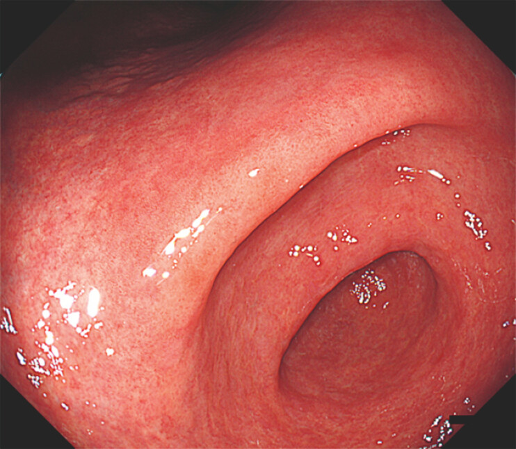 Fig. 1
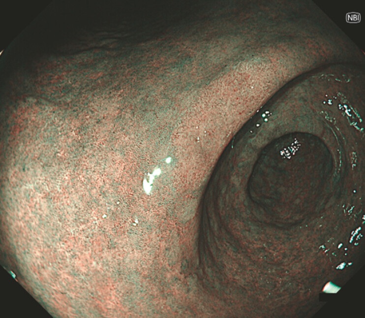 Fig. 2
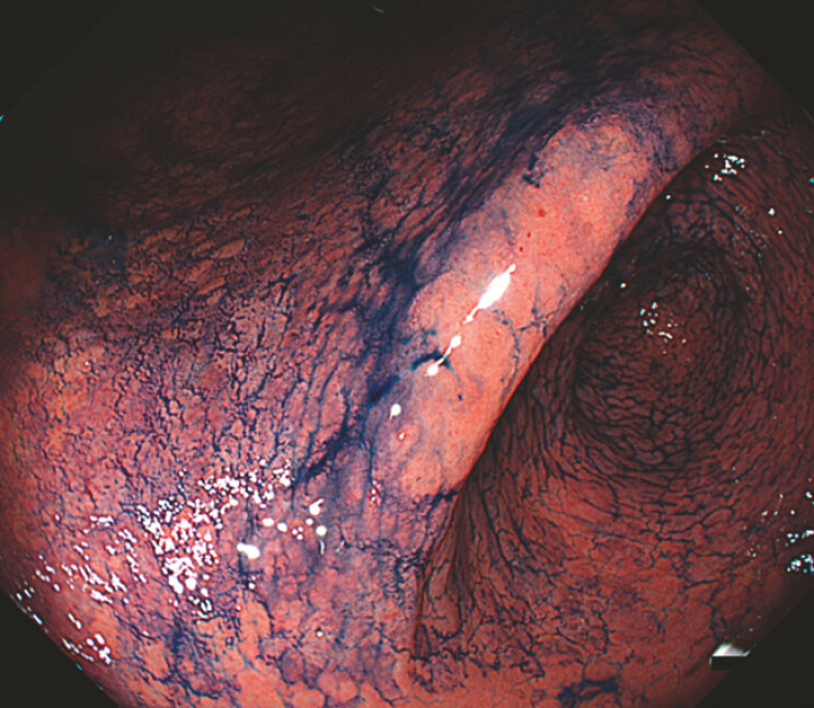 Fig. 3
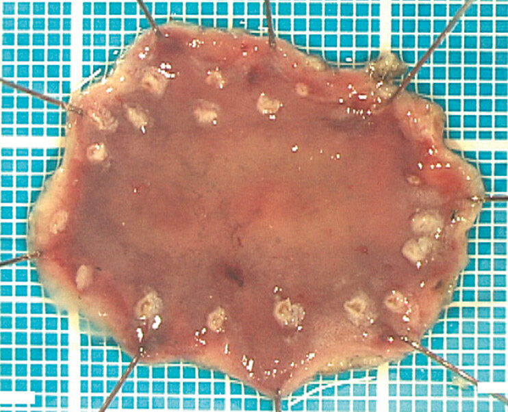 Fig. 4
Peer Reviews
No public reviews on file for this paper yet. If you reviewed it on a platform where reviews are public (OpenReview, ICLR, NeurIPS, ICML), you can paste yours below so the community can read it here.
Videos
No videos yet. Explain this paper in a talk, walkthrough, or lecture? Add one.
Taxonomy
TopicsGastrointestinal motility and disorders · Esophageal Cancer Research and Treatment · Helicobacter pylori-related gastroenterology studies
A 76-year-old man with a history of diabetes mellitus, hypertension, hyperlipidemia, and prostate cancer underwent annual surveillance esophagogastroduodenoscopy after successful Helicobacter pylori eradication. During routine endoscopy, a subtle lesion was suspected at the lesser curvature of the gastric angle, and the patient was referred for further evaluation.
Esophagogastroduodenoscopy revealed a flat, approximately 20-mm dysplastic lesion. Under white light imaging, the lesion showed only a faint whitish discolouration without surface irregularities, making recognition challenging ( Video 1 ; Fig. 1 ). Narrowband imaging revealed a brownish area ( Fig. 2 ), and magnifying observations demonstrated a clear demarcation line with irregular microvascular and microsurface patterns consistent with gastric dysplasia. To improve lesion detection and margin delineation, an acetic acid–indigocarmine mixture (AIM) was sprayed ( Fig. 3 ). The mixture consisted of 15 mL of distilled water containing dimethicone, 15 mL of 1.5% acetic acid solution, and 10 mL of indigocarmine. The surrounding mucosa exhibited acetowhitening with diffuse blue indigocarmine dye pooling on the mucosal surface, whereas the dysplastic epithelium repelled the dye and appeared as a distinct pink area, sharply contrasting with the background mucosa.
White-light imaging demonstrated a subtle flat lesion with a minimal colour change at the lesser curvature of the gastric angle, followed by narrow-band imaging and acetic acid–indigocarmine mixture chromoendoscopy, which clearly delineated the lesion margins.Video 1
White-light esophagogastroduodenoscopy showing a flat lesion at the lesser curvature at the gastric angle. The lesion demonstrates only a faint whitish discolouration without surface irregularity, making recognition challenging under white-light imaging.
Narrow-band imaging reveals a brownish area corresponding to the flat dysplastic lesion, allowing visualization.
Chromoendoscopy using an acetic acid–indigocarmine mixture. The surrounding non-neoplastic mucosa shows acetowhitening with diffuse blue indigocarmine dye pooling on the mucosal surface, whereas the dysplastic epithelium repels the dye and appears as a distinct pink area with sharp margin delineation.
Endoscopic submucosal dissection was performed, achieving en bloc resection with negative horizontal and vertical margins (HM0 and VM0), confirming curative treatment ( Fig. 4 ).
Resected specimen after endoscopic submucosal dissection, demonstrating complete en bloc resection with negative horizontal and vertical margins (HM0 and VM0). Upon gross inspection, the lesion appears as a slightly whitish area.
The acetic acid–indigocarmine mixture enhances mucosal contrast by combining acetowhitening and differential surface dye retention 1 2 3 . Normal gastric mucosa allows the indigocarmine dye to pool on the mucosal surface and appears diffusely blue, whereas neoplastic epithelium tends to repel the dye and shows reduced acetowhitening, resulting in a pink appearance. This optical contrast is particularly advantageous for detecting flat gastric lesions with minimal baseline colour changes. This case highlights the utility of AIM-assisted chromoendoscopy for the accurate identification and margin delineation of flat gastric dysplasia.
Endoscopy_UCTN_Code_CCL_1AB_2AD_3AB Endoscopy_UCTN_Code_TTT_1AO_2AB
The reference list from the paper itself. Each links out to its DOI / PubMed record.
- 1Kawahara Y Takenaka R Okada H Novel chromoendoscopic method using an acetic acid-indigocarmine mixture for diagnostic accuracy in delineating the margin of early gastric cancers Dig Endosc 200921141910.1111/j.1443-1661.2008.00824.x 19691795 · doi ↗ · pubmed ↗
- 2Kono Y Takenaka R Kawahara Y Chromoendoscopy of gastric adenoma using an acetic acid indigocarmine mixture World J Gastroenterol 2014205092509710.3748/wjg.v 20.i 17.509224803824 PMC 4009546 · doi ↗ · pubmed ↗
- 3Yamamoto S Shafazand M Acetic acid-indigocarmine mixture for evaluating the margins of sessile serrated adenomas/polyps Dig Endosc 20172981781810.1111/den.1294728816376 · doi ↗ · pubmed ↗