The role of PAQR3 in cancer progression—Molecular regulation, signaling pathways, and clinical implications: A review
Yan Lv, Dan Li, Xiao-Fei Ren, Qiang Guo, Qiao-Ya Ren

TL;DR
PAQR3 is a tumor suppressor protein that inhibits cancer growth by regulating key signaling pathways and could serve as a potential biomarker for cancer prognosis.
Contribution
This review systematically summarizes PAQR3's molecular regulation, signaling interactions, and clinical relevance in cancer progression.
Findings
PAQR3 is frequently downregulated in cancers, possibly due to promoter methylation, and low expression correlates with poor survival.
PAQR3 inhibits tumor growth by suppressing ERK and PI3K/AKT pathways and modulating EMT.
PAQR3 interacts with multiple upstream regulators and signaling pathways, including NF-κB/p53 and EGF/β-catenin.
Abstract
Progesterone and adiponectin receptor 3 (PAQR3) is a Golgi-localized seven-transmembrane protein that anchors rapidly accelerated fibrosarcoma kinase (Raf) and suppresses rat sarcoma/rapidly accelerated fibrosarcoma/mitogen-activated protein kinase kinase/extracellular signal-regulated kinase (Ras/Raf/MEK/ERK) signaling, thereby influencing cellular proliferation, differentiation, and metastasis. This review aims to summarize the expression patterns, regulatory mechanisms, key downstream pathways, and clinical significance of PAQR3 in cancer. We synthesized findings from published clinical and experimental studies, including in vitro assays and nude mouse xenograft models, that evaluate PAQR3 expression, function, and signaling interactions across various tumor types. Overall, PAQR3 is frequently downregulated in many cancers, potentially due to promoter methylation, and low expression…
Genes, proteins, chemicals, diseases, species, mutations and cell lines named across the full text — each resolved to its canonical identifier and authoritative record.
Click any figure to enlarge with its caption.
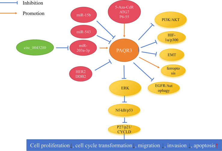 Figure 1
|
|
|
|
|
|
|
|
|---|---|---|---|---|---|---|
| Osteosarcoma | 60 | Down | Down | SOSP-9607, SAOS-2, MG63, U2OS | hFOB | [ |
| Thyroid cancer | 60 | Down | - | - | - | [ |
| Glioma | 11 | Down | Down | U251, U87, LN-18 | 1800 | [ |
| Lung cancer | 106 | Down | - | - | - | [ |
| Lung cancer | 60 | Down | - | - | - | [ |
| Lung cancer | 20 | Down | Down | SK-MES-1, A549, SPCA-1, H1229 | BEAS-2B | [ |
| Hepatocellular carcinoma | 60 | Down | Down | HepG2, Hep3B, Bel7402, SMMC-7721 | HL-7792 | [ |
| Hepatocellular carcinoma | 194 | Down | - | - | - | [ |
| Cervical cancer | 40 | Down | - | - | - | [ |
| CRC | 54 | Down | - | - | - | [ |
| CRC | 62 | Down | - | - | - | [ |
| DLBCL | 46 | Down | Down | SUDHL4, OCI-LY19, U2932, OCI-LY10 | HMy2.CIR | [ |
| ALL | 43 | Down | - | - | - | [ |
| Breast cancer | 60 | Down | Down | MDA-MB-231, BT474, SKBR3, MCF7 | MCF-10A | [ |
| Breast cancer | 82 | Down | - | - | - | [ |
| Breast cancer | 46 | Down | - | - | - | [ |
| ESCA | 40 | Down | Down | KYSE150, ECA-109, TE-1 | HEsEpiCs | [ |
| ESCA | 80 | Down | - | - | - | [ |
| ESCA | - | - | Down | EC9706, TE13, ECA109 | HEEC | [ |
| Gastric cancer | 166 | Down | Down | HGC27, SGC7901 | GES-1 | [ |
| Gastric cancer | 300 | Down | - | - | - | [ |
| RCC | 31 | Down | - | - | - | [ |
|
|
|
|
|
|
|
|
|---|---|---|---|---|---|---|
| Osteosarcoma | MG63 | Inhibition | - | - | - | [ |
| Glioma | U251, U87 | Inhibition | - | - | - | [ |
| Lung cancer | A549, H1299 | Inhibition | Inhibition | Inhibition | Promotion | [ |
| Lung cancer | A549, H1299 | Inhibition | - | Inhibition | Promotion | [ |
| Hepatocellular carcinoma | HepG2 | Inhibition | - | - | - | [ |
| Hepatocellular carcinoma | Hep-3B | Inhibition | Inhibition | - | - | [ |
| Cervical cancer | MS751, HeLa | Inhibition | - | - | - | [ |
| Colorectal cancer | SW-480 | Inhibition | Inhibition | - | - | [ |
| DLBCL | SUDHL4, U2932 | Inhibition | - | - | - | [ |
| ALL | CEM-C1, Jurkat | Inhibition | - | - | Promotion | [ |
| Breast cancer | MCF7, SKBR3, MDA-MB-231, MDA-MB-468, MDA-MB-453 | Inhibition | Inhibition | - | - | [ |
| Breast cancer | MCF-7 | - | Inhibition | [ | ||
| ESCA | ECA-109, TE-1 | Inhibition | Inhibition | Inhibition | - | [ |
| ESCA | ECA-109 | Inhibition | [ | |||
| Gastric cancer | HGC27 | Inhibition | Inhibition | - | - | [ |
| Gastric cancer | AGS | Inhibition | Inhibition | - | - | [ |
| RCC | HUVEC | Inhibition | - | - | - | [ |
| Lung cancer | HCC827 | Inhibition | Inhibition | - | - | [ |
| Prostate cancer | PC3, DU145 | Inhibition | Inhibition | - | - | [ |
| Gastric cancer | BGC-823, SGC-7901 | Inhibition | - | - | - | [ |
| Gastric cancer | AGS | Inhibition | Inhibition | - | - | [ |
| Colon Cancer | HCT116, HCT115 | Inhibition | Inhibition | - | - | [ |
|
|
|
|
|
|
|
|
|---|---|---|---|---|---|---|
| Osteosarcoma | MG63 | Inhibition | Inhibition | - | - | [ |
| Glioma | U251, U87 | Inhibition | Inhibition | - | - | [ |
| Hepatocellular carcinoma | HepG2 | - | Inhibition | - | - | [ |
| Cervical cancer | MS751, HeLa | Inhibition | Inhibition | Inhibition | - | [ |
| Breast cancer | MDA-MB-231 | Inhibition | Inhibition | - | - | [ |
| Breast cancer | MCF7, SKBR3, MDA-MB-231, MDA-MB-468, MDA-MB-453 | Inhibition | - | Inhibition | - | [ |
| Breast cancer | MCF-7 | - | Inhibition | - | - | [ |
| ESCA | ECA-109, TE-1 | - | Inhibition | - | - | [ |
| ESCA | ECA-109 | Inhibition | Inhibition | - | - | [ |
| Gastric cancer | HGC27 | Inhibition | Inhibition | - | [ | |
| Gastric cancer | AGS | Inhibition | - | Inhibition | - | [ |
| RCC | HUVEC | Inhibition | - | Inhibition | Inhibition | [ |
| Prostate cancer | PC3, DU145 | Inhibition | - | Inhibition | - | [ |
| Gastric cancer | BGC-823, SGC-7901 | - | Inhibition | Inhibition | - | [ |
| Gastric cancer | AGS | Inhibition | - | Inhibition | - | [ |
| Gastric cancer | AGS | Inhibition | - | - | - | [ |
| Colon Cancer | HCT116, HCT115 | Inhibition | - | - | - | [ |
|
|
|
|
|
|---|---|---|---|
| Glioma | U251 | Inhibition | [ |
| Cervical cancer | - | Inhibition | [ |
| CRC | SW-480 | Inhibition | [ |
| DLBCL | U2932 | Inhibition | [ |
| ESCA | ECA109 | Inhibition | [ |
| ESCA | ECA109 | Inhibition | [ |
| Lung cancer | HCC827 | Inhibition | [ |
| Gastric cancer | AGS | Inhibition | [ |
|
|
|
|
|
|---|---|---|---|
| Osteosarcoma | p-ERK | - | [ |
| Glioma | PI3K/AKT | - | [ |
| Lung cancer | Nf-kB/p53 | - | [ |
| Lung cancer | PI3K/AKT | - | [ |
| Hepatocellular carcinoma | - | miR-543/PAQR3↓ | [ |
| Cervical cancer | EMT, p-AKT, p-ERK | circ_0043280/miR-203a-3p/PAQR3↑ | [ |
| CRC | EGF/p-ERK, EGF/β-catenin | - | [ |
| DLBCL | LDLR/PI3K/AKT, ferroptosis | - | [ |
| ALL | Nrf2/ferroptosis | - | [ |
| Breast cancer | - | miR-15b/PAQR3↓ | [ |
| Breast cancer | - | HER2/PAQR3↓ | [ |
| Breast cancer | - | 5-Aza-CdR/PAQR3↑ | [ |
| ESCA | P27/p21/CYCLD, p-ERK | 5-Aza-CdR/PAQR3↑ | [ |
| ESCA | EMT, p-AKT | - | [ |
| ESCA | EMT, p-ERK | - | [ |
| Gastric cancer | TGF-β/Smad/EMT | - | [ |
| Gastric cancer | p-AKT, p-ERK, EMT | - | [ |
| RCC | VEGF/ERK, HIF-1α/p300 | - | [ |
| Lung cancer | BECN1/Autophagy, EGFR/Autophagy | ATG7/PAQR3↑ | [ |
| Prostate cancer | p-AKT, p-ERK, EMT | - | [ |
| Gastric cancer | EMT | miR-15b-5p/PAQR3↓ | [ |
| Gastric cancer | EGF/ERK, AKT, insulin | DDB2/PAQR3↓ | [ |
| Gastric cancer | Twist1/EMT | - | [ |
| Colon cancer | PI3K/AKT | P6-55/PAQR3↑ | [ |
|
|
|
|
|
|
|---|---|---|---|---|
| Osteosarcoma | Down | - | Metastasis↑ | [ |
| Thyroid cancer | Down | - | Extrathyroidal-extension | [ |
| Lung cancer | Down | OS↓ | Pathological stages↑, tissue differentiation↓, metastasis↑ | [ |
| Lung cancer | Down | - | Tumor size↑, diagnosis | [ |
| Hepatocellular carcinoma | Down | OS↓, DFS↓ | AFP↑, tumor size↑, tumor grade↓, recurrence↑ | [ |
| CRC | Down | - | Tissue differentiation↓, lymph node metastasis↑, depth of invasion↑ | [ |
| CRC | Down | - | Gender, tumor grade↓ | [ |
| DLBCL | Down | OS↓ | - | [ |
| Breast cancer | Down | OS↓, DFS↓ | Tissue differentiation↓, pathological stage↑, Her2 status | [ |
| ESCA | Down | - | Pathological stage↑, lymph node metastasis↑ | [ |
| ESCA | Down | OS↓, DFS↓ | Nationality, tumor size↑, lymph node metastasis↑, recurrence↑ | [ |
| Gastric cancer | Down | OS↓ | Helicobacter pylori, venous invasion↑, invasion depth↑, lymph node metastasis↑, pathological stage↑ | [ |
| Gastric cancer | Down | DFS↓ | Age, Helicobacter pylori, tumor size↑, tumor differentiation↓, venous invasion↑, lymph node metastasis↑, invasion depth↑, pathological stage↑, distant metastasis↑ | [ |
Peer Reviews
No public reviews on file for this paper yet. If you reviewed it on a platform where reviews are public (OpenReview, ICLR, NeurIPS, ICML), you can paste yours below so the community can read it here.
Videos
No videos yet. Explain this paper in a talk, walkthrough, or lecture? Add one.
Taxonomy
TopicsMechanisms of cancer metastasis · Chromatin Remodeling and Cancer · Circular RNAs in diseases
Introduction
Numerous studies have demonstrated that abnormal gene expression is associated with cancer cell growth, metastasis, and poor prognosis in cancer patients [1–6]. Progestin and AdipoQ Receptor 3 (PAQR3) is a gene located on human chromosome 4 that encodes a membrane protein characterized by a unique seven-transmembrane domain architecture. This protein is predominantly localized in the Golgi apparatus and plays a critical role in intracellular signal transduction. It functions by anchoring Raf kinase to the Golgi membrane, thereby exerting a negative regulatory effect on the Ras/Raf/MEK/ERK signaling cascade. PAQR3 influences essential cellular processes, including proliferation and differentiation, highlighting its significance as a key regulator in maintaining normal cellular physiology through Rat sarcoma (Ras)/Rapidly Accelerated Fibrosarcoma (Raf)/mitogen-activated protein kinase kinase (MEK)/extracellular signal-regulated kinase (ERK) signaling [6]. Recent research has confirmed that the downregulation of PAQR3 is linked to various aspects of cancer biology, such as cell proliferation, migration, and invasion across multiple cancer types [7–36]. Despite significant advancements in cancer treatment, the complexity of cancer cell biology, particularly the role of specific genes like PAQR3, continues to present challenges in developing more effective and targeted therapies. This review provides an update and expansion relative to existing synthesizations, including recent findings from 2023–2025 that were not addressed by Guo et al., and encompasses a broader range of literature than the meta-analysis conducted by Zhai et al. [34, 35]. The aim of this review is to summarize the roles, mechanisms, and clinical significance of PAQR3 in cancer, offering new insights and potential therapeutic targets for clinical practitioners and researchers.
Expression levels of PAQR3 significantly decrease in cancer
The expression levels of PAQR3 are significantly reduced in various cancer tissues and cell lines [7–27]. Specifically, compared to normal tissues, PAQR3 expression is notably lower in tissues from osteosarcoma, thyroid cancer, glioma, lung cancer, hepatocellular carcinoma, cervical cancer, colorectal cancer, diffuse large B-cell lymphoma, acute lymphoblastic leukemia, breast cancer, esophageal cancer, gastric cancer, and renal cell carcinoma (Table 1). This decline in expression is also observed in osteosarcoma, glioma, lung cancer, hepatocellular carcinoma, diffuse large B-cell lymphoma, breast cancer, esophageal cancer, and gastric cancer cells (Table 1). The downregulation of PAQR3 in cancer cells may be attributed to epigenetic modifications. Specifically, DNA methylation of the PAQR3 promoter region has been implicated in certain cancer types, leading to reduced gene transcription and subsequently lower protein expression levels [10, 22]. Preliminary evidence suggests that PAQR3 may act as a tumor suppressor gene in cancer progression.
Overexpression of PAQR3 inhibits cancer cell growth and metastasis
Research has confirmed that inhibiting oncogene expression or promoting tumor suppressor gene expression can suppress cancer growth and metastasis [34–36]. PAQR3 is frequently downregulated in cancer tissues and cells, and its activation can inhibit cancer cell growth (Table 2). Specifically, PAQR3 can suppress the proliferation of osteosarcoma, glioma, lung cancer, hepatocellular carcinoma, cervical cancer, colorectal cancer, diffuse large B-cell lymphoma, acute lymphoblastic leukemia, breast cancer, esophageal cancer, gastric cancer, renal cell carcinoma, colon cancer, and prostate cancer cells [7, 9, 11–15, 17–19, 21, 23, 25–32, 36]. Moreover, PAQR3 overexpression suppresses colony formation in lung cancer, hepatocellular carcinoma, colorectal cancer, breast cancer, esophageal cancer, gastric cancer, colon cancer, and prostate cancer cells [11, 14, 17, 21–23, 26, 27, 29, 30, 32, 36]. Additionally, PAQR3 overexpression can inhibit cell cycle progression in lung cancer and esophageal cancer cells [11, 12, 23]. Furthermore, PAQR3 promotes apoptosis in lung cancer and acute lymphoblastic leukemia cells [11, 12, 19].
Activation of PAQR3 expression can also suppress cancer cell metastasis (Table 3). Specifically, results from Transwell and wound healing assays indicate that PAQR3 overexpression can inhibit the migration of osteosarcoma, glioma, cervical cancer, breast cancer, esophageal cancer, gastric cancer, renal cell carcinoma, colon cancer, and prostate cancer cells [7, 9, 15, 20–23, 25–33, 36]. Additionally, PAQR3 overexpression inhibits the invasion of osteosarcoma, glioma, hepatocellular carcinoma, cervical cancer, breast cancer, esophageal cancer, and gastric cancer cells [7, 9, 13, 15, 22, 23, 25, 31]. Notably, PAQR3 overexpression inhibits the tube formation ability in renal cell carcinoma cells [28]. Based on this information, it can be preliminarily concluded that PAQR3 functions as a tumor suppressor in vitro, and enhancing PAQR3 expression may delay cancer cell growth and metastasis.
Activation of PAQR3 expression in glioma, cervical cancer, colorectal cancer, diffuse large B-cell lymphoma, esophageal cancer, lung cancer, and gastric cancer cells, followed by the establishment of tumorigenic models in athymic nude mice, demonstrated that PAQR3 overexpression significantly inhibits tumor growth and proliferation in these models [9, 15, 17, 18, 23, 25, 29, 33]. This finding suggests that PAQR3 functions as a tumor suppressor in vivo (Table 4), and enhancing PAQR3 expression could represent a viable therapeutic strategy in clinical settings. However, further research is necessary to develop safe and effective methods to specifically upregulate PAQR3 expression in human cancer patients without causing significant side effects.
PAQR3 involves the signaling mechanisms
Upstream regulation of PAQR3 in cancer
PAQR3 may be inhibited by upstream circular RNA/microRNAs (miRNAs), thereby affecting the function of the target genes of PAQR3 [13, 15, 20, 31]. For instance, Yu et al. reported that miR-543 is overexpressed in hepatocellular carcinoma tissues, while PAQR3 is downregulated, establishing a negative correlation and a targeted regulatory relationship between the two. miR-543 can promote the proliferation and invasion of hepatocellular carcinoma cells by specifically targeting and suppressing PAQR3 expression [13]. Zhang et al. noted a significant decrease in circ_0043280 levels in cervical cancer; circ_0043280 can competitively bind to miR-203a-3p, thereby promoting PAQR3 expression and suppressing the growth and metastasis of cervical cancer [15]. Qi et al. demonstrated a targeted regulatory relationship between miR-15b and PAQR3, where miR-15b inhibits PAQR3 expression, promoting progression in breast and gastric cancers [20, 31]. Additionally, PAQR3 may be regulated by factors such as Human epidermal growth factor receptor 2 (HER2), 5-Aza-2′-deoxycytidine (5-Aza-CdR), Autophagy related 7 (ATG7), P6-55, and Damage-specific DNA binding protein 2 (DDB2) [19, 21–23, 29, 32, 36]. For example, Li et al. reported that PAQR3 can inhibit the proliferation, colony formation, and migration of breast cancer cells. Trastuzumab, which inhibits HER2 expression, can promote PAQR3 expression, thereby enhancing its inhibitory effects on breast cancer [21]. Chen et al. demonstrated that treatment with 5-Aza-CdR significantly stimulates PAQR3 expression in breast and esophageal cancer cells, thereby enhancing PAQR3’s efficacy against cancer cells [22, 23].
Downstream effects of PAQR3 in cancer
The PAQR3/Ras/Raf/MEK/ERK signaling axis
ERK, a member of the Mitogen-Activated Protein Kinase (MAPK) family, plays a pivotal role in the signaling network governing cell growth, development, and division [37]. PAQR3 has been shown to inhibit the growth and metastasis of various cancers, including osteosarcoma, cervical cancer, esophageal cancer, gastric cancer, renal cell carcinoma, colorectal cancer, and prostate cancer, by suppressing ERK signaling [7, 15, 17, 23, 25, 27, 28, 30, 32], as illustrated in Table 5 and Figure 1. For example, Ma et al. demonstrated that overexpression of PAQR3 suppresses the phosphorylation of ERK (p-ERK), thereby inhibiting the proliferation and invasion of osteosarcoma cells. Notably, the MEK inhibitor U0126 completely negates the effects of PAQR3 silencing on osteosarcoma cell proliferation and invasion [7]. Bai et al. reported that PAQR3 overexpression reduces p-ERK protein levels in esophageal cancer cells and enhances the expression of downstream ERK proteins, such as Cyclin-dependent kinase inhibitor 1B (CDKN1B) and Cyclin-dependent kinase inhibitor 1A (CDKN1A), while inhibiting cyclin D1, Cyclin-dependent kinase 4 (CDK4), and Cyclin-dependent kinase 2 (CDK2), thus suppressing esophageal cancer growth [23, 25]. Additionally, PAQR3 overexpression can inhibit downstream signaling of Nuclear Factor Kappa B (NF-κB) and Protein 53 (p53) via ERK, consequently delaying the growth of lung cancer cells [11].
PAQR3-centered signaling network. Schematic summary of reported upstream regulators and downstream effectors of PAQR3 in cancer, with emphasis on the PAQR3/Ras/Raf/MEK/ERK axis discussed in the text. Upstream non-coding RNAs (circ_0043280; miR-15b, miR-543, miR-203a-3p) and protein/epigenetic regulators (HER2/ERBB2, DDB2; 5-Aza-2′-deoxycytidine (5-Aza-CdR), ATG7, p6-55) modulate PAQR3 expression/activity. Functionally, PAQR3 attenuates ERK signaling (including reduced ERK phosphorylation reported in multiple tumor models) and influences downstream modules such as NF-κB/p53 and cell-cycle regulators (p21/CDKN1A, p27/CDKN1B, cyclin D1), thereby restraining tumor cell proliferation, cell-cycle progression, migration and invasion, while promoting apoptosis. PAQR3 also intersects with additional pathways implicated in tumor biology, including PI3K/AKT, HIF-1α/p300, EMT, EGFR-linked autophagy, and ferroptosis (context-dependent). Blue blunt-ended lines indicate inhibition; orange arrows indicate activation/promotion. Abbreviations: PAQR3: Progestin and adipoQ receptor family member 3; circ_0043280: Circular RNA circ_0043280; miR: MicroRNA; HER2/ERBB2: Human epidermal growth factor receptor 2; DDB2: Damage-specific DNA binding protein 2; 5-Aza-CdR: 5-Aza-2′-deoxycytidine; ATG7: Autophagy related 7; EGFR: Epidermal growth factor receptor; ERK: Extracellular signal-regulated kinase; NF-κB: Nuclear factor kappa B; p53: Tumor protein p53; CDKN1A/p21: Cyclin-dependent kinase inhibitor 1A; CDKN1B/p27: Cyclin-dependent kinase inhibitor 1B; PI3K: Phosphatidylinositol 3-kinase; AKT: AKT serine/threonine kinase; HIF-1α: Hypoxia-inducible factor-1 alpha; EP300/p300: Histone acetyltransferase p300; EMT: Epithelial–mesenchymal transition.
The PAQR3/phosphatidylinositol 3-kinase (PI3K)/AKT serine/threonine kinase (AKT) signaling axis
The PI3K/AKT pathway is a well-established signaling cascade that promotes cell survival and insulin secretion [38, 39]. Research has elucidated a significant relationship between PAQR3 and the PI3K/AKT signaling pathway [9, 12, 15, 18, 24, 27, 30, 36]. For instance, Li et al. found that PAQR3 overexpression inhibits PI3K/AKT signaling by reducing PI3K phosphorylation (p-PI3K) and AKT phosphorylation (p-AKT) without significantly affecting the overall protein levels of PI3K and AKT, thus inhibiting the growth of lung cancer cells A549 and H1299 [12]. In diffuse large B-cell lymphoma U2932 cells, PAQR3 may impede cell progression through the Low-Density Lipoprotein Receptor (LDLR)/PI3K/AKT signaling pathway by modulating the expression levels of LDLR, p-AKT, and p-PI3K [19]. Moreover, studies indicate that PAQR3 expression is associated with insulin signaling mechanisms [32]. PAQR3 inhibits insulin-stimulated phosphorylation of p-AKT and Glycogen Synthase Kinase 3 Beta (GSK3β), thereby influencing the growth and metastasis of gastric cancer [32].
The PAQR3/Epithelial-mesenchymal transition (EMT) signaling axis
The EMT process is critically linked to cancer metastasis [40]. PAQR3 contributes to the metastasis of cervical cancer, esophageal cancer, gastric cancer, and prostate cancer through the EMT process [15, 24, 25, 27, 30, 31, 33]. For instance, Huang et al. reported that PAQR3 overexpression suppresses vimentin expression during EMT while promoting E-cadherin and Zonula Occludens-1 (ZO-1) expression, thereby inhibiting migration of prostate cancer cells PC3 and DU145 [30]. Bai et al. noted that PAQR3 overexpression enhances E-cadherin expression while inhibiting N-cadherin expression, consequently reducing migration of esophageal cancer cells [24, 25]. Wu et al. demonstrated that PAQR3 inhibits the expression levels of snail, vimentin, Transforming Growth Factor Beta 1 (TGF-β1), Phosphorylated SMAD Family Member 2 (p-SMAD2), and Phosphorylated SMAD Family Member 3 (p-SMAD3) within the TGF-β/SMAD/EMT signaling pathway, thereby promoting E-cadherin expression and inhibiting gastric cancer progression [26].
Other downstream signaling mechanisms
Dysregulation of PAQR3 may also impact ferroptosis through interactions with nuclear factor erythroid 2-related factor 2 (Nrf2), Epidermal Growth Factor (EGF)/β-catenin signaling, and autophagy, thereby influencing cancer progression [17–19, 26, 28, 29, 36]. For example, Wang et al. reported that PAQR3 inhibits nuclear accumulation of β-catenin in colorectal cancer SW-480 cells, thus delaying tumorigenic potential [17]. Additionally, it may promote ferroptosis in diffuse large B-cell lymphoma and acute lymphoblastic leukemia [18, 19]. Specifically, Song et al. observed that PAQR3 overexpression suppresses glutathione (GSH) levels in diffuse large B-cell lymphoma cells while promoting Malondialdehyde (MDA), Reactive Oxygen Species (ROS), and Fe2+ levels [18]. Jin and Tong found that PAQR3 overexpression enhances MDA, Dichlorofluorescein (DCF), and Fe2+ levels in acute lymphoblastic leukemia cells [19]. Furthermore, PAQR3 can inhibit the Hypoxia-Inducible Factor 1a (HIF-1α)/E1A Binding Protein p300 (p300), Beclin 1 (BECN1)/autophagy, and Epidermal Growth Factor Receptor (EGFR)/autophagy pathways to suppress progression in lung cancer and renal cell carcinoma [28, 29].
Decreased PAQR3 expression as a biomarker for poor prognosis in cancer patients
Table 6 presents indicators related to the prognosis and pathological features associated with PAQR3 overexpression in cancer patients. Notably, overexpression is linked to favorable characteristics concerning metastasis, pathological stage, tumor size, and diagnosis [7, 8, 10, 11, 14, 16–18, 21, 23, 24, 26, 27]. Specifically, decreased PAQR3 expression levels correlate significantly with shorter overall survival (OS) in patients with lung cancer, hepatocellular carcinoma, diffuse large B-cell lymphoma, breast cancer, esophageal cancer, and gastric cancer [10, 14, 18, 21, 24, 26]. Furthermore, reduced PAQR3 levels are associated with shorter disease-free survival (DFS) in patients with hepatocellular carcinoma, breast cancer, esophageal cancer, and gastric cancer [14, 21, 24, 27]. Additionally, diminished PAQR3 expression correlates significantly with pathological staging, subtype, tissue differentiation, metastasis, tumor size, and diagnosis in lung cancer patients [10, 11]. It is also linked to factors such as Helicobacter pylori, venous invasion, invasion depth, lymph node metastasis, pathological stage, age, tumor size, tumor differentiation, and distant metastasis in gastric cancer patients [26, 27]. The relationship between PAQR3 expression and patient prognosis may be confounded by other factors, including co-existing genetic mutations, the patient’s immune status, and concurrent medication use. Future studies should consider these variables to accurately assess the prognostic value of PAQR3.
Conclusion
Research has established that PAQR3 functions as a tumor suppressor gene in cancer, and activation of PAQR3 expression may enhance patient prognosis. This is linked to various signaling mechanisms, including PI3K/AKT, EMT, ferroptosis, and Ras/Raf/MEK/ERK pathways, and is regulated by miR-543, miR-203a-3p, miR-15b, HER2, and 5-Aza-CdR, thereby influencing cancer cell growth and metastasis (Table 5). The anti-tumor mechanisms of PAQR3 exhibit significant heterogeneity. In certain cancer types, such as diffuse large B-cell lymphoma, PAQR3 enhances ferroptosis through the PI3K/AKT pathway [18]. In contrast, in other cancers, such as gastric cancer, it may operate via different pathways. These differences may be attributed to cancer-specific expression patterns, tissue-specific microenvironments, mutation backgrounds, and upstream regulatory factors. Furthermore, while epigenetic drugs like 5-Aza-CdR can demethylate and activate PAQR3 expression, their clinical application faces challenges, including strong toxicity and the development of drug resistance, underscoring the urgent need for more precise epigenetic intervention strategies.
Despite the progress, studies on PAQR3 remain in preliminary stages. Future research should focus on the upstream and downstream mechanisms of PAQR3 to deepen the understanding of its pathogenesis and ultimately impede disease progression. Additionally, anti-cancer treatments targeting PAQR3 encounter various challenges. First, the heterogeneity of the underlying mechanisms necessitates the design of specific treatment strategies tailored to different cancer types, alongside precise regulation of the PAQR3 pathway in conjunction with tumor molecular typing and microenvironment characteristics. Second, the development of drugs targeting PAQR3 must address critical issues, including low delivery efficiency and poor stability. Moreover, the PAQR3 pathway interacts with multiple signaling networks, raising concerns about potential off-target effects from targeted therapies. Enhancing treatment safety through highly selective delivery systems (e.g., nanocarriers or conditionally activated gene editing tools) is essential. Ultimately, the combined targeting of upstream regulatory factors (e.g., HER2 inhibitors) or downstream effector pathways (e.g., ferroptosis inducers) may represent a crucial approach to improve efficacy and overcome drug resistance. Future research could utilize patient-derived organoids to more accurately replicate the in vivo tumor microenvironment and investigate the roles of PAQR3. Overall, this review summarizes the mechanisms and clinical significance of PAQR3, providing a novel theoretical foundation and direction for cancer treatment.
The reference list from the paper itself. Each links out to its DOI / PubMed record.
- 1Xu J Zhang Z Qian M Wang S Qiu W Chen Z Cullin-7 (CUL 7) is overexpressed in glioma cells and promotes tumorigenesis via NF-κB activation J Exp Clin Cancer Res 202039159 https://doi.org/10.1186/s 13046-020-01553-73225280210.1186/s 13046-020-01553-7PMC 7132976 · doi ↗ · pubmed ↗
- 2Lin C Wu Y Qian Y Li J He Y Yu HSATB 2 promotes radiation resistance of esophageal squamous cell carcinoma by regulating epithelial-to-mesenchymal transition via the Wnt/β-catenin pathway Front Oncol 2025151543426 https://doi.org/10.3389/fonc.2025.15434264007819410.3389/fonc.2025.1543426 PMC 11896856 · doi ↗ · pubmed ↗
- 3Zhang Z Mei Y Hou M Knockdown RBM 15 inhibits colorectal cancer cell proliferation and metastasis via N 6-methyladenosine (m 6A) modification of My D 88 m RNA Cancer Biother Radiopharm 2022371097686 https://doi.org/10.1089/cbr.2021.02263484245710.1089/cbr.2021.0226 · doi ↗ · pubmed ↗
- 4Yan Q Sun Q Feng Y Hu Q Zhu JATP 1B 3 may promote glioma proliferation and migration through MAPK/NF-κB signaling pathway Front Oncol 2025151537687 https://doi.org/10.3389/fonc.2025.15376874002713010.3389/fonc.2025.1537687 PMC 11868815 · doi ↗ · pubmed ↗
- 5Guo Q Ke XX Liu Z Gao WL Fang SX Chen C Evaluation of the prognostic value of STEAP 1 in lung adenocarcinoma and insights into its potential molecular pathways via bioinformatic analysis Front Genet 202011242 https://doi.org/10.3389/fgene.2020.002423226598510.3389/fgene.2020.00242 PMC 7099762 · doi ↗ · pubmed ↗
- 6Lei L Ling ZN Chen XL Hong LL Ling ZQ Characterization of the Golgi scaffold protein PAQR 3, and its role in tumor suppression and metabolic pathway compartmentalization Cancer Manag Res 20201235362 https://doi.org/10.2147/CMAR.S 2109193202144810.2147/CMAR.S 210919 PMC 6970510 · doi ↗ · pubmed ↗
- 7Ma Z Wang Y Piao T Li Z Zhang H Liu Z The tumor suppressor role of PAQR 3 in osteosarcoma Tumour Biol 2015365331924 https://doi.org/10.1007/s 13277-014-2964-z 2551067010.1007/s 13277-014-2964-z · doi ↗ · pubmed ↗
- 8Gao J Ma XP Deng FS Jiang L Jia WD Li M Associations of the BRAF V 600E mutation and PAQR 3 protein expression with papillary thyroid carcinoma clinicopathological features Pathol Oncol Res 2020263183341 https://doi.org/10.1007/s 12253-019-00779-x 3175840810.1007/s 12253-019-00779-x · doi ↗ · pubmed ↗