Assessing Age‐Associated Influences on Paramagnetic and Diamagnetic Susceptibility Maps in Postmortem Human Brains
José Henrique Monteiro de Azevedo, Maria Concepción Garcia Otaduy, Andre Avanzine, Roberta Diehl Rodriguez, Fábio Seiji Otsuka, Fernando Barbosa, Chunlei Liu, Carlos Ernesto Garrido Salmon

TL;DR
This study explores how brain aging affects magnetic susceptibility maps in postmortem human brains, revealing age-related changes in iron and myelin distribution.
Contribution
The study introduces postmortem validation of susceptibility maps using APART-QSM, linking them to biological factors like iron and myelin in aging brains.
Findings
Diamagnetic susceptibility declines with age in basal ganglia regions, possibly due to shared biological factors.
Putamen's paramagnetic susceptibility increases with age, aligning with in vivo observations and showing a moderate correlation with iron concentration.
Diamagnetic susceptibility in white matter correlates with fractional anisotropy, suggesting a myelin contribution.
Abstract
Maps of paramagnetic and diamagnetic components of magnetic susceptibility can provide insights into the distribution of iron and myelin during brain aging. Postmortem validation is essential to ensure that these maps accurately reflect in vivo biological processes. In this study, we applied the APART‐QSM method for susceptibility separation to in situ (intracranial) postmortem MRI data from 47 subjects (ages 31–91) to investigate how age affects magnetic susceptibility components, comparing the results with previously reported in vivo associations. Linear regression was used to assess age‐related associations with susceptibility values in 17 deep gray matter (DGM) and white matter (WM) regions. Diamagnetic susceptibility showed a consistent age‐related decline in DGM basal ganglia regions, which appeared to result from a shared underlying factor across these areas. Based on the…
Genes, proteins, chemicals, diseases, species, mutations and cell lines named across the full text — each resolved to its canonical identifier and authoritative record.
Click any figure to enlarge with its caption.
 Figure 1
 Figure 2
 Figure 3
 Figure 4
 Figure 5
 Figure 6
 Figure 7
 Figure 8
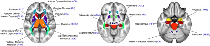 Figure 9
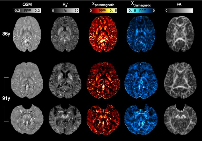 Figure 10
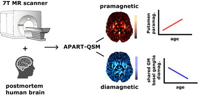 Figure 11
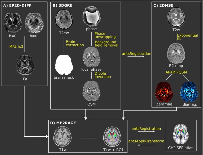 Figure 12
 Figure 13
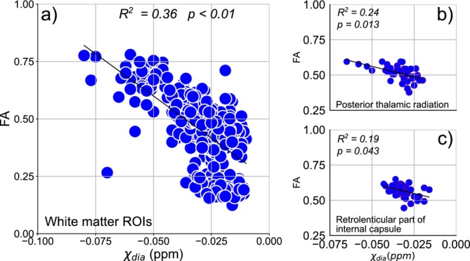 Figure 14
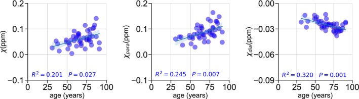 Figure 15
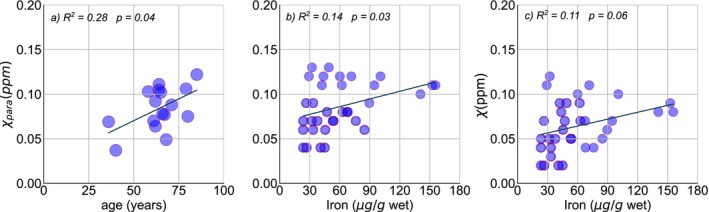 Figure 16
Peer Reviews
No public reviews on file for this paper yet. If you reviewed it on a platform where reviews are public (OpenReview, ICLR, NeurIPS, ICML), you can paste yours below so the community can read it here.
Videos
No videos yet. Explain this paper in a talk, walkthrough, or lecture? Add one.
Taxonomy
TopicsAdvanced MRI Techniques and Applications · Functional Brain Connectivity Studies · Advanced Neuroimaging Techniques and Applications