Learning from multiple readings for axial spondyloarthritis classification of the sacroiliac joints
Amir Jamaludin, Rhydian Windsor, Sarim Ather, Gregory Ligozio, Aimee Readie, Pedro M. Machado, Timor Kadir

TL;DR
This paper introduces an automated machine learning system to classify MRI lesions in the sacroiliac joints of axial spondyloarthritis patients, aiming to reduce variability and improve consistency.
Contribution
A novel multi-reader machine learning framework is proposed to model variability and automate lesion classification in axial spondyloarthritis MRI evaluations.
Findings
The automated system achieved AUCs between 0.85 and 0.99 for classifying five lesion types in sacroiliac joints.
Contouring accuracy showed 95% of test cases had errors below 2.76mm, comparable to expert inter-reader agreement.
Performance was validated on multiple clinical datasets including MEASURE-1, PREVENT, and SURPASS.
Abstract
Magnetic resonance imaging (MRI) is a cornerstone in the evaluation and monitoring of axial spondyloarthritis (axSpA), a chronic inflammatory condition primarily affecting the sacroiliac joints (SIJs), spine, entheses, and peripheral joints. Accurate quantification of axSpA-related changes on MRI is critical for effective research and patient management; however, current lesion detection and grading approaches suffer from substantial intra- and inter-reader variability, limiting their consistency and reliability. To address these challenges, we propose a fully automated machine learning system for SIJ delineation and lesion classification on coronal MRI. The end-to-end pipeline automatically extracts SIJ contours using a vector-field—based open-contour model and classifies the presence or absence of five lesion types (bone marrow oedema, ankylosis, sclerosis, erosions, and fatty…
Genes, proteins, chemicals, diseases, species, mutations and cell lines named across the full text — each resolved to its canonical identifier and authoritative record.
Click any figure to enlarge with its caption.
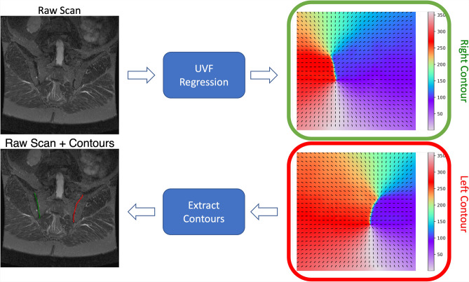 Figure 1
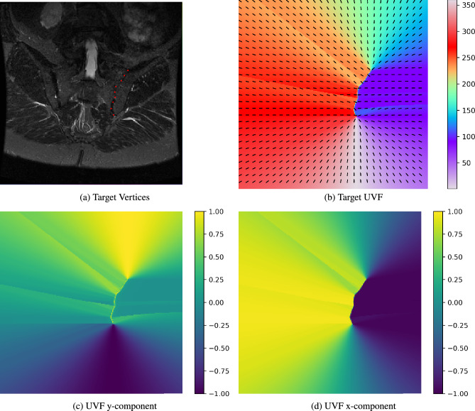 Figure 2
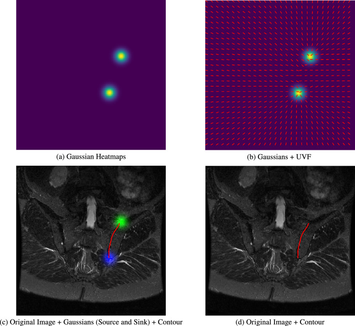 Figure 3
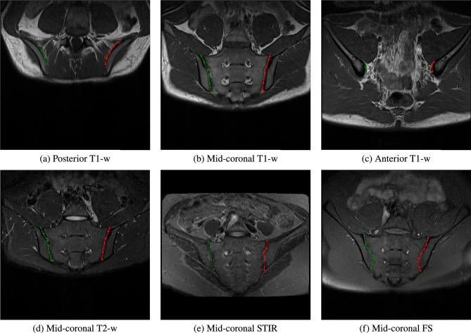 Figure 4
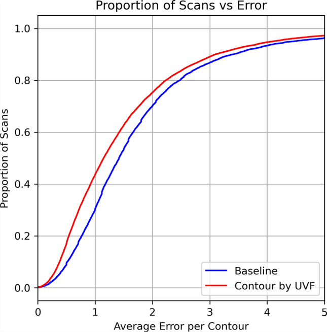 Figure 5
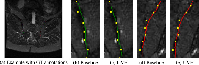 Figure 6
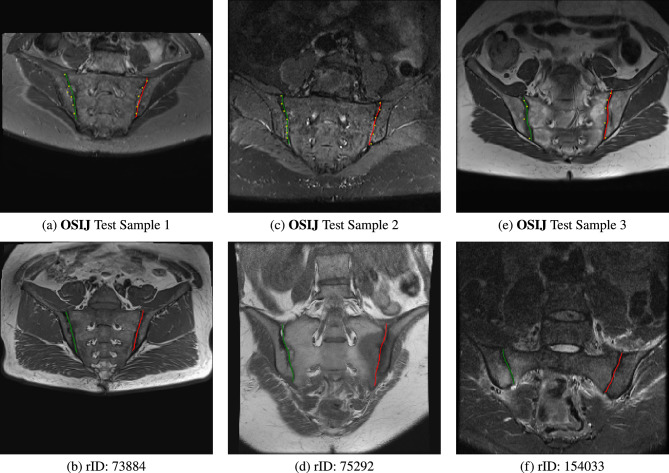 Figure 7
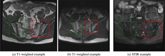 Figure 8
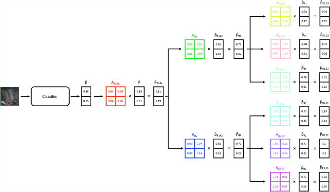 Figure 9
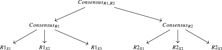 Figure 10
Peer Reviews
No public reviews on file for this paper yet. If you reviewed it on a platform where reviews are public (OpenReview, ICLR, NeurIPS, ICML), you can paste yours below so the community can read it here.
Videos
No videos yet. Explain this paper in a talk, walkthrough, or lecture? Add one.
Taxonomy
TopicsSpondyloarthritis Studies and Treatments · Medical Imaging and Analysis · Spine and Intervertebral Disc Pathology