Stop, neighbor! KLU–PREs positional signaling restricts female germline fate in Arabidopsis
Jiajun Wang

Abstract
Genes, proteins, chemicals, diseases, species, mutations and cell lines named across the full text — each resolved to its canonical identifier and authoritative record.
Click any figure to enlarge with its caption.
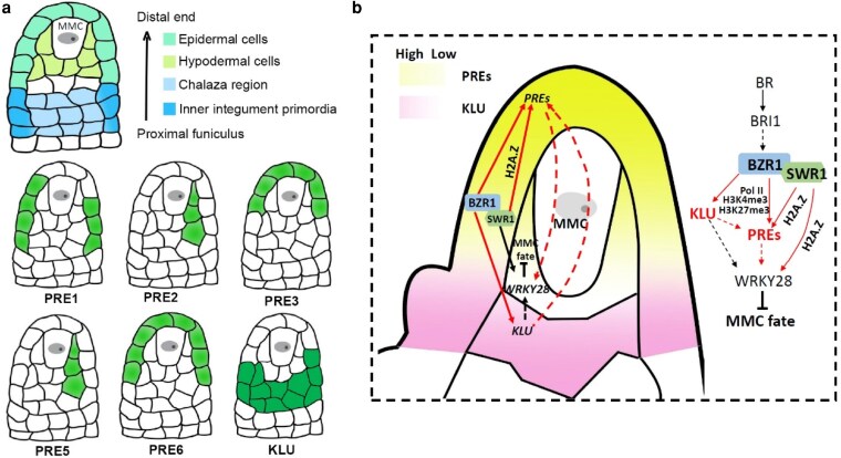 Figure 1
- —National Natural Science Foundation of China10.13039/501100001809
Peer Reviews
No public reviews on file for this paper yet. If you reviewed it on a platform where reviews are public (OpenReview, ICLR, NeurIPS, ICML), you can paste yours below so the community can read it here.
Videos
No videos yet. Explain this paper in a talk, walkthrough, or lecture? Add one.
Taxonomy
TopicsPlant Molecular Biology Research · Plant Reproductive Biology · Plant-Microbe Interactions and Immunity
The ability to specify a single megaspore mother cell (MMC) within the ovule primordium, while preventing neighboring somatic cells from acquiring germline fate, is essential for successful sexual reproduction in plants. This tightly regulated process establishes the female germline and sets the developmental trajectory for subsequent female gametophyte formation in flowering plants (Huang et al. 2024). While complex signaling networks ensure the singular MMC fate, how surrounding somatic nucellar cells maintain their identity remains unclear. In new work, Hanyang Cai and colleagues (Cai et al. 2026) elucidate a crucial positional signaling module involving KLU and the PRE family that is orchestrated by the brassinosteroid-responsive transcription factor BZR1 enforces this developmental fidelity (Fig. 1).
The PACLOBUTRAZOL-RESISTANCE (PRE) gene family encodes 6 atypical, likely non–DNA-binding basic helix–loop–helix (bHLH) proteins (PRE1–PRE6) that integrate multiple signaling pathways to regulate cell elongation as well as floral organ growth and development (Shin et al. 2019). Cai et al. found that generating a PRE quintuple mutant (pre-quin) via CRISPR-mediated knockout of PRE3 in a pre1 pre2 pre5 pre6 artificial microRNA knockdown background (pre-amiR) resulted in sterility, with approximately 35% of ovules exhibiting a multiple MMC-like specification phenotype. In contrast to KLU, a cytochrome P450 enzyme that is primarily expressed in the inner integument primordia and the chalaza region of the proximal funiculus, PRE1/2/3/5/6 are preferentially expressed in the epidermis (PRE1/3/6) and hypodermis (PRE2/5) of distal nucellar cells surrounding the MMC. This complementary expression pattern is essential for restricting MMC fate. In this work, ectopic expression of PRE1/2/3/5/6 in the inner integument primordia and chalaza region, but not in other regions, significantly increased the proportion of hypodermal cells that differentiated into MMC-like cells. Conversely, ectopic expression of KLU in the distal epidermis and hypodermal MMC-surrounding cells also induced the formation of extra MMC-like cells. These results indicate that the spatially restricted expression of PREs in distal epidermal and hypodermal cells, together with KLU expression in the inner integument primordia and chalaza region, is crucial for limiting distal nucellar cell fate to a single MMC.
Interestingly, PRE proteins have previously been found to spread to neighboring cells during early ovule development (Lu et al. 2018). The authors therefore investigated why PRE proteins normally fail to extend into the inner integument primordia and chalaza region. In klu mutants, the spatiotemporal expression patterns of PREs were unaffected; however, PRE proteins spread ectopically from distal epidermal or hypodermal MMC-surrounding cells into the inner integument primordia and chalaza region. KLU directly interacts with PREs and likely inhibits PRE protein spread into these proximal domains by acting on the conserved M8 motif of PREs. Artificially confining PREs to their native domain rescued the multiple-MMC phenotype of klu mutants, confirming the functional importance of this spatial restriction. Consistently, knocking down PRE1/2/5/6 in the klu mutant background significantly increased the proportion of ovules producing multiple MMC-like cells and resulted in sterility.
To investigate the upstream mechanisms controlling the distinct positional expression of PREs and KLU in the ovule primordium, the authors focused on BZR1-family transcription factors. Brassinosteroids have previously been implicated in female germline specification, and the bzr1 bes1 beh1 beh3 beh4 quintuple mutant (qui-1) displays more than 70% of ovules containing multiple MMCs (Cai et al. 2022). The authors showed that BZR1 binds directly to E-box motifs in the promoters of PRE1/2/3/5/6 and KLU, thereby activating their transcription. Introducing a klu loss-of-function mutation into 5 different combinations of BZR1-family quadruple mutants significantly increased the frequency of ovules containing enlarged MMC-like cells compared with either the respective quadruple mutants or the klu single mutant.
In addition, BZR1 was found to physically interact with SWC6, a subunit of the SWR1 chromatin remodeling complex, and SWC6 and KLU genetically interact to suppress supernumerary MMC specification. BZR1 binding to the PRE1 promoter was shown to be partially dependent on SWC6 and KLU. Similar to the BZR1-family quintuple mutant (qui-1), the swc6 klu double mutant exhibited markedly reduced RNA polymerase II occupancy, H3K4me3 levels, and H2A.Z deposition near the transcription start sites of PRE1/2/3/5/6, accompanied by a pronounced increase in H3K27me3. These chromatin changes correlated with a strong reduction in PRE1/2/3/5/6 transcription in the swc6 klu background, indicating that BZR1-dependent PRE activation partially relies on SWC6- and KLU-mediated chromatin regulation.
In summary, by integrating detailed cytological observations with molecular and genetic analyses, the authors uncover a hierarchical regulatory network in which the precise spatial expression of PREs and KLU in the ovule primordium suppresses germline potential in surrounding somatic nucellar cells. BZR1 executes this control through 2 parallel pathways: it promotes PRE transcription in distal epidermal and hypodermal cells in a manner partially dependent on SWC6 and KLU, while simultaneously activating KLU expression in the inner integument primordia and chalaza region to prevent PRE protein spread into this domain. This coordinated spatial regulation ensures the robust restriction of MMC fate to a single cell during ovule development. Collectively, this work elegantly integrates hormone signaling, transcriptional control, chromatin dynamics, and protein mobility to explain a fundamental patterning event in plant reproduction.
Recent related articles in The Plant Cell
Jiang et al. (2025) revealed that a precise balance between DNA methylation and demethylation, rather than absolute methylation levels, maintains the specification of a single megaspore mother cell (MMC) by ensuring differential mCHH hypomethylation in the MMC precursor compared to its somatic neighbors. Wang et al. (2024) demonstrated that brassinosteroid signaling spatially restricts the expression of the AGAMOUS-activated gene ZINC FINGER PROTEIN 11 in the chalaza and nucellus via the transcription factor BZR1, thereby orchestrating integument development and controlling ovule number to ensure proper female reproductive structure formation. Shi et al. (2024) identified that the adaptor protein ECAP forms a transcriptional activator complex with the corepressor LEUNIG and the transcription factor BEH3 that epigenetically regulates SPOROCYTELESS expression to control archesporial cell specification and microsporocyte generation during early anther development.
The reference list from the paper itself. Each links out to its DOI / PubMed record.
- 1Cai H et al 2022. Brassinosteroid signaling regulates female germline specification in Arabidopsis. Curr Biol. 32:1102–1114 e 5. 10.1016/j.cub.2022.01.022.35108524 · doi ↗ · pubmed ↗
- 2Cai H et al 2026. The KLU-PRE module provides positional cues that maintain somatic cell identity around the megasporocyte cell in Arabidopsis. Plant Cell. 38:koag 008. 10.1093/plcell/koag 008.41546620 · doi ↗ · pubmed ↗
- 3Huang Y et al 2024. Revisiting the female germline cell development. Front Plant Sci. 15:1525729. 10.3389/fpls.2024.1525729.39877734 PMC 11773337 · doi ↗ · pubmed ↗
- 4Jiang T et al 2025. The DNA methylation-demethylation balance prevents development of multiple megaspore mother cells in Arabidopsis. Plant Cell. 37:koaf 023. 10.1093/plcell/koaf 023.39899470 PMC 11827614 · doi ↗ · pubmed ↗
- 5Lu K-J, De Rybel B, van Mourik H, Weijers D. 2018. Regulation of intercellular TARGET OF MONOPTEROS 7 protein transport in the Arabidopsis root. Development. 145:dev 152892. 10.1242/dev.152892.29358212 · doi ↗ · pubmed ↗
- 6Shi L et al 2024. The adaptor protein ECAP, the corepressor LEUNIG, and the transcription factor BEH 3 interact and regulate microsporocyte generation in Arabidopsis. Plant Cell. 36:2531–2549. 10.1093/plcell/koae 086.38526222 PMC 11218778 · doi ↗ · pubmed ↗
- 7Shin K et al 2019. PACLOBUTRAZOL-RESISTANCE gene family regulates floral organ growth with unequal genetic redundancy in Arabidopsis thaliana. Int J Mol Sci. 20:869. 10.3390/ijms 20040869.30781591 PMC 6412927 · doi ↗ · pubmed ↗
- 8Wang X et al 2024. Brassinosteroid signaling represses ZINC FINGER PROTEIN 11 to regulate ovule development in Arabidopsis. Plant Cell. 36:5004–5022. 10.1093/plcell/koae 273.39373565 PMC 11638486 · doi ↗ · pubmed ↗