Functional and structural basis of a hypermorphic TRPC3 variant
Briar Bell, Angela M. Jaramillo-Granada, Luis O. Romero, Irene A. Gutierrez, Venkata K.P.S. Mallampalli, Guizhen Fan, Sameer Varma, Matthew L. Baker, Irina I. Serysheva, Valeria Vásquez, Julio F. Cordero-Morales

TL;DR
This study explains how a faulty TRPC3 protein causes cerebellar ataxia by staying open and leading to cell death, and identifies a potential drug target to treat it.
Contribution
The paper reveals the structural and functional basis of a TRPC3 mutation in ataxia and identifies a new druggable site for treatment.
Findings
A TRPC3 mutation stabilizes the channel in an open state, causing calcium-dependent cell death in cerebellar neurons.
Cryo-EM and simulations show the structural changes in the TRPC3 variant and reveal an allosteric inhibitory site.
Expressing a calcium pump in Purkinje cells improves cell viability, and the TRPC3 variant causes neurodegeneration in C. elegans.
Abstract
Cerebellar ataxias are characterized by impaired motor coordination resulting from neuronal dysfunction within the cerebellum. The mechanisms underlying this pathology and its cerebellar-specific neurodegeneration remain unknown. We uncover how a gain-of-function canonical transient receptor potential member 3 (TRPC3) mutation, coupled with a cerebellum-specific isoform, stabilizes the channel’s open state, resists the leading inhibitor Pyr3, and drives calcium-dependent cell death. Restoring calcium homeostasis by expressing a Purkinje cell calcium pump improves cell viability. Transgenic expression of the TRPC3 hypermorphic variant in Caenorhabditis elegans induces neurodegeneration, confirming its pathogenicity across species. Cryo–electron microscopy and molecular simulations reveal the structural basis for the stabilization of the cerebellar-specific TRPC3 variant in its open state…
Genes, proteins, chemicals, diseases, species, mutations and cell lines named across the full text — each resolved to its canonical identifier and authoritative record.
Click any figure to enlarge with its caption.
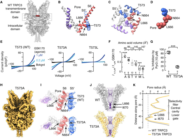 Figure 1
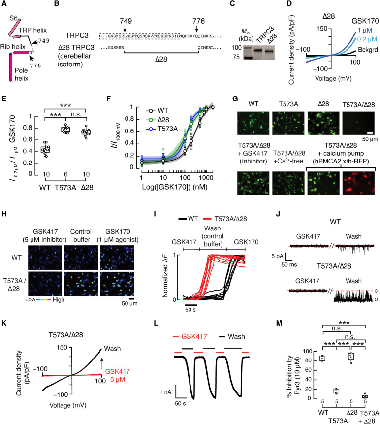 Figure 2
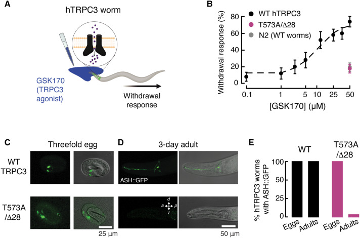 Figure 3
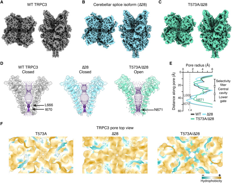 Figure 4
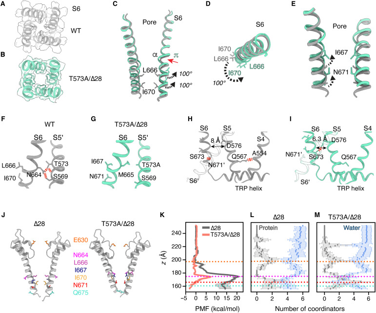 Figure 5
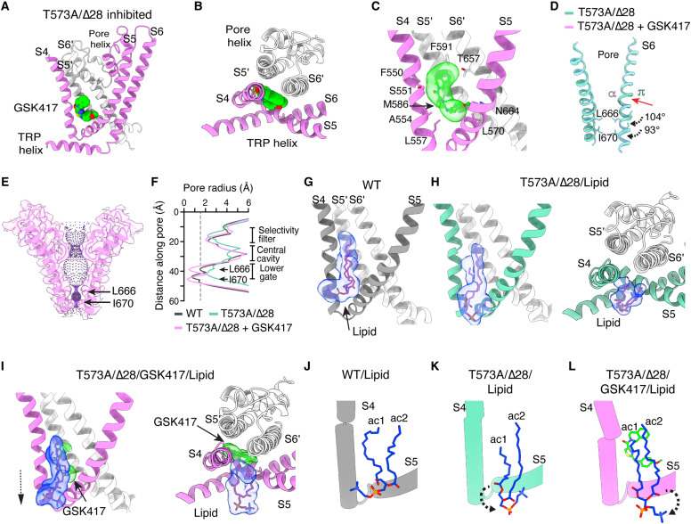 Figure 6
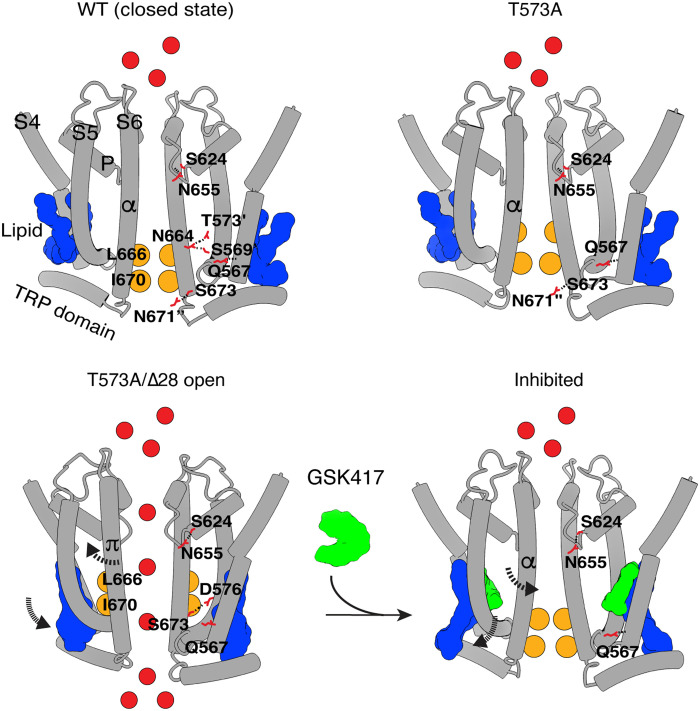 Figure 7
Peer Reviews
No public reviews on file for this paper yet. If you reviewed it on a platform where reviews are public (OpenReview, ICLR, NeurIPS, ICML), you can paste yours below so the community can read it here.
Videos
No videos yet. Explain this paper in a talk, walkthrough, or lecture? Add one.
Taxonomy
TopicsIon Channels and Receptors · Genetic Neurodegenerative Diseases · Hearing, Cochlea, Tinnitus, Genetics