miniMTI: minimal multiplex tissue imaging enhances biomarker expression prediction from histology
Zachary Sims, Sandhya Govindarajan, Kaoutar Ait-Ahmad, Cigdem Ak, Marigold Kuykendall, Gordon B. Mills, Sebnem Eksi, Young Hwan Chang

TL;DR
miniMTI combines H&E histology with a few measured markers to accurately predict large multiplex tissue imaging data, improving biomarker prediction and preserving biological and clinical information.
Contribution
miniMTI introduces a minimal set of molecular markers combined with H&E to reconstruct full multiplex imaging data, enabling scalable and interpretable virtual staining.
Findings
miniMTI reduces a 40-marker MTI assay to H&E plus three measured markers while preserving cellular phenotypes and spatial architecture.
The method accurately recovers withheld markers and disease-associated molecular programs like Gleason grade-linked signatures.
miniMTI outperforms H&E-only virtual staining by integrating sparse molecular data with histology context.
Abstract
Virtual multiplexing from routine histology has advanced rapidly, yet morphology alone provides limited access to molecular state, imposing an intrinsic ceiling on H&E-only inference. Here, we introduce miniMTI, a molecularly anchored virtual staining framework that determines the minimal set of experimentally measured markers required, alongside H&E, to accurately reconstruct large multiplex tissue imaging (MTI) panels while preserving biologically and clinically relevant information. miniMTI learns from paired same-section H&E–MTI data using a unified multimodal generative model that can condition on arbitrary combinations of measured marker channels, coupled with an iterative panel selection strategy to rank informative molecular anchors. Across colorectal and prostate cancer cohorts spanning two MTI platforms and over 40 million cells, miniMTI reduces a 40-marker MTI assay to H&E…
Genes, proteins, chemicals, diseases, species, mutations and cell lines named across the full text — each resolved to its canonical identifier and authoritative record.
Click any figure to enlarge with its caption.
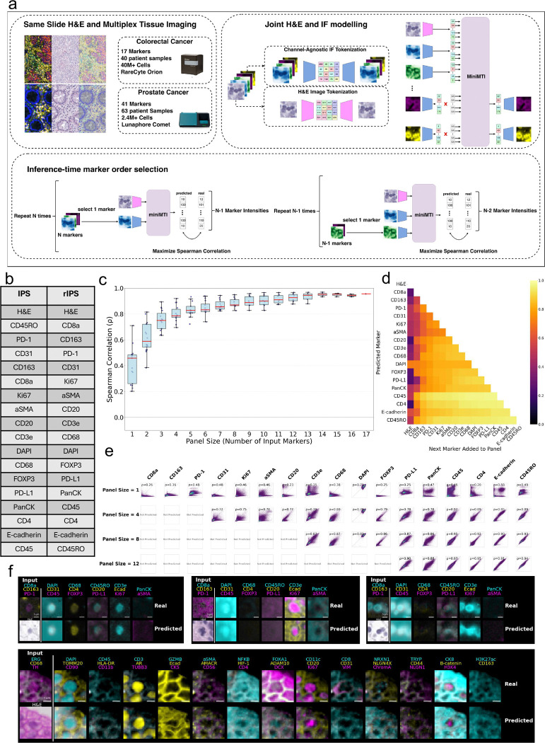 Figure 1
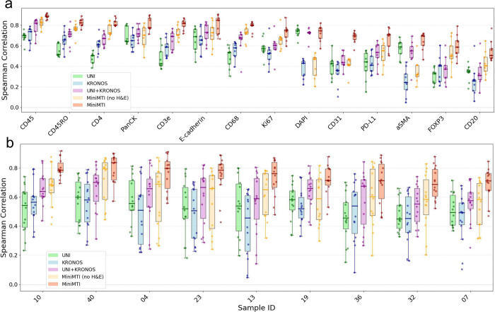 Figure 2
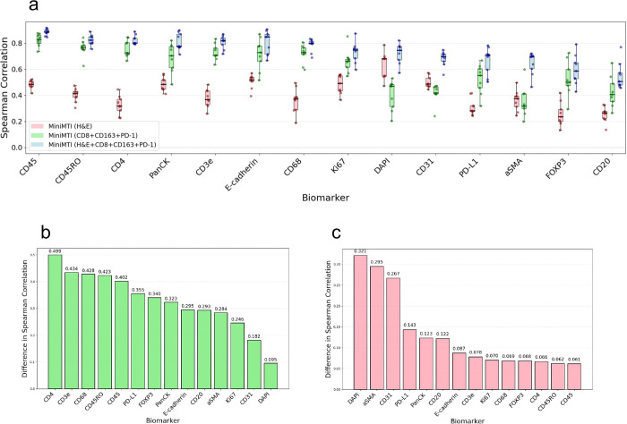 Figure 3
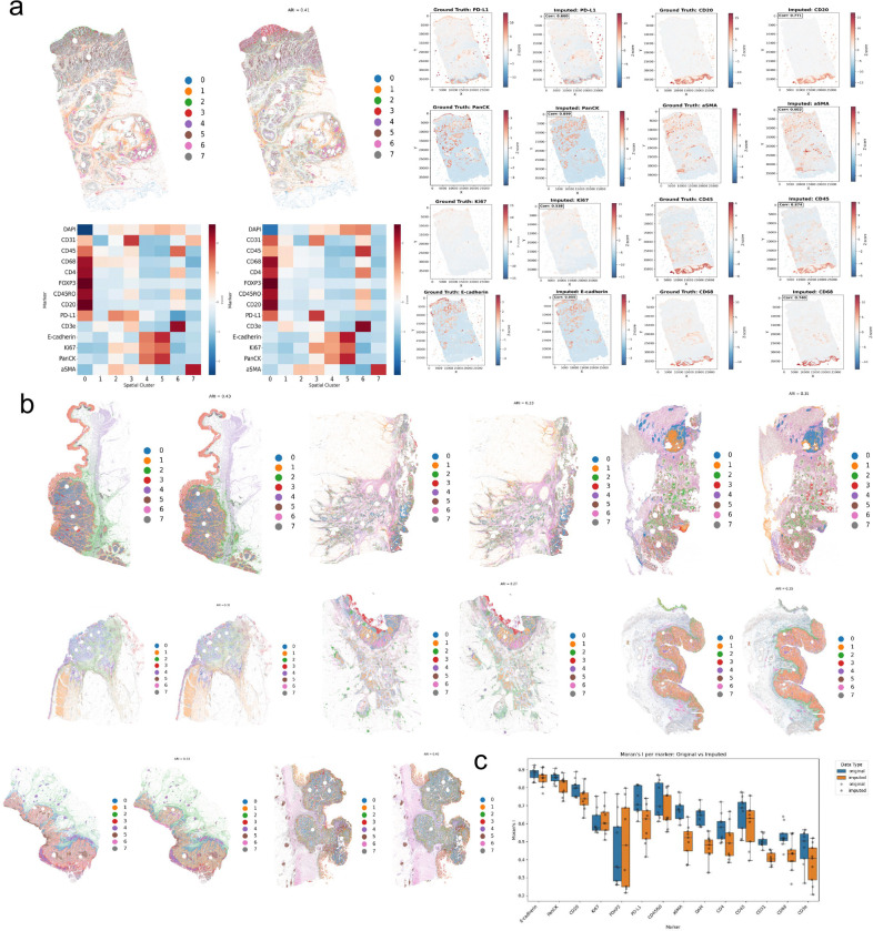 Figure 4
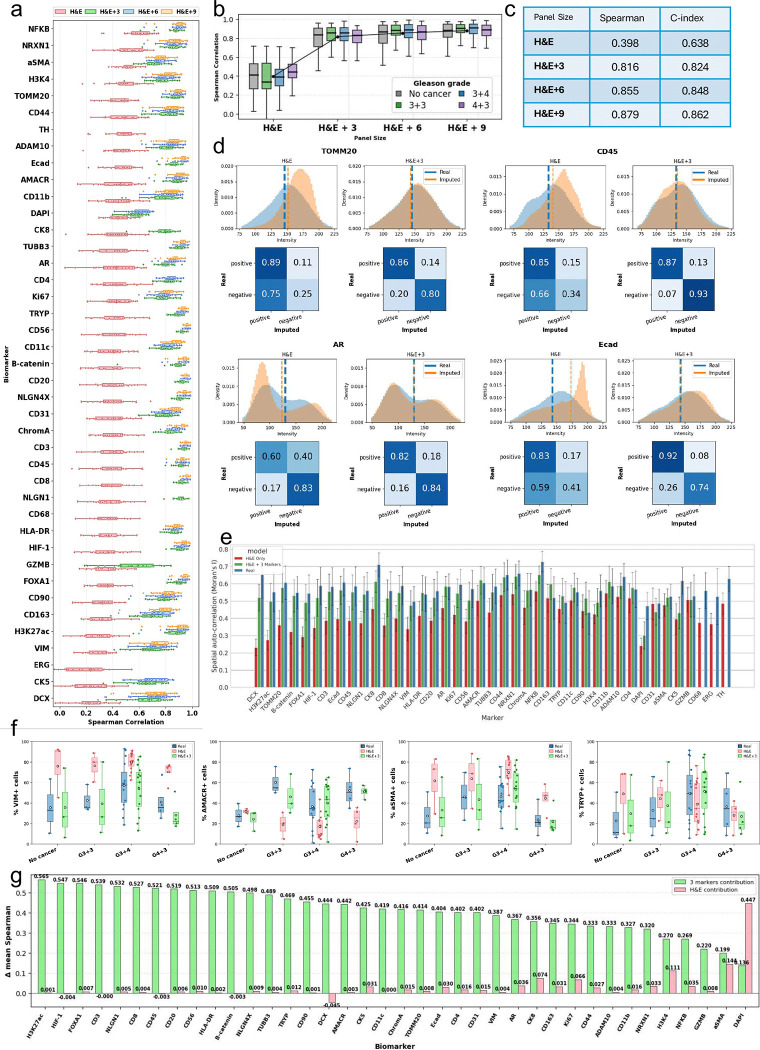 Figure 5
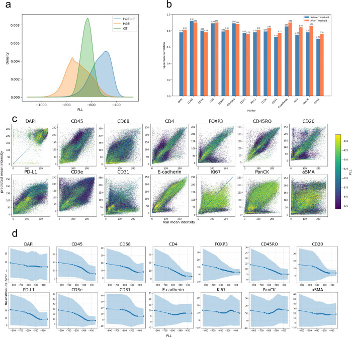 Figure 6
Peer Reviews
No public reviews on file for this paper yet. If you reviewed it on a platform where reviews are public (OpenReview, ICLR, NeurIPS, ICML), you can paste yours below so the community can read it here.
Videos
No videos yet. Explain this paper in a talk, walkthrough, or lecture? Add one.
Taxonomy
TopicsSingle-cell and spatial transcriptomics · Cell Image Analysis Techniques · Cancer Genomics and Diagnostics