Microcomputed tomography analysis of root canal morfology of hypomineralized permanent molars compared with healthy teeth: unveiling hidden anatomical variations
Hazal Karslıoğlu, Didem Sakaryalı Uyar, Mert Ocak, Hakan Hamdi Çelik

TL;DR
This study uses microcomputed tomography to compare the root canal structure of teeth with a developmental enamel defect to healthy teeth, revealing differences in minor apical foramina and cervical width.
Contribution
The study provides new insights into root canal morphology variations in hypomineralized molars using µCT, which is critical for endodontic treatment planning.
Findings
Hypomineralized teeth had significantly more minor apical foramina in the middle third of the root compared to healthy teeth.
The cervical width was significantly greater in hypomineralized molars than in healthy molars.
No significant differences were found in cervical thickness, canal area, or isthmus distribution between the two groups.
Abstract
Molar–incisor hypomineralization (MIH) is a prevalent developmental enamel defect that frequently affects permanent molars and is associated with increased dentin permeability, hypersensitivity, and a higher risk of pulpal inflammation. These clinical features may necessitate endodontic treatment at an early age; however, information regarding potential variations in root canal morphology in MIH-affected molars remains limited. This study aimed to investigate the root canal morphology of hypomineralized permanent molars and compare it with that of healthy molars via microcomputed tomography (µCT). A total of sixty extracted permanent molars were included in this study, comprising thirty hypomineralized teeth and thirty healthy control teeth. All samples were scanned using a SkyScan 1172 µCT system. Root canal configurations were classified according to the Sert and Bayırlı…
Genes, proteins, chemicals, diseases, species, mutations and cell lines named across the full text — each resolved to its canonical identifier and authoritative record.
Click any figure to enlarge with its caption.
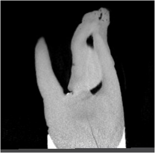 Figure 1
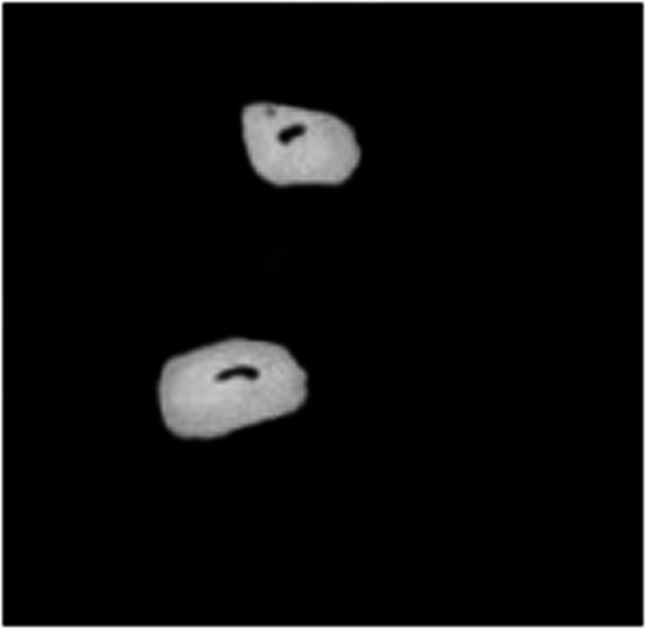 Figure 2
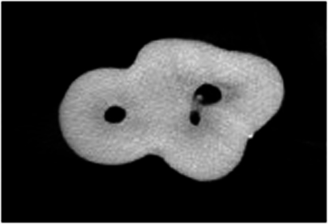 Figure 3
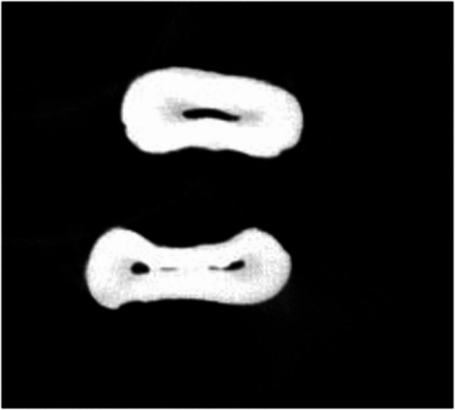 Figure 4
Peer Reviews
No public reviews on file for this paper yet. If you reviewed it on a platform where reviews are public (OpenReview, ICLR, NeurIPS, ICML), you can paste yours below so the community can read it here.
Videos
No videos yet. Explain this paper in a talk, walkthrough, or lecture? Add one.
Taxonomy
TopicsBone and Dental Protein Studies · Endodontics and Root Canal Treatments · dental development and anomalies