Variance reduction with synaptic density imaging in Parkinson’s disease using direct-4D PET image reconstruction
Paul Gravel, Jean-Dominique Gallezot, Kathryn Fontaine, David Matuskey, Richard E Carson

TL;DR
This study shows that direct-4D PET image reconstruction reduces noise and variability in Parkinson’s disease scans compared to traditional methods.
Contribution
First demonstration that direct-4D reconstruction lowers variability and bias in both within- and between-subject analyses for Parkinson’s disease.
Findings
DR reduced within-subject variability for K1 and VT compared to IR across all count levels.
Between-subject variability was significantly lower with DR than IR for K1 and VT.
DR at 5% count level matched IR at 20% count level in variability for K1 and VT.
Abstract
Objective. Direct reconstruction (DR) of parametric images from dynamic positron emission tomography data has been shown to provide substantial noise reduction compared to the conventional indirect reconstruction (IR) approach where frames are first reconstructed and then voxel time-activity curves are fitted to a kinetic model. The main goal was to compare DR and IR, on both within-subject and between-subject variability. Approach. This work evaluated the Parametric motion-compensation OSEM List-mode algorithm for resolution-recovery-1T DR method, using multiple scans of Parkinson’s disease patients with [11C]UCB-J, a radioligand for synaptic vesicle glycoprotein 2A (SV2A), a marker for synaptic density. This was achieved by comparing K1, k2, and VT parametric images estimated, at full- and lower-count levels (20%, 10%, and 5%), between DR and IR. Main Results. DR delivered…
Genes, proteins, chemicals, diseases, species, mutations and cell lines named across the full text — each resolved to its canonical identifier and authoritative record.
Click any figure to enlarge with its caption.
 Figure 1
 Figure 2
 Figure 3
 Figure 4
 Figure 5
 Figure 6
 Figure 7
 Figure 8
 Figure 9
 Figure 10
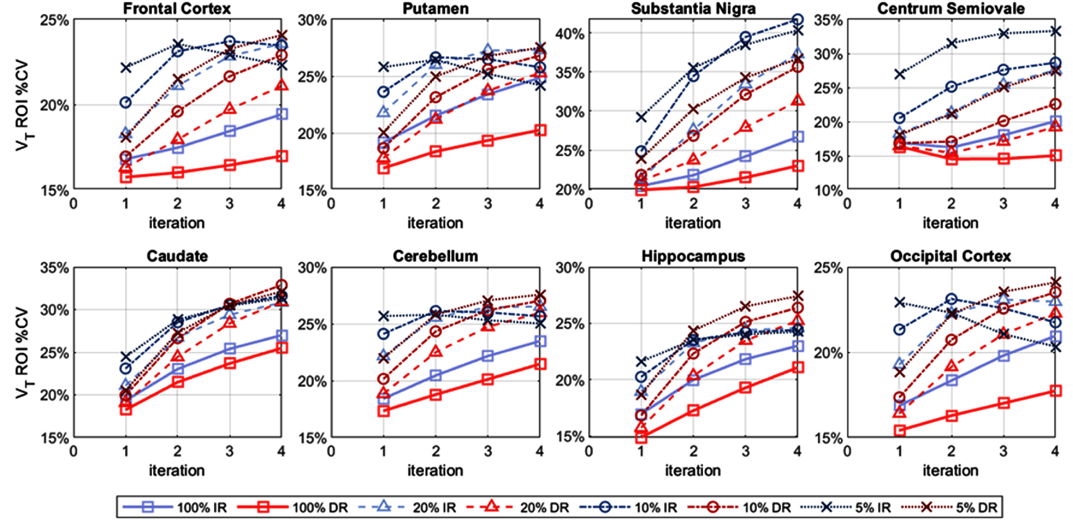 Figure 11
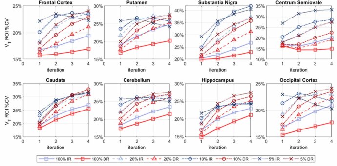 Figure 12
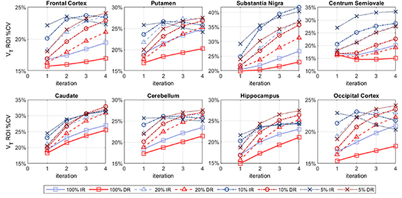 Figure 13
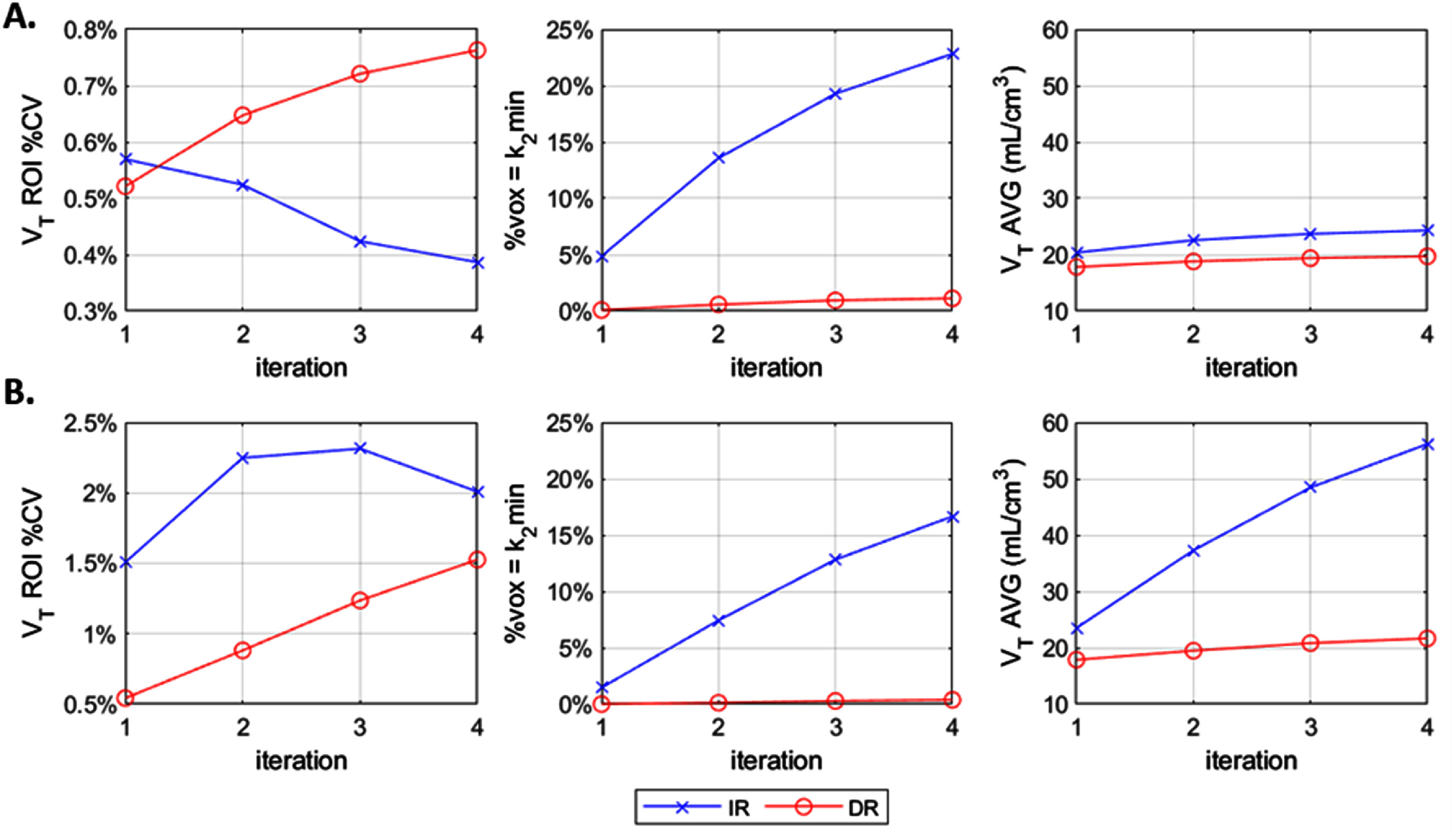 Figure 14
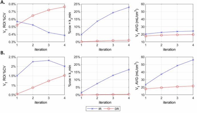 Figure 15
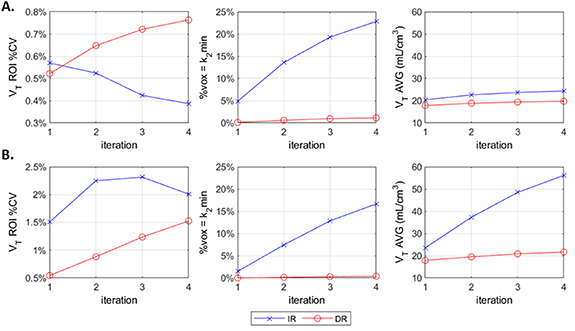 Figure 16
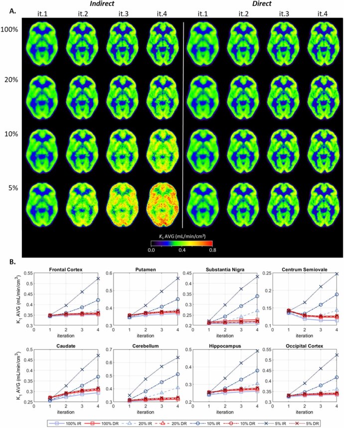 Figure 17
 Figure 18
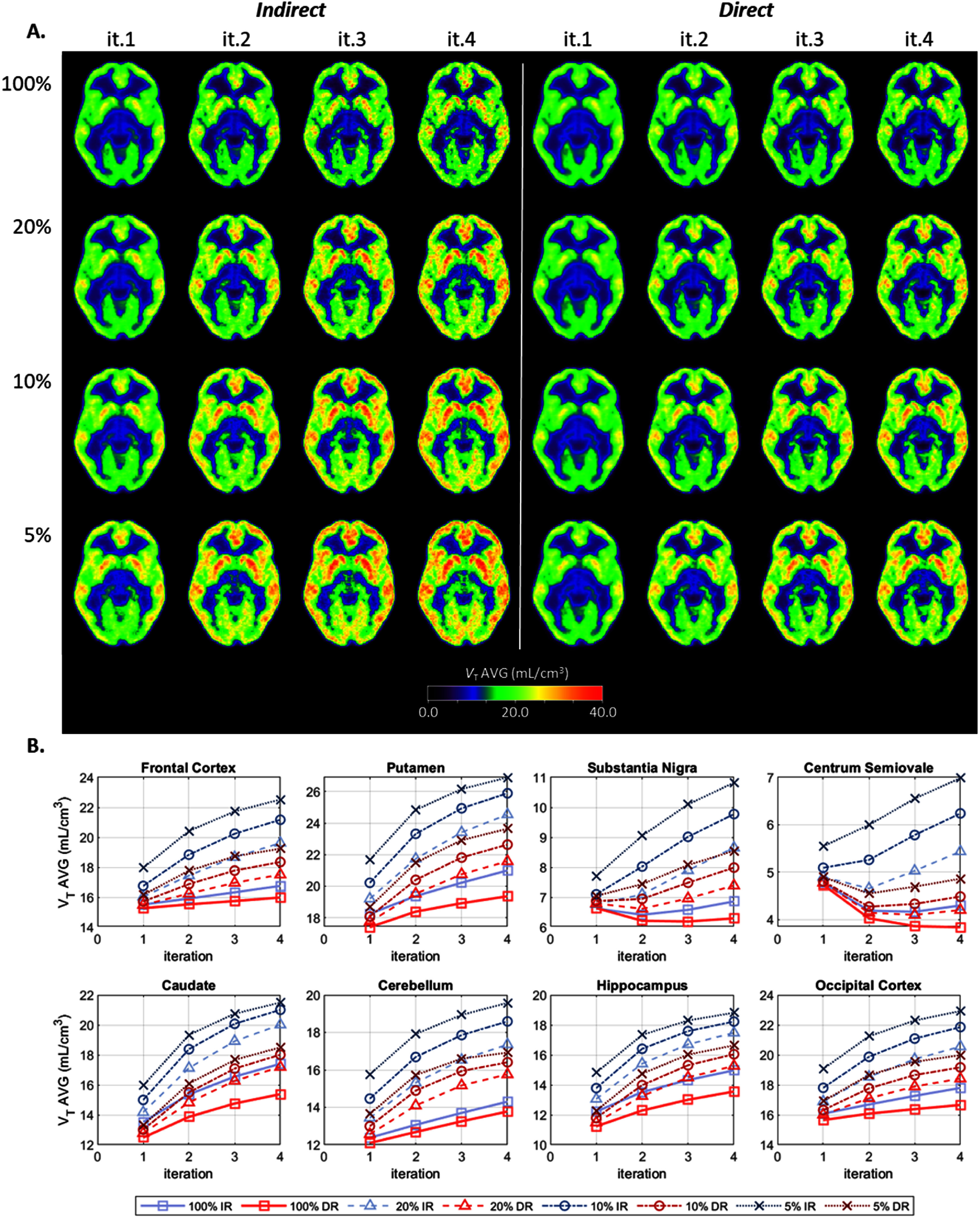 Figure 19
 Figure 20
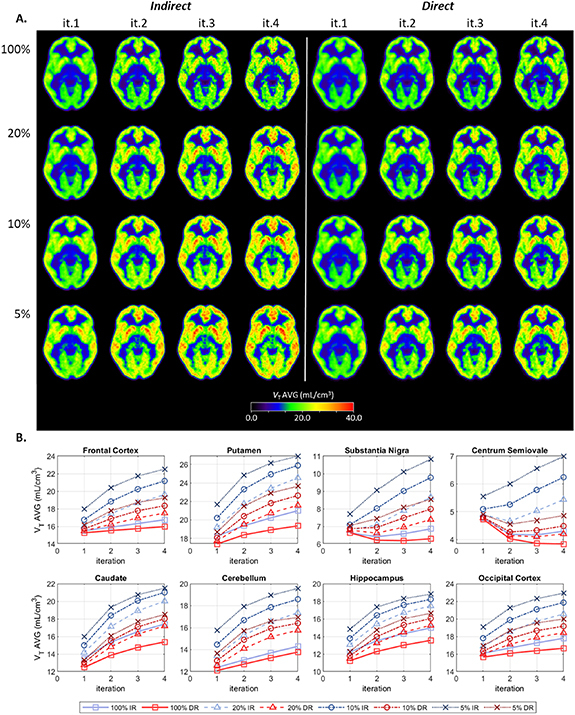 Figure 21
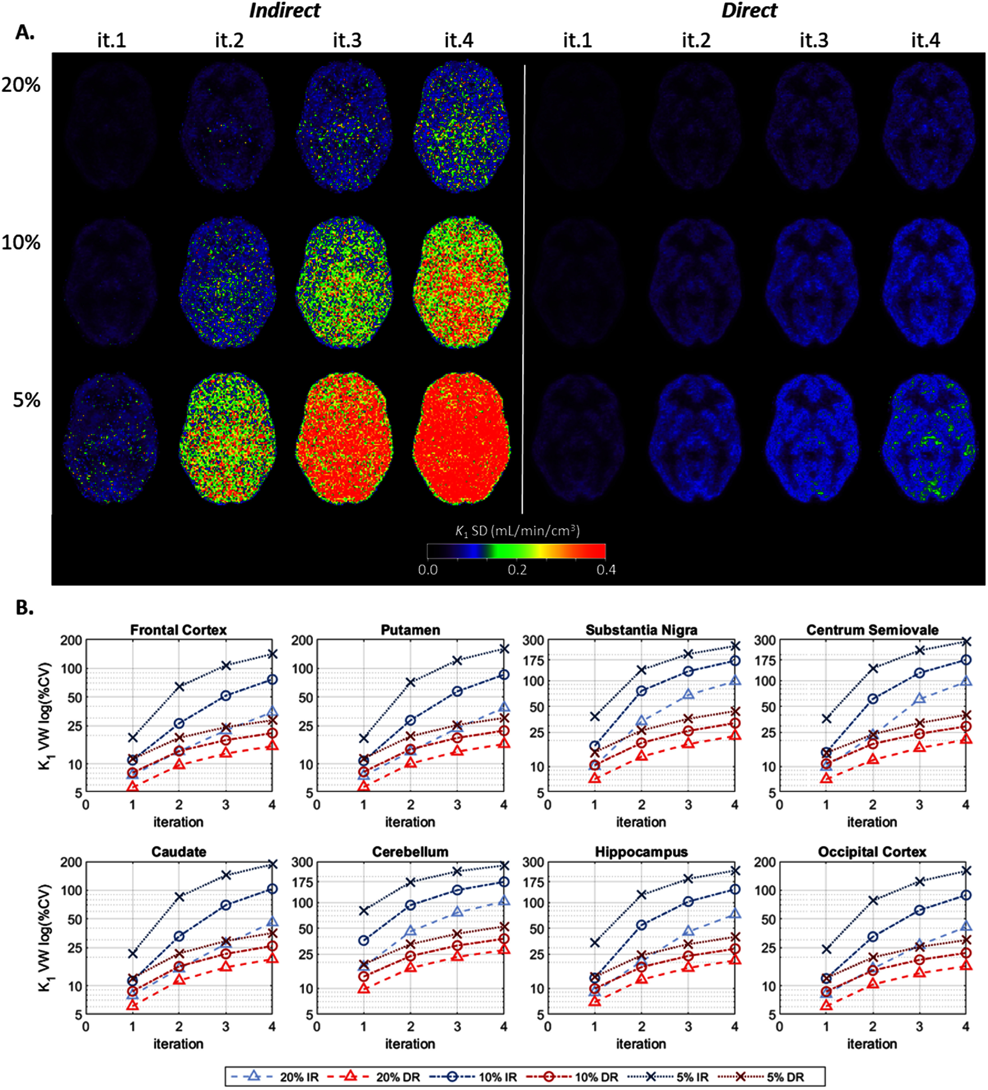 Figure 22
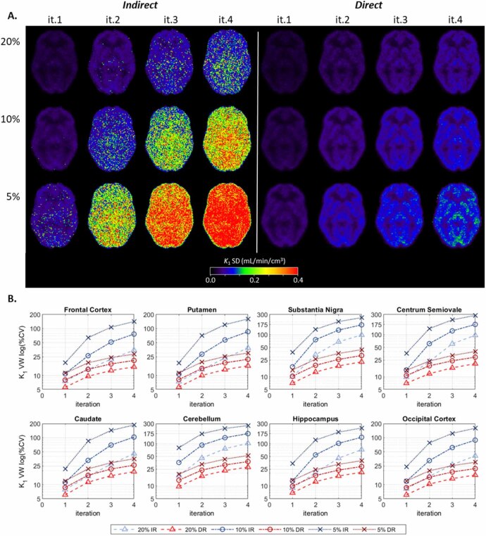 Figure 23
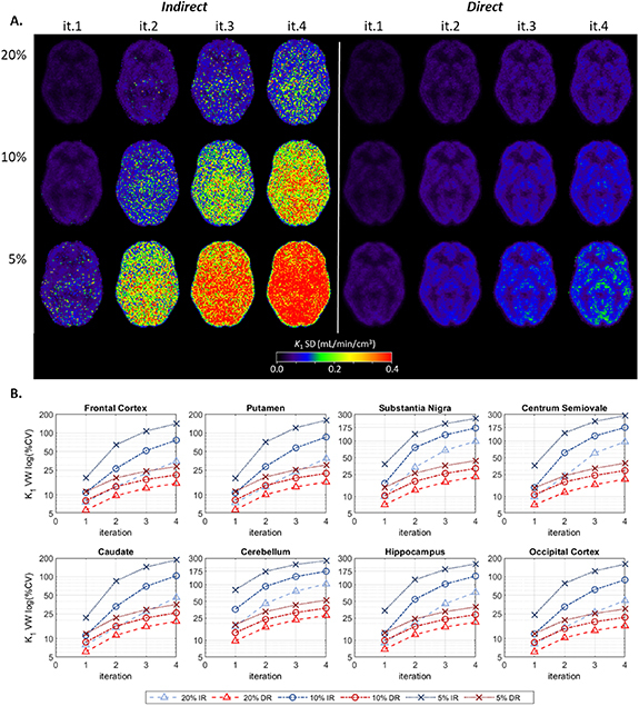 Figure 24
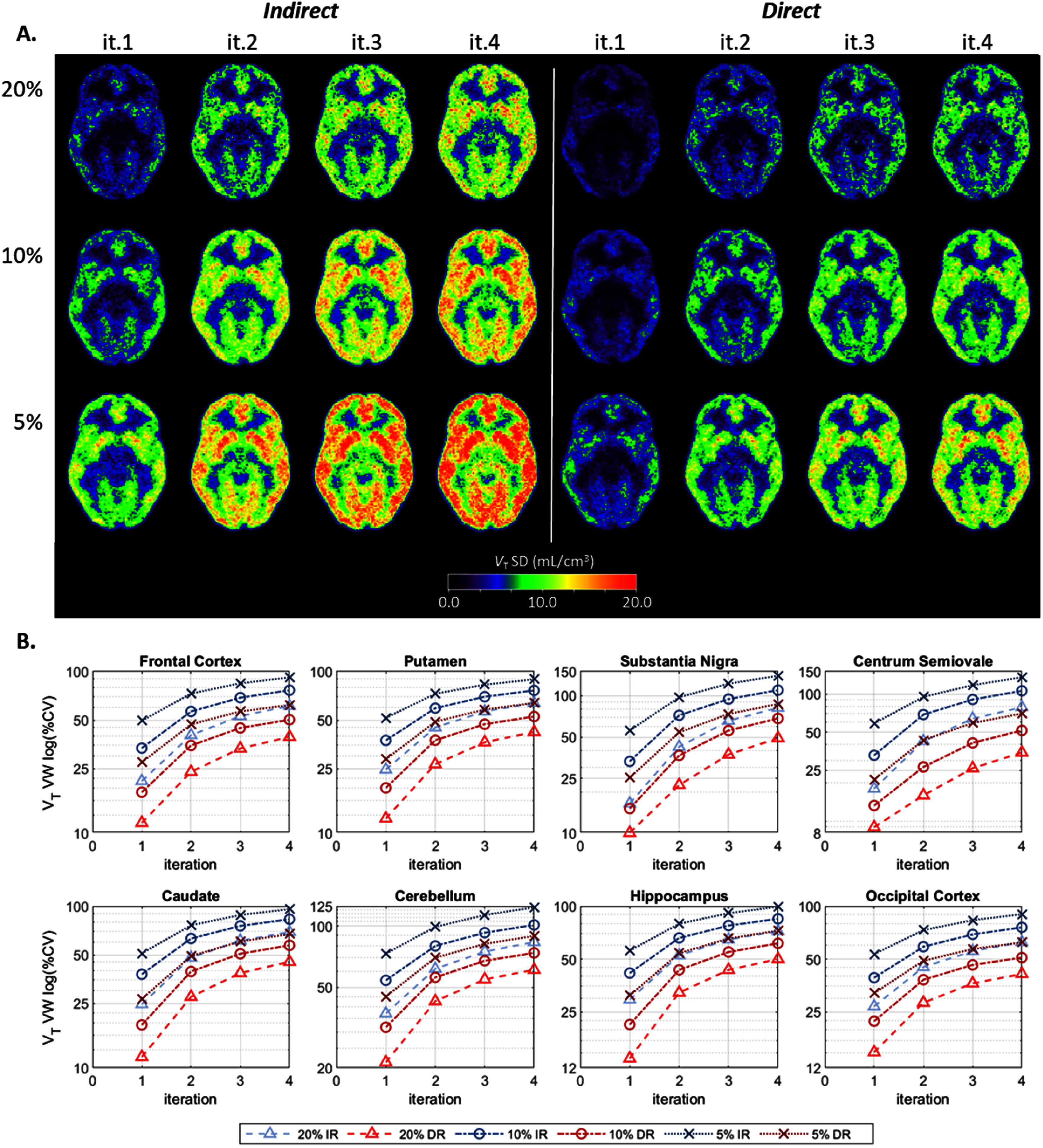 Figure 25
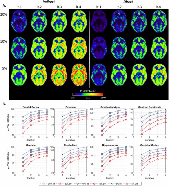 Figure 26
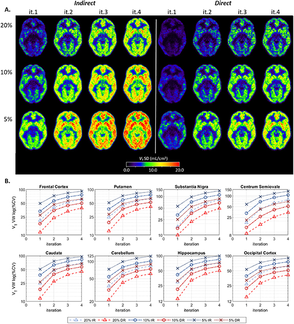 Figure 27
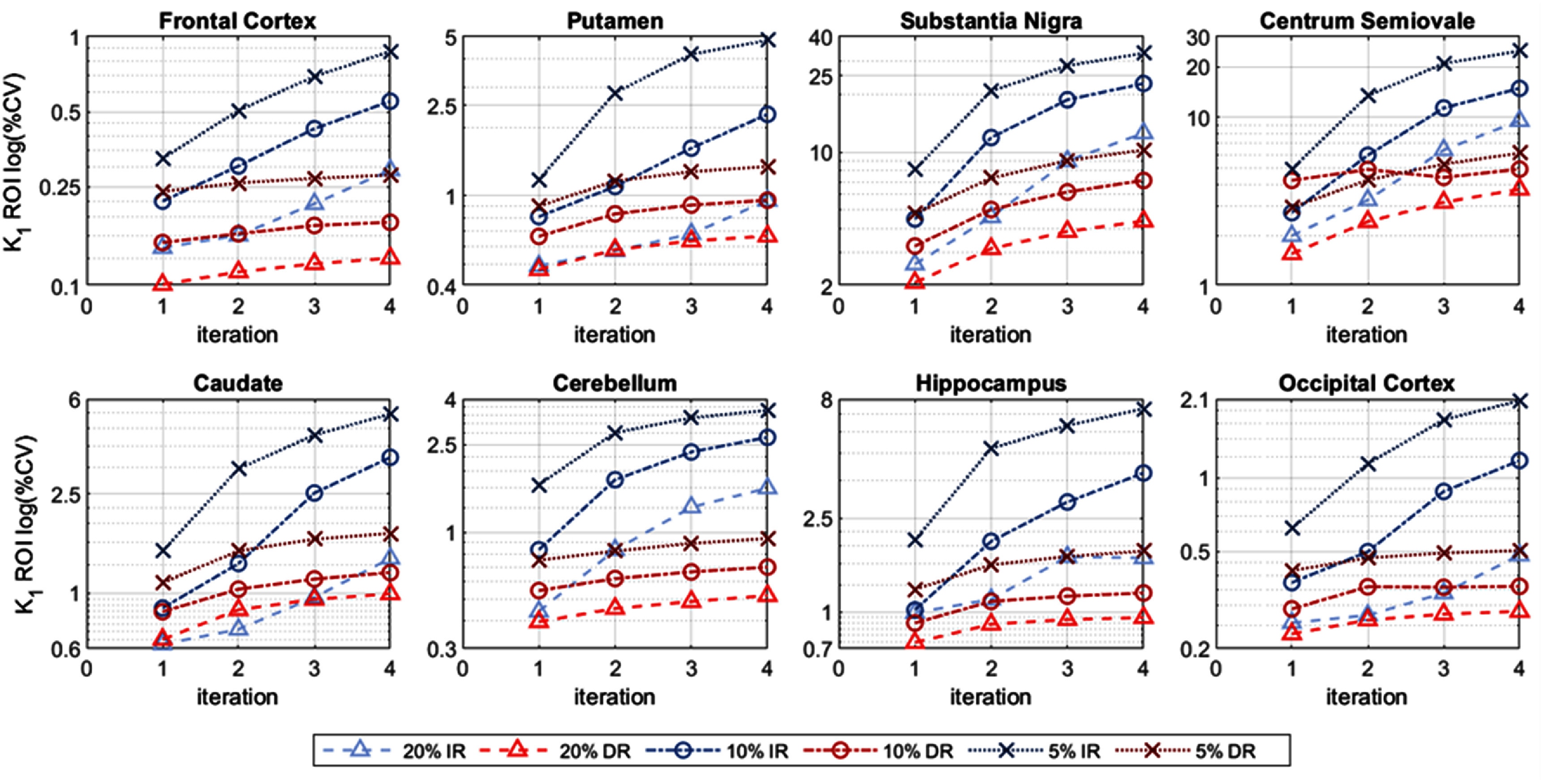 Figure 28
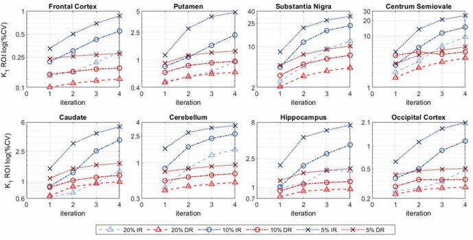 Figure 29
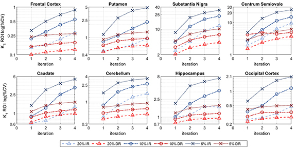 Figure 30
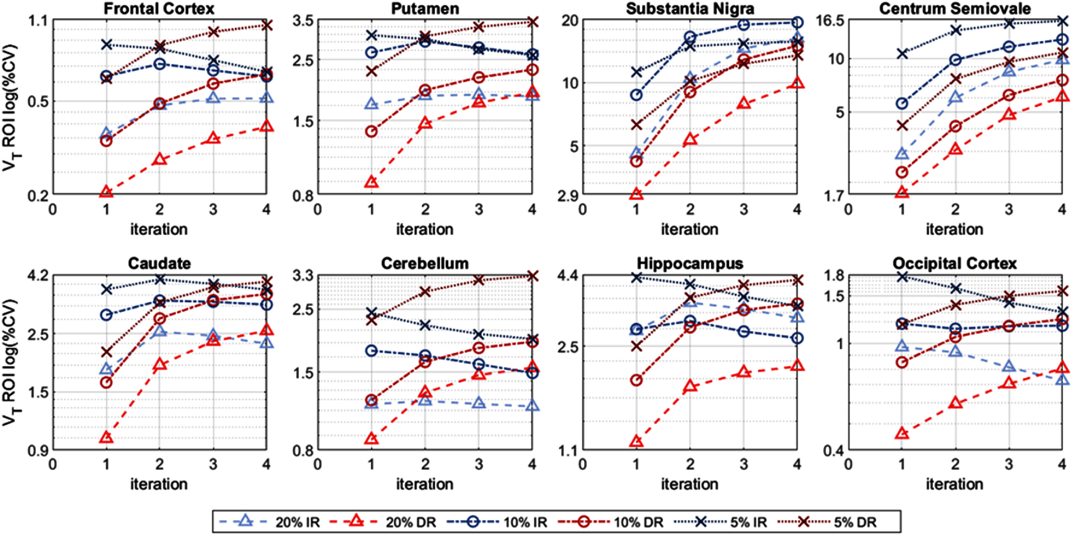 Figure 31
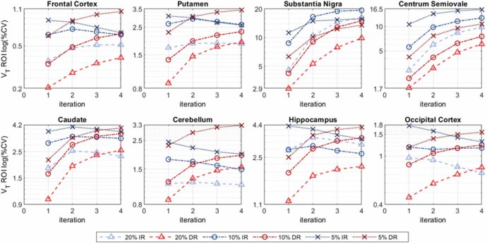 Figure 32
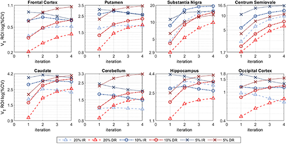 Figure 33
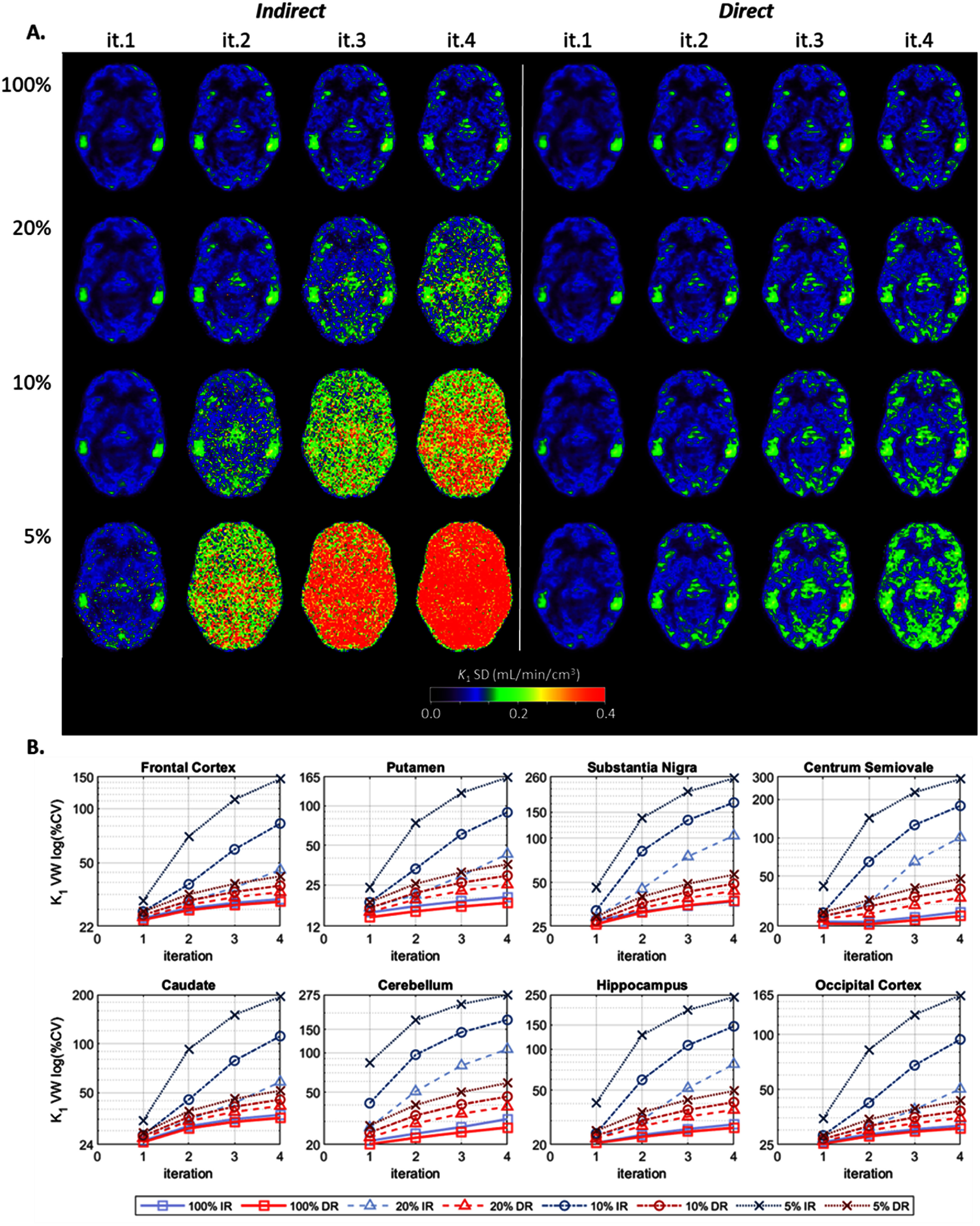 Figure 34
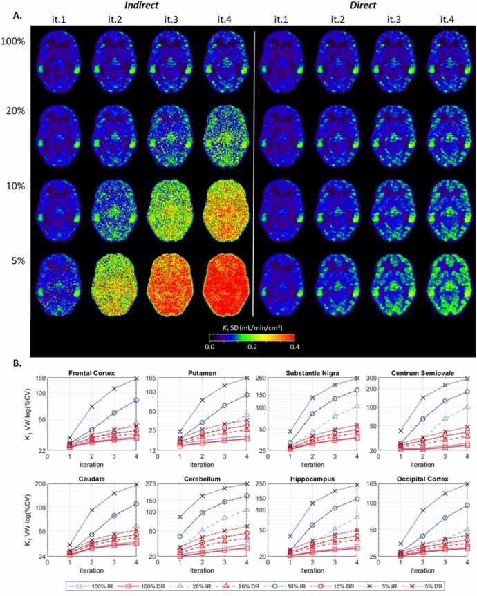 Figure 35
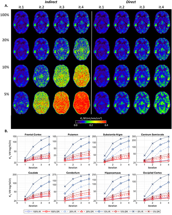 Figure 36
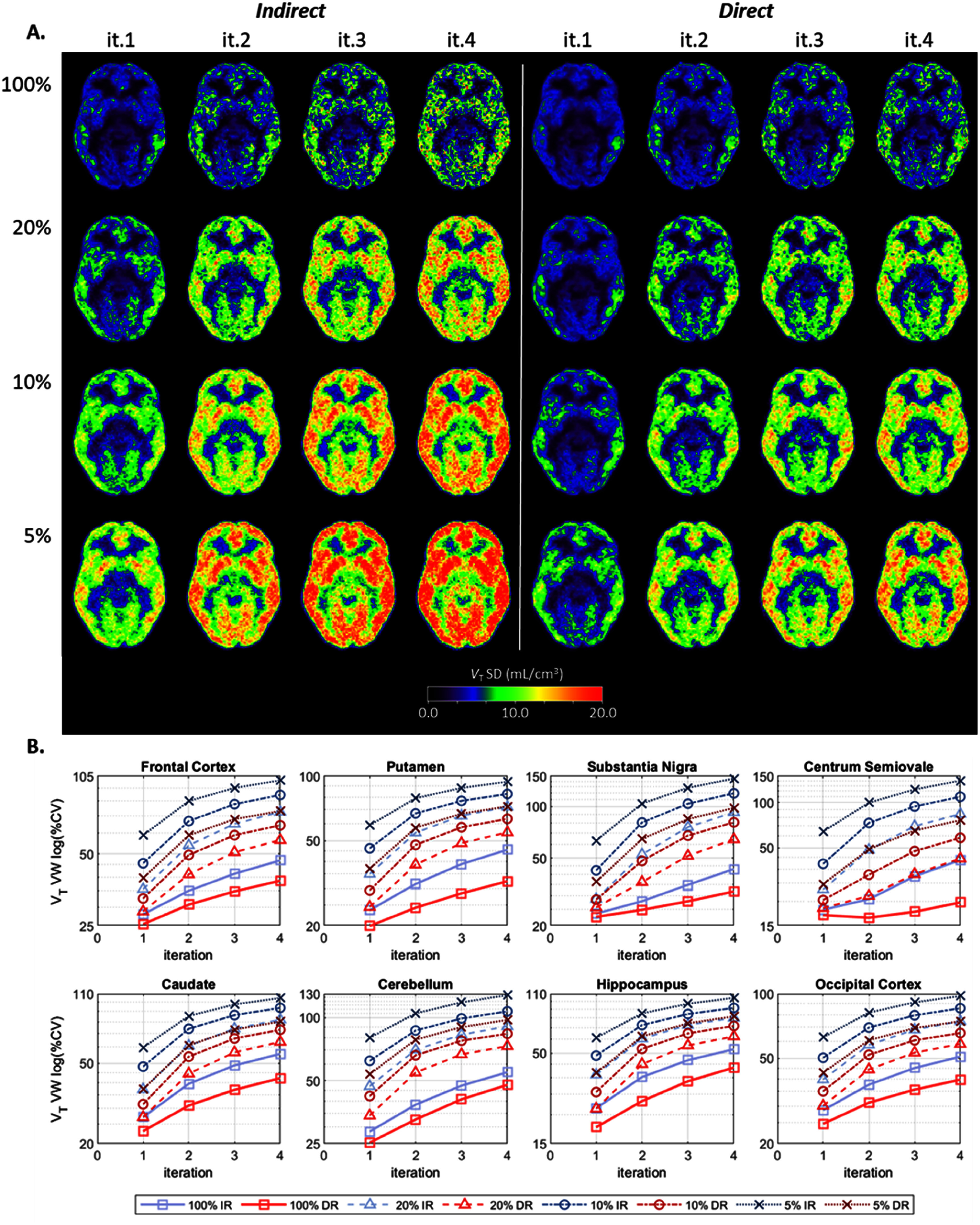 Figure 37
 Figure 38
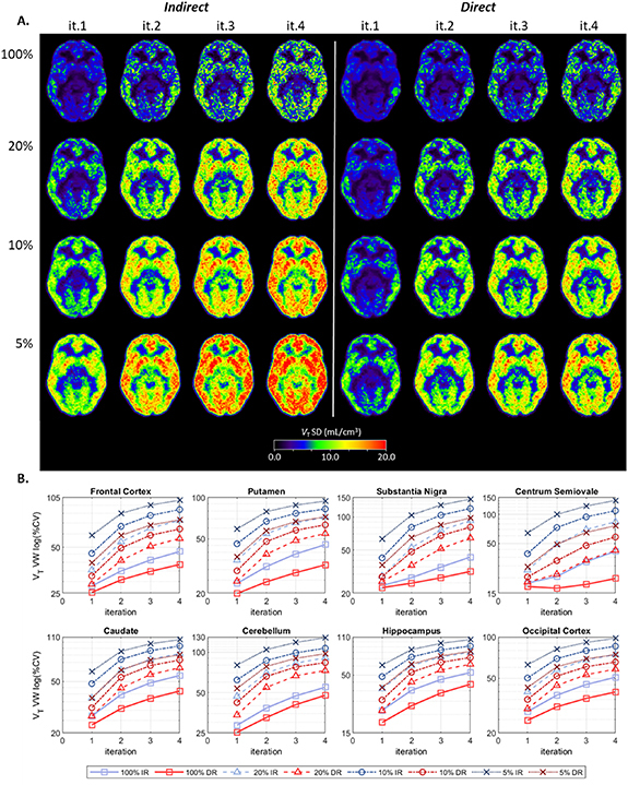 Figure 39
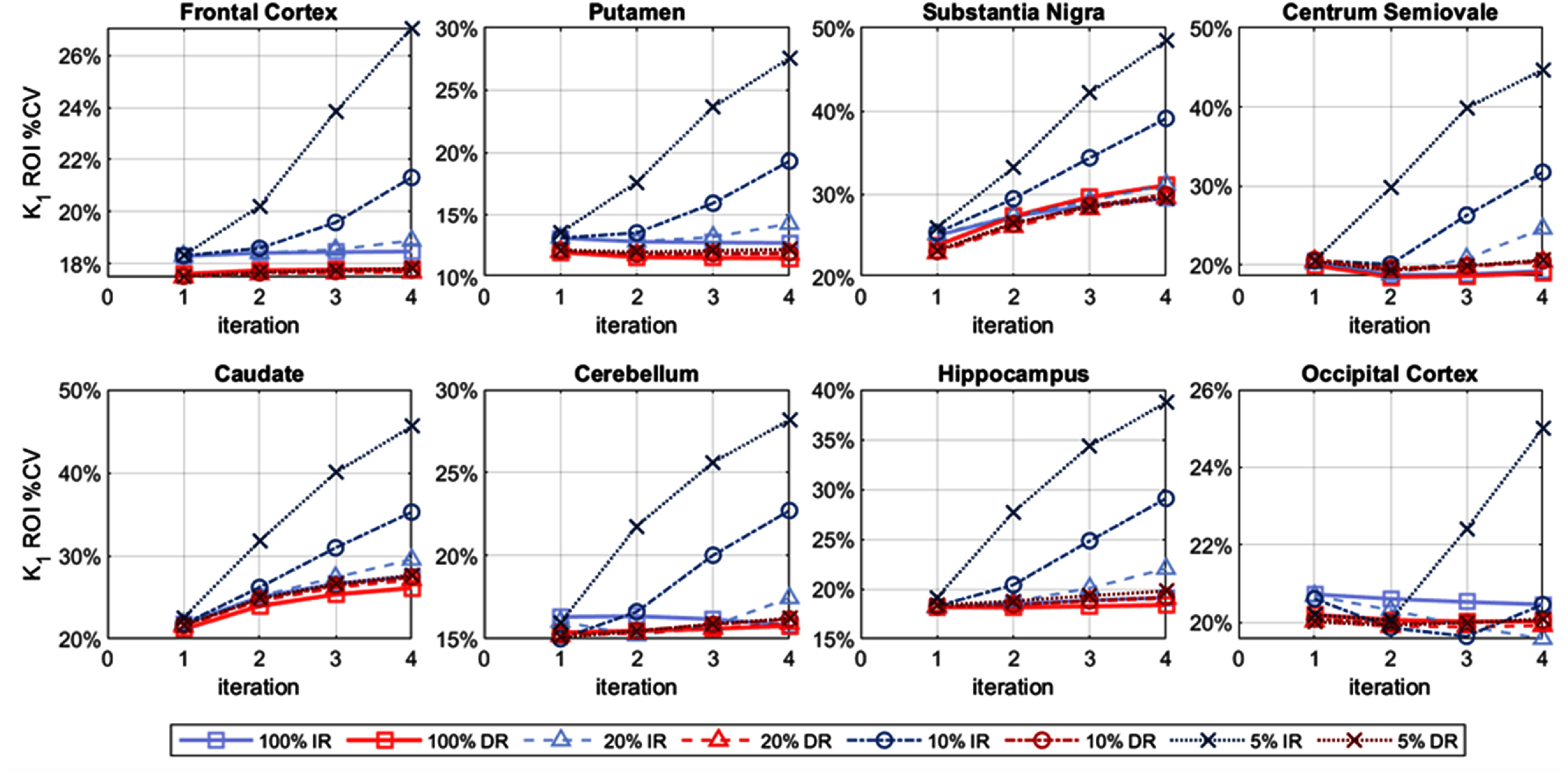 Figure 40
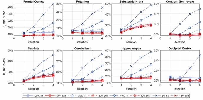 Figure 41
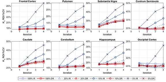 Figure 42
 Figure 43
 Figure 44
 Figure 45
 Figure 46
 Figure 47
 Figure 48
 Figure 49
 Figure 50
Peer Reviews
No public reviews on file for this paper yet. If you reviewed it on a platform where reviews are public (OpenReview, ICLR, NeurIPS, ICML), you can paste yours below so the community can read it here.
Videos
No videos yet. Explain this paper in a talk, walkthrough, or lecture? Add one.
Taxonomy
TopicsParkinson's Disease Mechanisms and Treatments · Medical Imaging Techniques and Applications · Neurological disorders and treatments