Predicting progression to proliferative diabetic retinopathy using automated versus manual quantification of retinal haemorrhages
Aditya Verma, Muneeswar G. Nittala, Roxan Mansoori Dara, Marius Facktor, Chaithanya A. Ramachandra, Malavika Bhaskaranand, Sandeep Bhat, Kaushal Solanki, Chaitra Jayadev, Swetha B. Velaga, Gavin Robertson, Bradley Yates, Rajiv Raman, SriniVas R. Sadda

TL;DR
This study compares automated and manual methods for measuring retinal haemorrhages in diabetic retinopathy and finds that automated detection can predict progression to a severe form of the disease.
Contribution
The study introduces automated quantification of retinal haemorrhages as a predictive tool for progression to proliferative diabetic retinopathy.
Findings
Automated measurements of retinal haemorrhages were significantly correlated with manual grading.
The distance of haemorrhages from the optic nerve was a significant risk factor for progression to proliferative diabetic retinopathy.
Automated detection of haemorrhages can predict disease progression despite detecting fewer lesions than manual grading.
Abstract
To compare automated and manual quantification of retinal haemorrhages in eyes with diabetic retinopathy (DR) and to analyse the risk of progression to proliferative DR (PDR). Retinal haemorrhages on ultra-widefield (UWF) pseudocolor images in eyes with non-proliferative diabetic retinopathy (NPDR) were manually segmented. DR severity was assessed within the seven ETDRS fields at baseline and 1-year follow-up. Lesions were also automatically segmented using EyeRead UWF software (Eyenuk) and the frequency and area of retinal haemorrhages and the average distance of haemorrhages from the optic nerve centre were computed. Manual and automated results were compared and correlated with progression to PDR at one year. Sixty-three eyes with NPDR at baseline were included, of which 29 progressed to PDR over one year. The automated measurements of total haemorrhage frequency, area and the…
Genes, proteins, chemicals, diseases, species, mutations and cell lines named across the full text — each resolved to its canonical identifier and authoritative record.
Click any figure to enlarge with its caption.
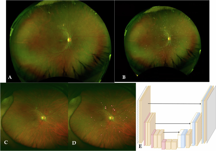 Figure 1
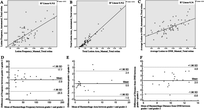 Figure 2
Peer Reviews
No public reviews on file for this paper yet. If you reviewed it on a platform where reviews are public (OpenReview, ICLR, NeurIPS, ICML), you can paste yours below so the community can read it here.
Videos
No videos yet. Explain this paper in a talk, walkthrough, or lecture? Add one.
Taxonomy
TopicsRetinal Diseases and Treatments · Retinal Imaging and Analysis · Retinal and Optic Conditions