Investigation of 3D choroidal components in myopic populations using ultra-widefield OCTA
Tengbo Rao, Jiarui Yang, Yanfeng Liao, Yi Ding, Yiwen Shi, Huijin Chen, Xuemin Li

TL;DR
This study uses advanced imaging to show how myopia affects the choroidal layers of the eye, revealing changes in blood vessels and tissue.
Contribution
The study introduces the use of ultra-widefield OCTA to analyze choroidal vascular and stromal components in different myopia severity levels.
Findings
Choroidal thickness decreases significantly with increasing myopia severity, especially in the sub-fovea and macular regions.
Choriocapillaris density increases with myopia severity in the macular region, while vascular volume declines.
Stromal volume changes are most notable between moderate and high myopia, and axial length correlates with stromal volume.
Abstract
This study investigates changes in vascular and stromal components of choroidal vascular layers using ultra-widefield optical coherence tomography angiography (OCTA). A cross-sectional study included 147 participants with varying myopia degrees, categorised as low myopia (70 eyes), moderate myopia (104 eyes), and high myopia (110 eyes) based on refractive status. The TowardPi Wide-field OCTA measured choroidal parameters, including thickness, choriocapillaris density, vascular volume, and stromal volume. Differences in these parameters were analysed among the three myopia groups. Choroidal thickness decreased with increasing myopia, notably at the sub-fovea (LM vs MM vs HM: 297.56 ± 83.19 μm vs 230.13 ± 77.35 μm vs 190.49 ± 70.24 μm, P < 0.001) and macular region (258.20 ± 67.40 μm vs 215.37 ± 56.07 μm vs 186.01 ± 49.10 μm, P < 0.001). Choriocapillaris density in the macular region…
Genes, proteins, chemicals, diseases, species, mutations and cell lines named across the full text — each resolved to its canonical identifier and authoritative record.
Click any figure to enlarge with its caption.
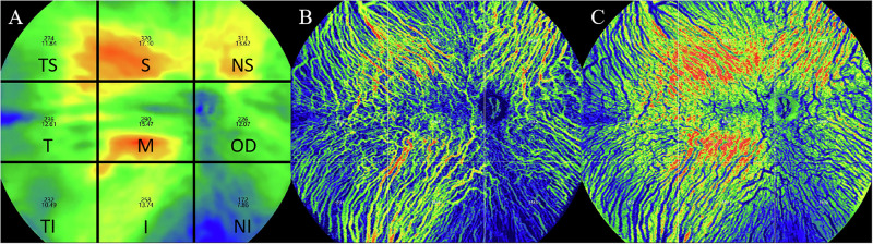 Figure 1
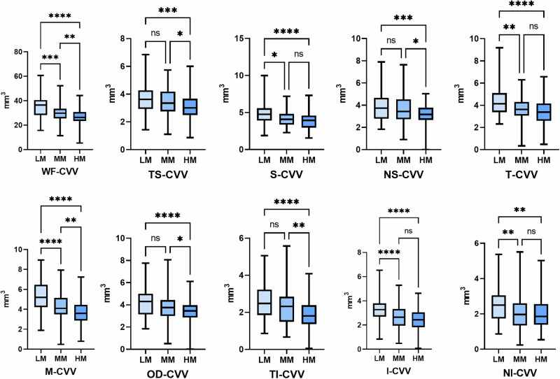 Figure 2
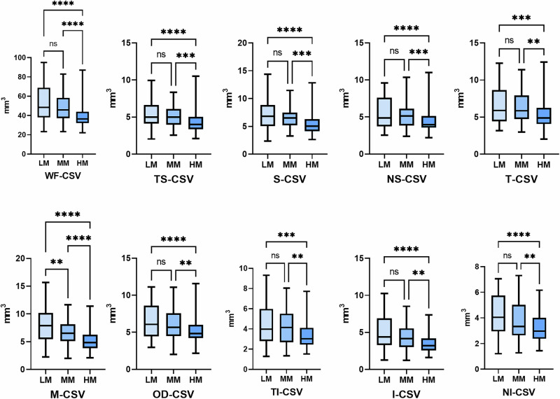 Figure 3
Peer Reviews
No public reviews on file for this paper yet. If you reviewed it on a platform where reviews are public (OpenReview, ICLR, NeurIPS, ICML), you can paste yours below so the community can read it here.
Videos
No videos yet. Explain this paper in a talk, walkthrough, or lecture? Add one.
Taxonomy
TopicsRetinal Diseases and Treatments · Optical Coherence Tomography Applications · Ophthalmology and Visual Impairment Studies